CHAPTER 30 Clinical history, examination, arthroscopic findings, and treatment of multidirectional instability
 Instability in more than one direction must occur in combination with symptomatic signs of hyperlaxity in order to obtain an accurate diagnosis for multidirectional instability (MDI).


Introduction
In 1980 Neer et al developed the concept of multidirectional glenohumeral instability and emphasized the importance of distinguishing this disorder from routine unidirectional instability. In order to address the underlying capsular redundancy, they introduced a one-incision, inferior capsular shift procedure designed to retensioning both the anterior and posterior portions of the capsuloligament complex.1 Understanding and classification of multidirectional instability (MDI) has continuously evolved since then. The original description of Matsen et al still lives in many orthopaedic textbooks, thanks to its easy to remember mnemonics, AMBRI (atraumatic multidirectional bilateral rehabilitation inferior capsular shift) and TUBS (traumatic unidirectional Bankart lesion surgery).2 With the evolution of imaging techniques and improved clinical understanding, there is consensus that the etiology of MDI is multifactorial, and the previous mnemonics may oversimplify the classification of shoulder instability. Although a sole traumatic event is a rare cause for glenohumeral instability in more than one direction, it may represent the first presentation of a ligamentously lax patient who will go on to develop multidirectional instability. In an effort to focus on underlying patient factors Gerber et al used their classification to stress the need to differentiate instability with and without coexisting hyperlaxity.3
Hyperlaxity, congenital or acquired, is multidirectional but not pathologic. Single or repetitive stresses on a hyperlax shoulder may lead to severe, symptomatic shoulder instability in a patient with underlying hyperlaxity. Even minor stresses may lead to the onset of chronic symptoms without the classic large traumatic event associated with unidirectional instability. Neurologic disorders such as suprascapular nerve entrapment or spinal root stenosis may account for further rare causes of global shoulder instability. It is critical to identify the small cohort of patients who voluntarily dislocate because of controlled abnormal muscle firing patterns. Their symptoms of multidirectional instability originate from abnormal muscle patterning as described and classified in the Stanmore triangle by Lewis et al (polar group III, nonstructural).4 These voluntary dislocators must be carefully separated from other patients who experience dislocations every time the shoulder is brought through a certain position or range of motion. Takwale et al described the latter as involuntary positional instability.5 True voluntary dislocators are poor candidates for any surgical intervention. The majority of patients with multidirectional instability can be managed successfully with a program of physiotherapy, so nonoperative measures should be employed for almost all patients after an initial evaluation.
Clinical history
Identification of the onset of symptoms is important. Rarely, the onset of symptoms may be associated with a clear traumatic event, but more frequently, repetitive stress or a minor injury may be more easily identified as causative factors. For patients who report multiple hospital admissions for shoulder relocation, voluntary dislocation must be excluded from the diagnosis if further treatment is to be successful. In this context, the value of a thorough history and physical examination cannot be overemphasized.
Physical examination
Reproduction of symptoms is crucial for an accurate diagnosis. Before focusing on laxity and instability assessment, the physical examination is started with a general evaluation of the entire shoulder girdle and cervical spine. Active and passive motion testing may present increased range of motion and often offers signs of the directions of the instability. Muscle patterning disorders, as described by Lewis et al,4 are explored by simple visual inspection from the front and the back.
Tests for laxity
Generalized joint laxity may be confirmed by looking for hyperextension of knee and elbow or the metacarpophalangeal joints. Tests for laxity of the shoulder are performed on the asymptomatic extremity first. Walch et al showed that the finding of passive external rotation of more than 90 degrees with the arm in an adducted position is a significant sign for hyperlaxity.6 The sulcus test evaluates inferior laxity. If a sulcus created anteriorly under the acromion is not changed with the addition of external rotation, hyperlaxity may be present. The likelihood of shoulder instability increases to a ratio of 6:1 when there is a sulcus sign of 2 cm or more7 (Fig. 30-1). The sulcus test in slight external rotation of the arm evaluates the integrity of the rotator interval, the area of the shoulder capsule between the tendons of the supraspinatus and subscapularis and the coracoid base. Harryman et al showed a significant increase in humeral head translation after sectioning of the rotator interval.8 The anterior and posterior drawer test, performed on the seated patient, is graded according to Hawkins (Grades 1 to 4) and may present increased translation of the humeral head.9 The hyperabduction test of Gagey is helpful to confirm inferior hyperlaxity.10 This maneuver is positive when passive abduction of the arm with a stabilized scapula is 105 degrees or more (Fig. 30-2). With the patient in the supine or lateral decubitus position the load-and-shift test assesses anterior, posterior, and inferior translation of the humeral head. In our hands, this test is only performed preoperatively with the patient already under anesthesia (Fig. 30-3).
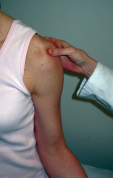
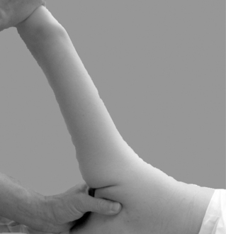
FIGURE 30-2 Hyperabduction test (Gagey test10). In a lateral decubitus position the physician’s one hand stabilizes the scapula while the other hand abducts the arm of the patient. A range of motion of more than 105 degrees is a sign for inferior laxity.
Related posts:
 Recurrent instability due to capsular deficiency
Open treatment of anterior instability—surgical technique
Arthroscopic treatment of multidirectional instability—surgical technique
Radiographic studies and findings
Examination and classification of instability
Imaging findings in posterior instability
Recurrent instability due to capsular deficiency
Open treatment of anterior instability—surgical technique
Arthroscopic treatment of multidirectional instability—surgical technique
Radiographic studies and findings
Examination and classification of instability
Imaging findings in posterior instability
Stay updated, free articles. Join our Telegram channel

Full access? Get Clinical Tree


