CHAPTER 31 Nonoperative management and scapular dyskinesis


Anatomy and biomechanics
From a biomechanical perspective, the GH joint is a closed chain mechanism comprised of bones, ligaments, and muscles that balances stability against excessive translations with mobility necessary to achieve positions and motions of the arm and hand to accomplish specific tasks.1 For almost all normal shoulder/arm functions, GH kinematics that result from this balance resemble a ball and socket arrangement.
The scapula, as the “G” of GH, is a key element in the closed chain mechanism. The scapula plays multiple roles in creating and maintaining the ball and socket kinematics. First, the glenoid must be dynamically positioned in three-dimensional space to maintain the “glenohumeral angle”—the orientation of the glenoid cavity and the long axis of the humerus from head to elbow—in a “safe zone” that minimizes glenohumeral shear,2 maximizes concavity-compression,3,4 and minimizes muscular activation.5 Jobe clinically estimated this angle to be +/− 30 degrees.5a This clinical observation has been verified by a biomechanical study that showed that muscle activation was most efficient in maintaining joint stability when the glenohumeral angle measured +/− 29.3 degrees.5 If the angle is maintained within these parameters, the resultant force vectors are directed within the glenoid cavity, shear forces are minimized, tension on the ligaments is minimized, and the muscle activation requirements are minimized, creating the most efficient joint conditions for stability. In this position, all of the intrinsic shoulder muscles of the rotator cuff can pull in relatively straight lines to maximize concavity-compression into the joint.
Achievement of this scapular position requires that the scapula be positioned in anticipation of arm and shoulder movements. There are several reasons for this anticipatory requirement. The speeds, forces, and motions around the shoulder are frequently too rapid and occur too quickly for sensory feedback to adjust muscle activation to move the scapula.1,5 Scapular movement by itself creates only up to 40% of the observed forces necessary for forward shoulder and arm acceleration.5 The majority of the forces developed through the kinetic chain activation sequence to move the arm forward come from the hip/trunk activation (core stabilization), which creates interactive moments to position and move the arm in space6,7 similar to the movement of the end of a whip. In normal shoulder movements, these anticipatory motions are part of a biomechanical closed chain that couples scapular and arm motions.1,5,6
The muscular activation sequences that allow this anticipatory bony positioning are learned, preprogrammed patterns, defined as force-dependent activation patterns,5,8 that integrate multiple muscles to move multiple joints.9–11 These patterns use feed-forward sensory information to position the bones and joints in the most efficient manner. They are highly developed and are quick to drop out with injury or disuse.
Typical muscle activation patterns involve stabilization of the contralateral hip and trunk extension as a base for scapular activity,12 anterior and posterior core stabilization for force development at the shoulder,13 sequential activation of contralateral, then ipsilateral abdominals before rotator cuff activation,14 and activation of scapular stabilizers before rotator cuff activation.15
The functional and observable result of the muscle activations producing dynamic positioning is scapulohumeral rhythm (SHR), the coupled synchronous movement of the arm and scapula. SHR has been likened to a “ball on a sea-lion’s nose,”16 describing the dynamic nature of the nose (the glenoid) actively moving in anticipation and response to movement of the ball (humerus) to keep the ball centered on the nose.
Second, the scapula is the point of origin for all of the intrinsic and extrinsic muscles that dynamically stabilize the GH joint in almost all ranges of motion. Muscles are responsible for GH stability through about 90% of the motions in all planes.1 A stable base is a requirement for maximal activation of all the rotator cuff and deltoid muscles.17–19 Demonstrated muscle strength can be improved by as much as 24% off a stabilized scapula.19 Maximal rotator cuff activation increases the compression of the humerus into the joint.
Third, optimal scapular position and motion is required to limit loads on the ligaments and other passive constraints in the joint. Increased scapular protraction creates excessive tensile loads on the anterior inferior GH ligament,20 increasing the risk of GH instability. Also, increased glenoid antetilting in protraction increases compression and shear loads on the posterior superior glenoid labrum, creating injury and decreasing the effectiveness of the labrum as a washer and a gasket to maximize GH stability.1,21
Alterations of the scapula associated with glenohumeral instability
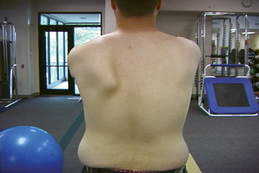
Alterations of static scapular position or dynamic scapular motion, collectively termed scapular dyskinesis (Fig. 31-1), are frequent in patients with demonstrated GH instability occurring in between 67% and 80% of patients.2,22,23 Scapular dyskinesis appears to alter normal shoulder biomechanics and joint stability by altering normal scapular kinematics. Type I (excessive anterior tilt) and type II (excessive lateral rotation) dyskinesis positions have the effects of increasing the glenohumeral angle beyond the “safe zone,” of increasing anterior shear, and of increasing tensile loads on the anterior band of the inferior glenohumeral ligament.20,24 Excessive scapular protraction, which results from type I or II patterns, also decreases maximum rotator cuff activation, decreasing the “compressor cuff” muscle function that establishes dynamic stability. Type III (lack of acromial elevation) position creates impingement on arm elevation, establishing the “instability/impingement” connection. However, no specific dyskinesis pattern is commonly associated with a specific type of GH instability. It appears that dyskinesis is primarily due to altered muscle flexibility, strength imbalance, and/or altered muscle activations.
In patients with instability due to repetitive microtrauma or labral injury, both frequently due to a process over time, weakness and inhibition of the lower trapezius and serratus anterior, coupled with inflexibility of the pectoralis minor, appear to be the main causative factors.2,23 In patients with MDI, inhibition of the subscapularis, lower trapezius, and serratus anterior, coupled with increased activation of pectoralis minor and latissimus dorsi, have been demonstrated to place the scapula in a protracted position.25–27
Physical examination
Scapular evaluation in glenohumeral instability
Related posts:
 Open surgical solutions for posterior instability of the shoulder
 Clinical history, examination, arthroscopic findings, and treatment of multidirectional instability
 Recognition and management of combined instability and rotator cuff tears
 Open treatment of multidirectional instability—surgical technique
 Humeral head defects—biomechanics, measurements, and treatments
 Nonoperative rehabilitation for traumatic and atraumatic glenohumeral instability
Stay updated, free articles. Join our Telegram channel

Full access? Get Clinical Tree


