Nonoperative Management and Rehabilitation of the Hip

CHAPTER 7 Nonoperative Management and Rehabilitation of the Hip


Introduction


The growing interest in musculoskeletal-related hip pathology is largely related to the evolution of hip arthroscopy and the recognition of previously unknown or underappreciated pathologies. Not only are new surgical interventions available for both intra- and extra-articular hip pathologies, but innovative conservative interventions have also been developed. An understanding of anatomic considerations, the recognition of pertinent clinical findings, and knowledge of available research are needed for the appropriate conservative management of individuals with hip pain. As compared with other body regions, there is a limited amount of information available regarding conservative management for individuals with musculoskeletal hip pathology. However, concepts that are commonly applied to the shoulder may prove to be useful in this area. The purpose of this chapter is to outline the evaluation process that we use to develop an intervention plan to conservatively manage individuals with musculoskeletal-related hip pathology.


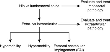
Evaluation algorithms and classification-based treatment systems are commonly used in the orthopedic community to assist with determining a diagnosis, prognosis, and intervention plan. An algorithm implemented during the patient evaluation allows for the systematic collection of information, whereas classification-based treatment defines subgroups of patients who are likely to respond to a specific treatment approach. A number of evaluation algorithms and classification-based treatment systems have been developed for various body regions; however, there are few that are specific to the hip. We have integrated an evaluation algorithm and classification-based treatment to help conservatively manage individuals with hip pain; this plan includes the consideration of the lumbosacral spine, the extra-articular soft tissue, and the intra-articular structures. Intra-articular pathologies are further divided to consider the issues of impingement, hypermobility, and hypomobility. An outline of our proposed algorithm can be found in Figure 7-1.


Lumbosacral spine


Similar to the evaluation process for the shoulder, in which radiating symptoms from the cervical spine should first be ruled out, pain radiating from the lumbosacral spine should first be considered as a possibility for individuals with hip pain. In addition, because of the kinetic relationship between the hip and the lumbosacral complex, conditions in these areas commonly coexist. This holds particularly true for more chronic cases in which muscle dysfunction (e.g., gluteus medius) causes gait deviations that negatively affect the lumbosacral spine. Therefore, the first step in our evaluation algorithm is to perform tests to rule out a lumbosacral spine contribution to the patient’s symptoms. Many tests that assess the lumbosacral region have been described. Examples of such tests include lumbar range of motion, the palpating of pelvic landmarks, standing flexion, long sit, prone knee flexion, flexion abduction external rotation test (FABER), and hip internal rotation range of motion. If any of these tests is positive for a lumbosacral disorder, treatment may be directed toward this area. The effect of this treatment on hip pain can be evaluated and modified accordingly. In our evaluation and treatment of lumbosacral problems, we use a classification system that was initially described by Delitto and colleagues. This approach has been modified slightly by the results of subsequent research as summarized by Fritz and colleagues. This classification system includes the categories of manipulation and mobilization, stabilization, specific direction preference exercises (e.g., flexion, extension, lateral shift), and traction.


The mobilization classification makes use of a treatment technique that is thought to generally mobilize the lumbosacral complex. In our experience, however, the technique is often directed toward the sacroiliac joint. Tests to assess for sacroiliac joint abnormalities include assessing pelvic landmarks for symmetry, the seated flexion test, the long sit test, the Gillet test, and the FABER test. Childs and colleagues developed a clinical prediction rule for the mobilization category that involves the following five factors: 1) the duration of the current symptoms being less than 16 days; 2) a score on the work subscale of the fear-avoidance belief questionnaire of less than 19; 3) hypomobility of the lumbar spine (assessed with posterior-to-anterior pressure); 4) internal rotation of one hip of more than 35 degrees; and 5) symptoms that do not extend distal to the knee. When four of these five factors were present, the patient was very likely to improve with a mobilization treatment. However, when two or fewer factors were present, the mobilization treatment was almost always associated with failure. These findings are consistent with Brown and colleagues, who found that individuals with limited hip internal rotation were 3.63 times more likely to have a hip disorder than a spine disorder.


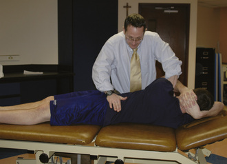
Examination and treatment with the mobilization of the lumbosacral complex are supported in the literature for individuals with hip pain. Cibulka and Delitto found that athletes with anterior or lateral hip pain and positive signs of sacroiliac joint dysfunction have a favorable treatment outcome with mobilization directed at the sacroiliac joint. The technique we use as an intervention for patients who we feel meet the criteria for the mobilization subgroup is depicted in Figure 7-2. The technique involves positioning the patient in a side-bending position toward and with rotation away from the painful side, with respect to the lumbar spine. A force directed anterior to posterior is applied to the ipsilateral anterior superior iliac spine with a Grade 5 thrusting maneuver. It should be noted that, before this manipulation is applied, contraindications for a thrust mobilization must be thoroughly cleared. The effect of this technique for reducing the patient’s hip pain is assessed. Depending on the amount of pain reduction and the results of reassessing previously positive signs, the evaluation can continue accordingly. In general, patients who fall into this category are also given lumbopelvic range-of-motion and stabilizing exercises. We find that patients with hip pain commonly have signs and symptoms that are consistent with this category and that they often positively respond to some degree to this mobilization technique.


Stabilization classification describes treating patients who are thought to have instability of the lumbar spine. Therefore, patients in the stabilization category appear to be generally very flexible, to have increased segmental lumbar movement, and to have weakness in the muscles that support the spine. Hicks and colleagues identified predictors for improvement with this treatment that included being less than 40 years old, having an average straight-leg raise of more than 91 degrees, and having aberrant movements present with lumbar flexion and extension performed when standing. Treatment emphasizes spinal stabilization through muscle strengthening and includes exercises to target the lumbopelvic musculature. We emphasize that these exercises are not meant to aggravate the patient’s symptoms and therefore will modify or omit them as appropriate.


Although the specific exercise and traction categories are not as commonly used as the mobilization and stabilization categories for individuals with hip pain, these categories are worthy of review. The specific exercise classification identifies subjects who preferentially respond to movements in one direction. Repeated or sustained lumbar flexion, extension, and side bending are performed to assess which of these movements produces a favorable response with regard to the patient’s symptoms; the movements that produce a favorable response are then incorporated into the patient’s intervention program. Those patients who best respond to an extension-specific exercise program are generally characterized by having symptoms that are distal to the buttocks, symptoms that centralize with lumber extension movements, and symptoms that spread distally down the lower extremity with lumber flexion. Those who may best respond to a flexion-specific exercise program are generally more than 50 years old, have imaging evidence of lumbar stenosis, and have pain that is relieved with sitting and increased with standing or walking. Those who may best respond to a side-bending–specific exercise program generally have a lateral shift in the frontal plane with visual deviation of the shoulder relative to the pelvis.


The traction classification seems to be the least common classification into which patients may fall, and it also has the least amount of evidence to support the interventions being used in treatment. The most common indication for this subgroup of patients is evidence of nerve root compression and symptoms that do not centralize with movements in one direction. When individuals meet the criteria for this subgroup, lumbar traction is applied as an intervention. This traction can be applied in the form of mechanical traction, deweighted treadmill, or aquatic exercises.


Individuals with symptoms below the knee are thought to primarily have lumbosacral disorders. These individuals are commonly categorized into either the exercise-specific or traction groups. Although hip pathology can occur concurrently with these groups, we find that the mobilization classification is the most common subgroup into which these patients fall, because the individual’s hip pain is either entirely or partially reduced with the use of the mobilization technique. Because lumbopelvic and hip pathologies can coexist, individuals with hip pain and positive signs of lumbosacral involvement often require an examination to determine the contribution of intra- and extra-articular soft-tissue pathology.


Extra-articular soft-tissue disorders


After the lumbosacral spine is evaluated and intervention applied as appropriate, extra-articular soft tissue and intra-articular structures need to be considered when the patient’s symptoms are not satisfactorily resolved. Examination to determine if the patient’s symptoms are of intra-articular origin includes the FABER, scour, and impingement tests. If these tests do not reproduce the patient’s symptoms, we consider the symptoms to be primarily from the extra-articular soft tissue that surrounds the hip. Common muscles that are involved include the adductor longus, the gluteus medius, the proximal hamstring, the psoas, and the abdominal musculature (i.e., sports hernia). The trochanteric bursa is another extra-articular soft-tissue structure that can cause pain and thus should be included in this discussion. Musculotendinous disorders including muscle strains and tendon disorders should be painful with palpation, stretching, and resisted movements directed at the involved muscle or tendon. If the source of pain is solely intra-articular in origin, then palpable pain is rarely present.


When treating soft-tissue disorders around the hip, an injury treatment program will depend on the phase of the injury, which can be defined as acute/inflammatory, subacute, or chronic/remodeling. Acute injuries are treated with modalities to promote healing and to decrease pain and inflammation, such as massage, submaximal isometric exercises, passive range-of-motion exercises, and lumbopelvic stabilizing exercises. It is important to emphasize that all of these intervention techniques should be pain free for the patient. Concentric movement through a full range of motion can be used as the criterion to progress to the subacute phase. Subacute injuries are treated with concentric exercises, including functional closed chain and weight-bearing exercises. Progression includes the addition of lumbopelvic stabilization activities, general flexibility, and progressive balance and stability exercises. Criteria to progress to chronic/remodeling phase can include a range of motion equal to that of the uninvolved side and strength of approximately 75% of that of the uninvolved side. Pain with resistance testing should be minimal. The remodeling phase should emphasize eccentric exercises and sport-specific training. Throughout this rehabilitation process, the strengthening of the lumbopelvic stabilizing muscles should be encouraged. However, we find educating the patient to engage these muscles during sport-specific activity to be critical. Lower-extremity biomechanics and muscle imbalances need to be carefully evaluated and corrected as appropriate. Our program often includes the use of foot orthoses or heel or shoe lifts as well as education in proper training techniques.


This generic program can be used to treat any musculotendinous disorder, including those that involve the gluteus medius, the proximal hamstring, the psoas, and the abdominals. However, the adductor longus may be the most commonly treated musculotendinous disorder. Holmich and colleagues found that, as compared with passive physical therapy of massage, stretching, and modalities, an 8- to 12-week strengthening program produced better outcomes. The strengthening program consisted of progressive resistive adduction and abduction, balance training, abdominal strengthening, and skating movements on a slide board. Both strength and flexibility issues have been found to play a role in the onset of musculotendinous injuries; however, Tyler and colleagues found that strength deficits play a larger role.


Athletic pubalgia (i.e., sports hernia) should also be addressed when discussing the treatment of musculotendinous disorders that can cause hip pain. The condition is described as chronic inguinal or pubic pain caused by the disruption of the inguinal canal components. Athletic pubalgia is often difficult to diagnose. The symptoms may include lower abdominal or groin pain that is usually made worse with sudden movements such as sprinting, kicking, side stepping, sneezing, or coughing. The symptoms generally begin slowly. We find that muscle imbalances between the psoas and the abdominals contribute to this problem. A tight and strong psoas can cause a tilting of the pelvis anteriorly, thereby stretching the weaker lower abdominal muscles. This muscle imbalance may eventually lead to small tears in the abdominal wall muscles. Individuals who engage in a lot of activity that requires prolonged forward bending (e.g., soccer, ice hockey) may be at risk for this disorder. Objectively, we find tenderness in the lower abdomen and at the top of the groin. The treatment progression can be similar to that of the generic program outlined for musculotendinous disorders. However, an emphasis is placed on reducing excessive anterior pelvic tilt. Therefore, we include exercises to stretch tight hip flexors and to strengthen muscles that promote a posterior pelvic tilt, as indicated.


When discussing extra-articular soft-tissue disorders around the hip, trochanteric bursitis with proximal iliotibial band (ITB) syndrome should be included in the discussion. Trochanteric bursitis can be caused by direct trauma, although it is more commonly related to biomechanical issues that produce an adductor moment on the ITB. We find that the most common cause of bursitis at the hip is a tight ITB. Trochanteric bursitis can also be associated with excessive adduction (i.e., pelvic drop) that results from weak hip abductors. Other causes include training errors, tight adductors, increased pronation, worn shoes, and leg-length discrepancy.


When present at the hip, ITB syndrome frequently manifests itself as a “snap,” and it is a subcategory of what has become known as snapping hip syndrome. Of course, there are other causes of snapping in the hip, and these must be entertained. First and foremost, it is necessary to determine whether the snapping sensation is intra- or extra-articular. Extra-articular snapping from a tight ITB commonly occurs during weight bearing with hip adduction and, internal and external hip rotation; this causes a taut ITB to move back and forth over the greater trochanter. Extra-articular snapping can also occur as a result of a tight iliopsoas as the tendon moves over the iliopectineal eminence during hip flexion and extension. The Ober and modified Thomas tests can be used to assess psoas and ITB flexibility. Intra-articular snapping or clicking that results from a labral tear may be reproduced during the FABER test.


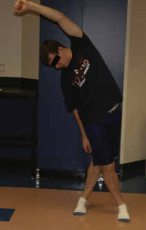
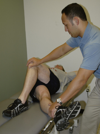
The treatment of trochanteric bursitis with proximal ITB syndrome can involve a generic progression of therapy: decrease inflammation, improve the range of motion, increase strength, and return to activity. Proper ITB stretching is done with adduction, slight extension, and slight external rotation, as outlined in Figure 7-3. We also find benefit from the manual stretching depicted in Figure 7-4. It makes sense to incorporate components of both hip flexion and hip extension into the adduction stretch because of the attachments of both the tensor and gluteus maximus to the ITB. Other manual stretching, deep soft-tissue mobilization and massage, and other modalities may be indicated. We find that the most successful treatments include interventions that address more than the lateral hip pain and that also correct any biomechanical abnormalities and muscle imbalances.


Intra-articular pathology


We include the FABER, scour, and impingement tests, as previously noted, to help determine whether the patient’s symptoms are of intra-articular origin. If these tests reproduce the patient’s symptoms, we consider the patient to have intra-articular pathology. After we determine that the origin of the patient’s symptoms is intra-articular, we assess for possible associated factors. Identifying factors associated with an intra-articular hip pathology may be similar to the identifying factors associated with shoulder pathology. We include tests for femoral acetabular impingement (FAI), capsular laxity, and articular cartilage degeneration to consider the issues of impingement, hypermobility, and hypomobility in our conservative intervention. There is little information about the conservative treatment of intra-articular lesions, including labral tears. However, if the experience is similar to that of glenohumeral labral tears, surgical intervention may be required for individuals who fail 4 to 6 weeks of conservative therapy and who want to maintain a high level of activity.


FAI is a common cause of hip pain. Patients with FAI generally report a pinching pain in the groin that occurs with sitting. A positive flexion adduction, internal rotation impingement test may indicate potential FAI. The treatment of FAI may be analogous to that of shoulder impingement; therefore, treatment should avoid movements that cause the abutment of the femoral head–neck junction with the acetabulum and address the movement dysfunction caused by weakness, restricted range of motion, and muscle imbalance. Therefore, we recommend that sagittal- and frontal-plane activities be performed within a limited pain-free range of motion. Just as scapular stabilization is important with shoulder impingement conditions, lumbopelvic stabilization may be important for individuals with FAI; we therefore include exercises to address this area. Again, all activities should be pain free to avoid joint irritation.


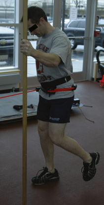
Capsular laxity of the hip may be comparable with capsular laxity of the shoulder. Generally, individuals with this condition have engaged in repetitive forceful rotational activities. We commonly find that repeated forceful external rotation to the end of the range of motion causes iliofemoral ligament insufficiency. The logroll test typically demonstrates an increase in motion on the involved side as compared with the uninvolved side in these individuals. Treatment generally emphasizes strengthening the surrounding musculature and performing neuromuscular training and proprioceptive exercises. Patients with anterior hip laxity commonly elicit symptoms of instability with external rotation of the hip (e.g., when swinging a golf club). Exercises that reproduce these movements are avoided, whereas closed-chain activities with internal rotation are encouraged. We commonly use an exercise that incorporates the use of a resistive rubber cord, as depicted in Figure 7-5. During this exercise, it is important to maintain a neutral and stable pelvis to work the internal rotators and to engage the gluteus medius and the lumbopelvic stabilizers.


< div class='tao-gold-member'>

Stay updated, free articles. Join our Telegram channel

Jul 24, 2016 | Posted by in MUSCULOSKELETAL MEDICINE | Comments Off on Nonoperative Management and Rehabilitation of the Hip

Full access? Get Clinical Tree

Get Clinical Tree app for offline access