Fig. 44.1
CT scan image. Needle biopsy of neurofibroma. Lytic, expanding, well-delimited mass in the sacrum
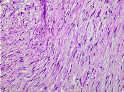
Fig. 44.2
Medium-power microscopic view of neurofibroma. Slender short spindle cells with long and wavy nuclei included in a collagenic matrix
Recommended Reading
Fawcett KJ, Dahlin DC. Neurilemmoma of bone. Am J Clin Pathol. 1967;47(6):759–66.PubMed
Related posts:


Stay updated, free articles. Join our Telegram channel

Full access? Get Clinical Tree


