(1)
Department of Pathology and Laboratory Medicine, Hospital for Special Surgery, New York, NY, USA
Abstract
Myeloma is a malignant tumor of plasma cell lineage; these cells proliferate and infiltrate the bone marrow, displacing the normal marrow and initiating secondary changes in the surrounding bone. Myeloma may be solitary at the outset (plasmacytoma or solitary myeloma) or it may present systemically (multiple myeloma, myelomatosis). There is a male predominance seen in most clinical series. Myeloma is a disease of older adults; most patients are in the sixth and seventh decades of life. Myeloma is most frequently in the distribution of red marrow. The axial skeleton is most frequently involved. Radiologically, myeloma may present as a solitary radiolucent lesion or multiple punched-out lesions without reactive or sclerotic borders. Histologically, punched-out or space-occupying lesions are comprised of cells having terminal plasma cell differentiation. Treatment options for multiple myeloma range from alkylating agents in combination with steroids to highly tailored combination drug regimens with or without marrow transplantation.
Keywords
Myeloma of boneBonePlasma cellDefinition
A malignant tumor of plasma cell lineage; these cells proliferate and infiltrate the bone marrow, displacing the normal marrow and initiating secondary changes in the surrounding bone.
Myeloma may be solitary at the outset (plasmacytoma or solitary myeloma) or it may present systemically (multiple myeloma, myelomatosis).
Since one of the differentiating clinical features of plasma cells is the production of immunoglobulins, unless a particular myeloma is extremely undifferentiated, it usually produces immunoglobulins like normal plasma cells. However, normal plasma cells produce immunoglobulins as antibodies in response to specific antigenic stimuli. In myeloma, because the tumor originally arises as an oncogenic event in a single cell, the malignant plasma cells are monoclonal. This means that the variable region of the immunoglobulin molecule that each malignant cell produces is chemically and structurally identical. As a consequence, the immunoglobulins are of a single class type and have identical charge, shape, molecular weight, and flow resistance, producing a dense and narrow band on a serum protein electrophoresis, a so-called monoclonal gammopathy. When immunoelectrophoresis is performed, the type of heavy chains and light chains can be determined, and in a large population of myeloma patients, the type of heavy and light chains produced by the tumors is proportionate to the frequency of the immunoglobulin amounts in a normal population. Thus, IgG-producing myelomas are more common than IgA myelomas, and IgA myelomas are more common than IgD and IgE myelomas. Normal immunoglobulins in myelomas are either hypermetabolized or decreased in production as the tumoral plasma cells become more dominant; and there is often compensatory hypoglobulinemia and altered serum immunity.
Multiple myeloma is distinguished from solitary myeloma which requires the presence of a histologically demonstrated plasmacytoma or greater than 10 % plasma cells in a bone marrow examination combined with monoclonal paraprotein in the serum or urine and organ dysfunction related to myeloma.
Synonyms
Multiple myeloma
Myelomatosis
Plasma cell myeloma
Solitary myeloma
Plasmacytoma
Kahler’s disease
Etiology
Myeloma is presumed to arise from the malignant transformation of a single plasma cell clone, but the cause of the transformation is not known.
Infection, genetic predisposition, and various environmental factors including radiation and chemical exposure have been implicated but not proven.
While the tumor may grow in perioral soft tissues and parenchymal organs, it usually presents either as a solitary osseous lesion (plasmacytoma) or multiple punched-out bone lesions or diffuse marrow infiltration of the skeleton.
If it is regarded as a primary bone tumor (rather than as a marrow tumor), myeloma easily outnumbers all other primary malignant tumors added together.
Clinical Features
Epidemiology
Myeloma has an incidence of 3–4/100,000 making it about four times more common than all primary malignant bone tumors combined.
Sex
There is a male predominance seen in most clinical series.
Age
Myeloma is a disease of older adults; most patients are in the sixth and seventh decades of life.
Some patients have a monoclonal immunoglobulin for many years prior to developing clinical myeloma.
Patients with solitary myeloma may have their disease localized for years although it usually eventuates in systemic disease.
In solitary myeloma, there is usually no monoclonal immunoglobulin detectable.
The type of immunoglobulin produced by the malignant clone of plasma cells parallels the type and distribution of normal plasma cells; i.e., IgG is the most frequent and IgE is the least frequent.
Malignant plasma cells may also overexpress the light-chain portions of immunoglobulins, resulting in Bence Jones proteinuria.
Sites of involvement
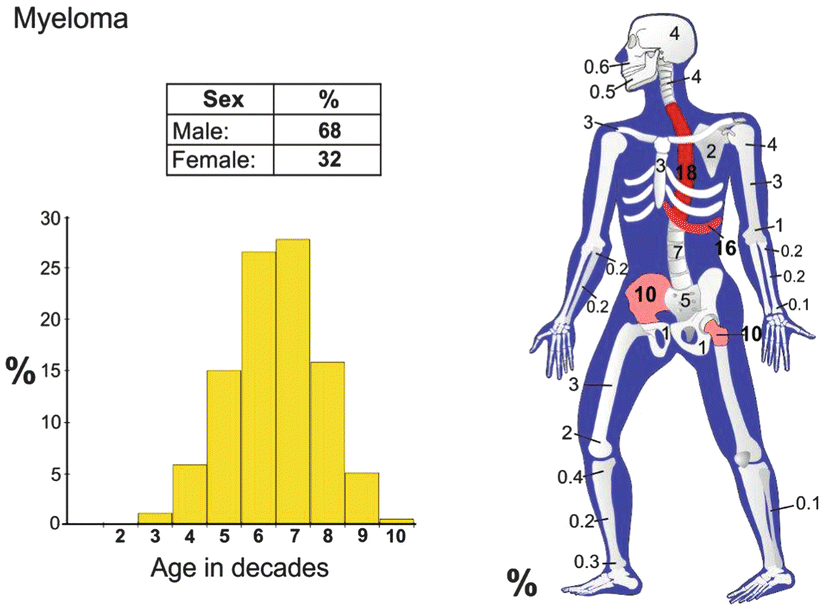
Myeloma is most frequently found in the distribution of red marrow.
The axial skeleton is most frequently involved.
The proximal appendicular skeleton is also involved.
Parenchymal organs, spleen, and lymph nodes may be involved, but it is usual for the bones to be involved first.
Clinical Symptoms and Signs
The most common presentation is bone pain, which is often back pain of relatively short duration.
In many cases, the disease is diagnosed serendipitously by nonspecific laboratory findings.
Patients may have weakness, weight loss, and even nausea.
Patients may present with recurrent infections (e.g., pneumonia) secondary to hypogammaglobulinemia.
Laboratory findings may include anemia of chronic disease, leukocytopenia, and thrombocytopenia. Most patients have monoclonal immunoglobulins on serum protein electrophoresis, and many have monoclonal light chains in their urine.
If the concentration of monoclonal protein is sufficient, patients may have rouleaux on peripheral blood smears and interference with renal function, which may be associated with hypercalcemia and hyperuricemia.
In very rare cases, myeloma may present as a peripheral polyneuropathy. There is a statistically higher incidence of this presentation with myelomas that are associated with osteosclerosis. The combination of polyneuropathy, organomegaly, endocrinopathy, myeloma monoclonal gammopathy, and sclerosing bone lesions defines the so-called POEMS syndrome; patients with this combination tend to have a shorter survival than those with usual myeloma.
Image Diagnosis
Radiographic Features
Myeloma most often presents on imaging as diffuse osteopenia.
Myeloma may present as a solitary radiolucent lesion or multiple punched-out lesions without reactive or sclerotic borders.
In less than 2 % of the cases, myeloma is associated with osteosclerosis.
Patients may present with pathologic fractures of peripheral bones or compression vertebral fractures.
Unless there are fractures, radionuclide scans are usually negative.
CT Features
CT is more sensitive than conventional radiographs and can demonstrate early bone destruction before it becomes visible on conventional radiographs.
CT demonstrates non-displaced fractures earlier than conventional radiography.
CT with many detectors is useful in the assessment of loss of bone and fracture risk.
MRI Features
Useful for evaluating small lesions that are not visible with radionuclide scans.
About 30–35 % of patients are understaged if radiographs are used instead of MRI.
While there are no signal-specific features, the lesions are usually hypointense to normal fat on T1-weighted images. Although myeloma is usually bright on T2-weighted images, it is difficult to distinguish from the bright T2 signal of fat. STIR and other fat suppression techniques are very sensitive in demonstrating marrow replacement by myeloma.
MRI is also useful to detect extension of lesions into soft tissue.
Image Differential Diagnosis
Metastatic Carcinoma
Patients are in the same age range.Related posts:

Stay updated, free articles. Join our Telegram channel

Full access? Get Clinical Tree


