Musculoskeletal Infection


Musculoskeletal Infection


Anthony A. Stans


Musculoskeletal infection may present in a myriad of clinical situations in all regions of the musculoskeletal system. Bone and joint infection may cause rapid destruction and permanent impairment if not treated urgently, so prompt diagnosis is imperative. Unfortunately, there is not a single clinical finding or a test that consistently allows rapid diagnosis. Trauma, neoplasm, inflammatory arthropathy, or synovitis may all present with a clinical picture similar to infection. In 20% or more of musculoskeletal infection cases, no organism is identified, making diagnosis and even definition challenging (1). To complicate the situation further, as a disease entity, musculoskeletal infection is continually changing. Over relatively short time periods, as immunization, antibiotics, and living conditions change, new infectious organisms causing clinically significant disease arise, and organisms previously responsible for infection become less prevalent. The most important recent change in musculoskeletal infection is the emergence of community-acquired methicillin-resistant Staphylococcus aureus (MRSA) infection in pediatric and adolescent patients, which is discussed in detail later in the chapter.

All of this would be of little interest to the orthopaedic surgeon if musculoskeletal infection in children was a rare condition, but, in fact, it is a relatively common disorder. These varied factors ensure that musculoskeletal infection will remain an important and a challenging pediatric orthopaedic disorder.


EPIDEMIOLOGY

Osteomyelitis is neither common nor rare, with the annual rate of acute hematogenous osteomyelitis (AHO) in children younger than 13 years estimated to be 1 in 5000 in the United States (5). Worldwide incidence estimates range from 1 in 1000 to 1 in 20,000 (6), and half of all cases of osteomyelitis occur in children younger than age 5 (7, 8). In childhood, septic arthritis occurs about twice as often as osteomyelitis and also tends to have its peak incidence in the early years of the first decade (9).

The epidemiologic patterns of musculoskeletal infection are continually changing. Atypical forms of infection such as subacute osteomyelitis are becoming more common, but several studies have suggested that the overall incidence of musculoskeletal infection may be declining (10, 11). These changes may be due to a variety of factors including immunization patterns and modification of clinical disease by antibiotics. At the Royal Hospital for Sick Children in Glasgow, Scotland, researchers noted a 44% decline in incidences of AHO when the period from 1970 to 1990 was compared to the period from 1990 to 1997 (12). Annual incidence dropped to a rate of 2.9 new cases per 100,000 population per year. Staphylococcus aureus remains the most common causative organism, occurring in 40% to 90% of cases of musculoskeletal infection (12, 13, 14, 15, 16 and 17). Other organisms commonly causing osteomyelitis or septic arthritis include coagulase-negative Staphylococcus, group A b-hemolytic Streptococcus, Streptococcus pneumoniae, and group B Streptococcus (17).

Since the development of routine vaccinations of infants against Haemophilus influenza, the incidence of musculoskeletal infection caused by H. influenza has dramatically decreased. In 1982, the University of Helsinki organized a prospective multicenter study of orthopaedic infection. In 1986, Finland began a large-scale immunization program against H. influenza. From 1982 to 1988, 36% of orthopaedic infections treated by the study group were caused by H. influenza, whereas from 1988 to 1998, there was not a single orthopaedic case of H. influenza infection, and the total number of childhood septic arthritis cases decreased by 30% (15). This change in epidemiology has resulted in modification of initial empiric antimicrobial therapy recommendations to cover primarily gram-positive cocci. Howard et al. (18) reported similar results following immunization for H. influenza in eastern Ontario, where H. influenza septic arthritis dropped from 41% of cases to 0% of cases following initiation of an H. influenza immunization program. Dramatic reduction in H. influenza infection following immunization has been confirmed by authors at other centers from around the world (13, 19).

Other organisms, such as Kingella kingae, are now recognized as being responsible for a greater percentage of musculoskeletal infections. In a study by Yagupsky and Dagan, K. kingae was the most common organism responsible for septic arthritis in children younger than 24 months (20). K. kingae is a fastidious, gram-negative bacillus that until relatively recently was thought to rarely cause clinical infection in children. Residing in the oropharynx of young children, K. kingae appears to be an opportunistic pathogen that gains access to the bloodstream during the course of upper respiratory infection. Kingella is transmitted from child to child and has been associated with outbreaks among day care attendees (21, 22).

Once in the bloodstream, K. kingae has a predilection for the heart and musculoskeletal system. Our greater appreciation of K. kingae as a clinically significant causative organism for musculoskeletal infection may in part be due to our improved understanding of how to culture this organism. Inoculation of a specimen into enriched blood culture media has considerably improved recovery rate (23, 24, 25 and 26). K. kingae is often resistant to Vancomycin and Clindamycin but sensitive to b-lactum antibiotics and typically responds well to appropriate antibiotic treatment with few sequelae (26, 27).

Musculoskeletal infection is much more likely to affect the lower extremity than the upper extremity or axial skeleton. In a recent study performed in Taiwan, 90% of septic arthritis cases occurred in the lower extremity. The hip was the most commonly involved joint, occurring in 54% of patients (17). Newton et al. (28) reported the hip and knee to be the joints most commonly affected in their series of 186 patients with septic arthritis. In a study by Khachatourians et al. (14) of 50 patients with septic arthritis and/or osteomyelitis, 70% of infections occurred in the lower extremities. Peltola et al. (29) reported 72% of osteomyelitis cases in their series to occur in the lower extremities.

Osteomyelitis and septic arthritis may occur simultaneously. Patients younger than 18 months have a blood supply to the chondroepiphysis, which predisposes infants to develop osteomyelitis and septic arthritis. These diseases can also occur in four locations in older children where the metaphysis lies within the joint: in the proximal femur, proximal humerus, distal lateral tibia, and proximal radius. Septic arthritis results when bacteria breach the metaphyseal periosteum and enter the joint. Perlman et al. (30) reported that signs of adjacent joint septic arthritis may be as high as 40%, and therefore careful evaluation of neighboring joints is important.

The most ominous change in musculoskeletal infection epidemiology is the emergence of MRSA. In a study of musculoskeletal infection treated at the University of Texas Southwestern, Gafur et al. (31) compared patients treated from 2002 to 2004, with patients treated at the same institution and reported in 1982. MRSA was isolated as the causative organism in 30% of children treated from 2002 to 2004 compared to no patient treated 20 years earlier.


ETIOLOGY

Bacteria travel through the circulatory and musculoskeletal systems daily and yet rarely cause clinical infection. For musculoskeletal infection to occur, several circumstances must be present. A virulent organism capable of causing infection is necessary, sufficient numbers of that organism for multiplying and
reaching a critical mass must be present, and the species and the number of bacteria present must overwhelm host defenses in the particular anatomic site in question. Although random chance may have a role in determining where and when bone and joint infection occurs, specific patterns of infection have been observed that can lead to no other conclusion than that specific factors influence where and in whom musculoskeletal infection occurs.

Metaphyseal bone adjacent to the physis is the most common site for AHO to develop. Hobo (32) described vascular loops present in the long bone metaphysis that take sharp bends and empty into venous lakes, creating areas of turbulence where bacteria accumulate and cause infection. Relative absence of tissue macrophages in metaphyseal bone adjacent to the physis appears to contribute to the predilection of osteomyelitis for this location. Others have suggested that gaps in the endothelium of growing metaphyseal vessels allow passage of bacteria (33) that may adhere to type I collagen in the hypertrophic zone of the physis. S. aureus surface antigens may play a key role in this local adherence (34).

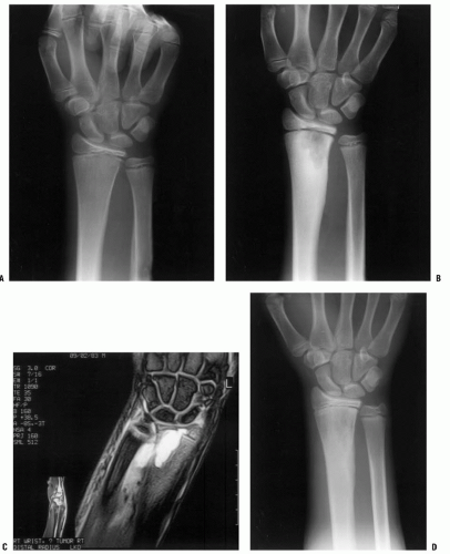
Among factors that have been implicated as contributing to the development of infection, none is as common as trauma (Fig. 12-1). Local trauma has been associated with AHO in 30% to 50% of reported cases (35, 36, 37, 38 and 39). The best evidence confirming the role of trauma has been established in an animal model by Morrissy and Haynes, who noted that intravenous injection of S. aureus caused infection in the metaphysis of an injured rabbit (40, 41). Interestingly, infection did not develop in fractures of the fibula diaphysis, indicating that fracture hematoma cannot be the explanation. Although rare, acute infection of fracture hematoma has occurred clinically. There are no similar clinical data for septic arthritis, but experimental models demonstrate the role of trauma in the production of the disease (42, 43). Therefore, the precise mechanism by which trauma reduces local host defenses and predisposes a particular location for infection has not been conclusively determined.

Naturally occurring situations confirm the importance of host defense mechanisms preventing bone and joint infection. Patients with conditions associated with decreased or altered immune response, such as the neonate, are known to be susceptible to infection. Varicella infection provides a portal for bacteria to enter the musculoskeletal system and also lowers the host immune system, making the host more susceptible to infection (44, 45). Other aspects of musculoskeletal infection etiology, such as the predilection for men and the lower extremity and peak age incidence, are less well understood and are yet to be explained.


CLINICAL FEATURES


Examination.

Much helpful information can be obtained while simply observing the child in the examination room while obtaining the history as part of the evaluation. If the child does not appear acutely ill or moribund, encourage the child to play independently while you are interviewing the parent. Unaware that he or she is being observed, the young child will often be more active than later in the structured segment of the physical examination. Refusal to bear weight on a lower extremity, a limp, or the disuse of an upper extremity gives important clues about the location of pathology.

Bone pain in a febrile child is osteomyelitis until proven otherwise. The importance of palpable bone pain in establishing the diagnosis of osteomyelitis cannot be overemphasized. Gentle, systematic palpation is often the best means available on physical examination to localize pathology in an irritable, uncooperative 2-year-old child who refuses to use an extremity. Allowing the child to remain in the arms of the parent and watching the child’s face, not the limb, while systematically palpating the limb often reveals the location of pathology. In the case of small children who cry at the mere presence of a stranger and panic at being touched, it is often beneficial to instruct the parent how to elicit the tender area. After showing the parent how to palpate the area, the physician should leave the room and allow the parent to first examine the unaffected part, then the affected part, and report the results.

In addition to establishing pain, the physician closely examines for increased warmth, erythema, or other skin changes. Erythema and swelling may appear as early as 24 to 36 hours following onset of pain and can progress rapidly. Skin changes are detectable earliest in bones or joints that are not covered by muscle. Visual comparison of the normal and affected limbs, symmetrically positioned, should always be done. Loss of normal concavities, contours, and normal skin wrinkles are other subtle clues that may be present. Severe limb swelling may indicate extensive underlying infection or deep venous thrombosis (DVT) (66).

Sympathetic joint effusion may occur adjacent to osteomyelitis but should not cause substantial joint irritability. Pain with passive joint motion is a hallmark sign of septic arthritis and is usually associated with decreased ROM as well. Palpation of joints often elicits tenderness, and joint effusion can frequently be demonstrated in joints that are not covered by large amounts of tissue. Joints of the axial skeleton, including the spine and pelvis, are less accessible for examination, and diagnosis is more dependent on findings such as pain with motion, percussion, and compression. The hip joint is also inaccessible to direct observation, but noting the position of thigh relative to the pelvis may be helpful. The patient often lies with the hip flexed, abducted, and externally rotated because internal rotation, extension, and adduction all tighten the hip capsule, causing pain in a distended and inflamed joint.

Other diagnoses included in the differential can also present with similar signs, but knowledge of characteristic patterns is helpful in establishing a diagnosis. Rheumatoid arthritis often presents as a joint that looks worse than it feels. The joint may be warm and markedly swollen with inflamed synovium and effusion but not be especially painful. Rheumatic fever has a tendency to appear just the opposite, with exquisite pain and markedly restricted motion in a joint having minimal effusion or swelling.


Laboratory.

Laboratory testing for suspected musculoskeletal infection should include complete blood count (CBC) with differential, blood culture, erythrocyte sedimentation rate (ESR), and C-reactive protein (CRP). None of these tests are specific for musculoskeletal infection. White blood cell (WBC) count is the least sensitive, being elevated in 25% to 73% of patients with osteomyelitis (2, 17, 29, 64, 67). Similar sensitivity has been reported for patients with septic arthritis (4, 35, 63, 64). Occasionally, patients with apparent AHO will have a low WBC or platelet count, which may indicate systemic sepsis or leukemia. If the diagnosis of musculoskeletal infection is in question, a manual differential count should be performed to look for atypical leukocytes and leukemia. In patients presenting with a clinical picture less clearly suggestive of bacterial sepsis, Lyme disease titer, antinuclear antibodies (ANAs), rheumatoid factor, and HLA-B27 antigen should also be considered.

The ESR and CRP are the two most common tests used to measure acute-phase response and are more useful than CBC. Acute-phase response is the increase or decrease in the levels of a variety of plasma proteins in response to cytokine production that occurs in acute or chronic inflammation. These proteins are responsible for many of the systemic symptoms seen in infection, such as fever, anorexia, lethargy, and anemia, and an increase in the levels of many of these proteins can be measured in the blood.

ESR is a nonspecific test that measures the rate at which an erythrocyte falls through plasma and is dependent on the
concentration of fibrinogen. The ESR result can be affected by the size, shape, and number of erythrocytes present, as well as by other proteins in plasma. Therefore, the ESR is less reliable in the neonate, in the presence of anemia, in patients with sickle cell disease, or when the patient is taking steroids (64, 65).

The ESR typically becomes elevated within 48 to 72 hours of the onset of infection and returns to normal over a period of 2 to 4 weeks after elimination of infection. The ESR is less reliable in the first 48 hours of infection than after 48 hours. Clinicians can expect the ESR to be elevated in 85% to 95% of cases of septic arthritis (4, 14, 17) and in 90% to 95% of osteomyelitis cases (63, 64). Although noted to be elevated just as often in patients with osteomyelitis, the ESR has been noted to be significantly higher in patients with septic arthritis (4).

One problem with the clinical usefulness of the ESR is that it continues to rise for 3 to 5 days after institution of successful therapy. Although a continuing rise beyond the 4th to 5th day of treatment can be an indication of treatment failure, it is because of this delayed response that the ESR is not a good means of assessing the resolution of sepsis during the first week of treatment (68).

The CRP is a substance found in the serum in response to inflammation and also trauma. The CRP may begin to rise within 6 hours of the triggering stimulus and then increases several hundredfold, reaching a peak within 36 to 50 hours. Because of the short half-life of the protein (47 hours), it also falls quickly to normal with successful treatment, in contrast to the ESR. This makes the CRP of greater value than the ESR, not only for earlier diagnosis of infection but also for determining resolution of the inflammation (69).

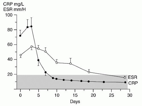
CRP is perhaps the most helpful laboratory test in the evaluation of musculoskeletal infection, its level being elevated in as many as 98% of patients with osteomyelitis compared to 92% of patients having elevated ESR (70). Peak CRP is typically noted on day 2 compared to peak ESR measured on days 3 through 5 (Fig. 12-2). Following initiation of treatment, it may take the ESR approximately 3 weeks to normalize, whereas the CRP typically returns to normal within 1 week. Failure of the CRP to rapidly normalize after initiation of treatment has been predictive of long-term sequelae (71). Therefore, the CRP is more likely to be helpful in diagnosing an early case of infection and is more useful in monitoring its resolution.

A CRP within the normal range is also a strong indicator that a patient does not have musculoskeletal infection. Levine et al. (72) reported that if the CRP is <1.0 mg/dL, the probability that a patient does not have septic arthritis is 87%. The presence of both osteomyelitis and adjacent septic arthritis also increases the likelihood that serologic testing will be abnormal. Khachatourians et al. (14) reported the ESR and CRP being elevated 100% of the time and the WBC count being elevated in 87% of patients with both septic arthritis and adjacent osteomyelitis.

As expected, the peak and the normalization of ESR and CRP are also affected by surgery (14). Khachatourians et al. (14) reviewed 50 patients with septic arthritis, osteomyelitis, or both. Twenty-five patients were treated with surgery, and 25 patients were treated with antibiotics alone. In the surgery group, it took twice as long for the CRP and ESR to reach peak values and then twice as long to normalize after initiation of treatment.


FIGURE 12-2. CRP reaches a peak value more precipitously and has a more rapid return to normal than does the ESR. The stippled area denotes the normal range of values. (Adapted from Unkila-Kallio L, Kallio MJT, Eskola J, et al. Serum C-reactive protein, erythrocyte sedimentation rate, and white blood cell count in acute hematogenous osteomyelitis of children. Pediatrics 1994;93:59-62.)

The question is often raised as to whether or not the CRP is useful in separating a musculoskeletal infection from an otitis media, which is commonly seen in children. Elevated CRP values are reported in 22% of patients with a bacterial otitis media and in 65% of those with a viral otitis media (73). Therefore, it would seem that CRP cannot reliably differentiate between musculoskeletal infection and otitis media.

The relatively low yield and delay in species identification associated with standard bacterial cultures has stimulated significant interest in molecular techniques for detection and speciation of bacterial and viral infections (16, 74). Molecular testing is appealing because it can be performed in an hour, does not depend on the presence of live bacteria for culture, and test results should not be affected if antibiotic treatment has already begun. Molecular testing techniques fall into two broad categories: nonamplified and amplified. In nonamplified techniques, direct binding of a target molecule is done with a labeled oligonucleotide probe or a monoclonal antibody, followed by the detection of the probe agent with radiolabeling, enzyme-linked immunosorbent assay (ELISA), or chemoluminescence.

When using amplification techniques, geometric amplification of a target molecule is achieved through enzyme-driven reactions. The most common technique is polymerase chain reaction (PCR). A target segment of bacterial DNA or RNA is chosen that is not present in human cells. A probe or primer specific to that segment of DNA or RNA is introduced, which
promotes binding of a polymerase that replicates the target segment in a series of temperature-dependent cycles. The amplification products are then identified by gel electrophoresis. PCR has produced some promising results in the diagnosis of periprosthetic infections and septic arthritis, but a high falsepositive rate has been reported (75). Recently, success has been reported performing molecular diagnosis of musculoskeletal K. kingae infection by specific, real-time PCR assay (76, 77 and 78). These authors report the K. kingae PCR assays to be reliable and especially helpful in identifying infection caused by this fastidious organism.

Blood cultures should always be included in the initial battery of tests obtained when one suspects musculoskeletal infection because, in both osteomyelitis and septic arthritis, blood cultures yield organisms in 30% to 60% of patients (5, 13, 64), allowing organism identification and facilitating optimal antibiotic therapy. The yield from both blood culture and aspirated material decreases with previous antibiotic therapy (4). Even with previous antibiotic treatment, however, the chances of obtaining positive cultures, when all sources (i.e., blood, bone, and joint fluid) are cultured, remain high (64).


Radiologic Features


Standard Radiographs.

Imaging should begin with standard radiographs (79); the sensitivity and specificity of radiographs range from 43% to 75% and from 75% to 83%, respectively (80). The role of radiography in the diagnosis of early bone and joint sepsis is often undervalued because clinicians often look only for changes seen in bone. Plain radiographs may show soft-tissue swelling and loss of tissue planes within 3 days of infection onset, whereas bone changes may not appear for 7 days or more (67, 81). Because the inflammation in the bone or joint produces edema in the soft tissues adjacent to the area of inflammation, there is swelling in this region, and enlargement of this muscle layer is detectable on the radiograph. Radiographs to detect deep soft-tissue swelling are of most value in suspected sepsis of the long bones. Symmetrically positioned views of the contralateral extremity may be helpful for comparison.

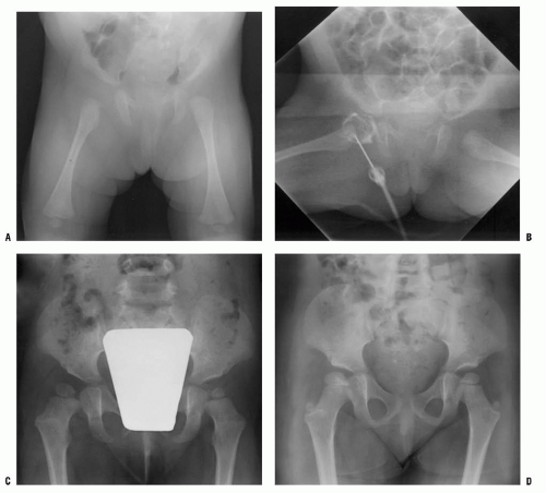
Septic arthritis may cause a large effusion that is most easily seen in peripheral joints such as the knee or elbow. At the hip, there may be asymmetric widening of the joint space compared to the uninvolved hip (Fig. 12-3). Although this may be seen frequently in the neonate, hip joint space widening is often lacking in older children. It is a late sign, and its absence is not to be interpreted as lack of sepsis (82). Untreated septic arthritis may result in joint destruction, narrowing of the joint space, or pathologic bone changes on both sides of the joint. Additional sequelae such as osteonecrosis of the femoral head may also be seen.

The time required before bone changes become visible on plain radiographs suggests that by the time bone changes are seen, osteomyelitis is already well established. While not entirely reliable, it is fair to suggest that when radiographic changes are present, surgical treatment of osteomyelitis is more likely to be necessary than if radiographic changes are not present. Although infection can appear in any bone and in any location, the most common radiographic presentation for osteomyelitis is a destructive, lytic, eccentric metaphyseal lesion, often associated with periosteal elevation and new bone formation. Bone destruction caused by osteomyelitis may appear aggressive, infiltrative, and ominous in appearance and may be mistaken for neoplasm (83, 84 and 85).


Bone Scan.

Radionucleotide technetium-99m diphosphonate bone scanning is an excellent test for localizing suspected musculoskeletal infection, with reported sensitivity of 89% to 94% and specificity of 94%, with overall accuracy of approximately 92% (13, 86). The bone scan consists of three phases: an angiogram, performed immediately after injection; immediately followed by the second or “blood pool” phase; and 2 to 3 hours later, the mineral phase, which reflects uptake in the bone. All three phases are helpful, especially in distinguishing cellulitis from osteomyelitis. The mechanism by which technetium-99 m bone scanning works is isotope uptake, which depends on vascularity and calcium phosphate deposition (87).

To obtain the highest quality and most sensitive images, the bladder should be empty at the time of the scan to prevent accumulated isotope from obstructing the sacrum and sacroiliac (SI) joints. Symmetrically positioned views of both sides should be obtained. Technetium scanning using pinhole-collimated views and single-photon emission computerized tomography can increase both sensitivity and specificity (88). Because most AHO occurs in the metaphysis adjacent to the physeal plate, such views are necessary to separate early metaphyseal changes from the large amount of uptake found in the physeal plate. These images are time consuming to obtain and may require that the child be sedated. It is therefore important that the physician communicate the desired areas of interest to the radiologist.

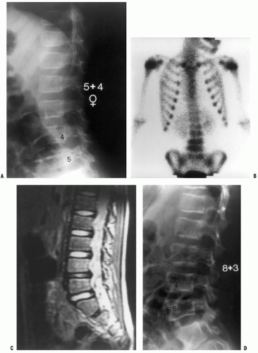
Technetium-99m scanning is most helpful when examining patients in whom the site of suspected musculoskeletal infection is unclear (Fig. 12-4) or when looking for multiple foci of bone involvement (89). Bone aspiration and initiation of treatment should not be delayed for fear of affecting bone scan results. Using an animal model, Canale et al. (90) demonstrated that if a bone scan is performed within 48 hours after bone aspiration, the bone aspiration does not cause a falsepositive scan result.

Whereas typically a bone scan is suggestive of osteomyelitis when “hot” or showing increased uptake, a “cold” bone scan may provide evidence of severe osteomyelitis and has been reported to have a positive predictive value of 100% (88, 91, 92) A “cold” scan results from infection causing an area of bone ischemia. Pennington et al. (93) at the Medical College of Wisconsin reviewed 81 patients evaluated with technetium bone scan for osteomyelitis. Seven of the 81 patients had a photopenic region defect, or cold scan, consistent with osteomyelitis. A control group of matched patients with hot scan osteomyelitis was compared to the cold scan group. Patients with cold scan osteomyelitis had statistically increased
temperature, resting pulse rate, ESR, length of hospital stay, and rate of surgical intervention compared to patients with hot scan osteomyelitis.


FIGURE 12-3. A: A 2-month-old infant presents following 3 days of increasing irritability, fever, and pseudoparalysis of the right leg. Anteroposterior pelvis radiograph demonstrates widening of the right hip joint space. B: The patient was brought emergently to the operating room, where the right hip was aspirated and an arthrogram was performed to document intraarticular position of the needle. Cell count of the hip joint aspirate was 65,000 per mL; open joint irrigation and debridement of septic arthritis were performed. Cultures later confirm group A Streptococcus infection. C: Two years following open surgical irrigation and drainage, the patient is asymptomatic but A-P pelvis radiograph demonstrates mild hip dysplasia on the right with acetabular index of 25 degrees compared to 22 degrees on the left, 50% femoral head coverage on the right compared to 70% coverage on the left, and widening of the right femoral neck. D: Four years following irrigation and debridement, the right hip dysplasia has improved, with the right acetabular index now measuring 21 degrees and with a femoral head coverage of 70%. Mild coxa magna and femoral neck widening persists.

Septic arthritis is suggested by equally increased uptake on both sides of a joint. Although bone scanning may correctly identify the site of joint sepsis in approximately 90% of infected joints, it does not separate bone from joint sepsis or differentiate infectious from noninfectious arthritis (86, 94). This is a particular problem in the hip, in which the differential diagnoses may include transient synovitis, septic arthritis, or osteomyelitis of the femoral neck.

Technetium-99m bone scanning does have its limitations. Technetium scanning is relatively nonspecific, and increased uptake may be caused by any process that increases vascularity

or deposition of calcium phosphate. Tumor, trauma, and bone resorption due to disuse may cause increased uptake. The scans may be negative in the first 24 hours of infection before stimulation of bone turnover, and there may be a 4% to 20% false-negative rate with technetium scanning (64). In neonatal infection, the reported sensitivity for technetium scanning has ranged from 30% to 86%, and standard radiography may be more helpful (6, 7, 95). Overall specificity and sensitivity are improved when the scan is interpreted with knowledge of the clinical findings and initial laboratory studies, compared to when the interpretation was a blind reading of the scan (96).


FIGURE 12-4. A 5-year-old child presents with an increasing limp over 48 hours and with suspected musculoskeletal infection. History and physical examination do not localize the process. ESR and CRP are elevated. A: The lateral (as well as the anteroposterior) radiograph of the spine is normal. B: Technetium bone scan shows increased isotope uptake in the L4 and L5 vertebral bodies suggestive of discitis, but neoplasm cannot be excluded. C: T2-weighted MRI helps confirm the diagnosis of discitis, demonstrating that the process is centered in the L4-L5 disc with no evidence of neoplasm, bone, soft tissue, or epidural abscess. Intravenous followed by oral antibiotic treatment was initiated, with complete resolution of symptoms after antibiotic therapy duration of 3 weeks. D: Final follow-up 3 years later demonstrates a normal lumbar spine radiograph in the asymptomatic patient.

Other radionucleotide imaging techniques have been less helpful in evaluating osteomyelitis in children. Gallium-67 citrate and indium-labeled leukocytes are more expensive, result in more radiation exposure, take longer to complete, and are not often useful in the evaluation of musculoskeletal infection in children (10). Indium-111-labeled WBC scanning may be helpful in the rare circumstance when infection is suspected but the technetium scan is normal. However, indium scanning requires preparation time and may take as long as 24 hours to perform (16). Granulocyte scintigraphy is an imaging technique performed with a technetium-99m-labeled monoclonal murine antibody (MoAb) against granulocytes and has been shown to be an effective and a specific method of imaging infection in adults. Unfortunately, in children, the same imaging technique was neither sensitive nor specific (97).


Magnetic Resonance Imaging.

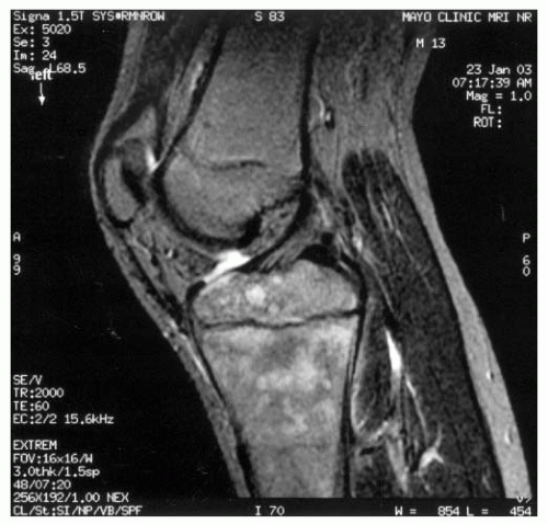
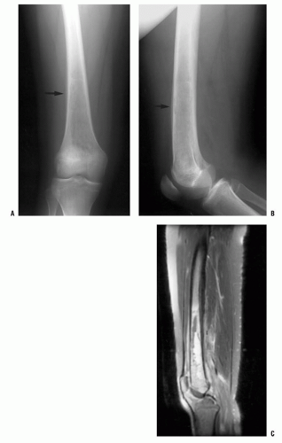
magnetic resonance imaging (MRI) is an increasingly valuable imaging tool used to evaluate musculoskeletal infection, with reported sensitivity ranging from 88% to 100%, specificity from 75% to 100%, and a positive predictive value of 85% (98, 99 and 100). MRI provides better soft-tissue resolution and can be used to identify abscesses as well as to help differentiate cellulitis from osteomyelitis. MRI is useful in visualizing marrow involvement and differentiating between malignant neoplasm and infection (Figs. 12-5 and 12-6). MRI findings of osteomyelitis include a decrease in the normally high marrow signal intensity on T1-weighted images caused by replacement of marrow fat by inflammatory cells and edema. The inflammatory cells and the edema appear as increased signal intensity on T2-weighted images (10). Grey has described a high signal intensity feature of the thin layer of granulation tissue that lines the abscess cavity on T1-weighted magnetic resonance (MR) images, calling it the “Penumbra Sign.” Subsequent authors have confirmed its value in differentiating osteomyelitis from neoplasm with a sensitivity of 73.3% and specificity of 99.1% for osteomyelitis (101).

Administration of gadolinium provides further assistance in differentiating infection from other pathologic processes such as neoplasm, fracture, or bone infarct. Acute bone infarcts demonstrated thin, linear rim contrast enhancement, whereas osteomyelitis caused more geographic and irregular marrow enhancement (102). Osteomyelitis cases may also demonstrate subtle cortical defects with abnormal signal crossing marrow and soft tissue.

Invasive community-acquired S. aureus musculoskeletal infection has been associated with a high incidence of extraosseous infection and other complications such as DVT that are effectively detected by MRI. In a series of 199 children with community-acquired S. aureus osteomyelitis treated at Texas Children’s Hospital, MRI was compared with bone scintigraphy (103). The sensitivity of MRI and bone scintigraphy for osteomyelitis was 98% and 53%, respectively. In all discordant cases, MRI was correct compared to bone scintigraphy. Extraosseous complications of community-acquired S. aureus osteomyelitis detected only by MRI included subperiosteal abscesses (n = 77), pyomyositis (n = 43), septic arthritis (n = 31), and DVT (n = 12). Therefore, MRI is the preferred imaging modality for the investigation of severe, pediatric community-acquired musculoskeletal infection because it offers superior sensitivity for osteomyelitis compared to bone scintigraphy and detects extraosseous complications that occur in a substantial proportion of patients.

MRI is very helpful when evaluating suspected sepsis involving the hip and axial skeleton. Yang et al. (104) used MRI when trying to differentiate between transient synovitis and septic arthritis. Septic arthritis was statistically more likely to have signal intensity abnormalities in adjacent bone marrow and contrast enhancement within surrounding soft tissue, while toxic synovitis was more likely to be associated with contralateral hip effusion. Karmazyn et al. (105) reported the utility of MRI when differentiating conditions such as pyomyositis, and sacroiliitis from septic arthritis of the hip, while McPhee et al. (106) reported the value of MRI when localizing infection in complex pelvis anatomy. MRI of suspected spinal infection allows visualization of pathology such as epidural or paraspinal abscesses as well as detection of the presence of softtissue masses that would be suggestive of neoplasm.

Whole-body MRI has recently been reported as a possible screening study that could be used to localize musculoskeletal infection in a manner similar to scintigraphy but with several potential advantages (107). Use of a moving tabletop and automatic direct realignment of the images after acquisition make whole-body MRI possible. The scan plane is coronal with additional planes being added depending on the indication and findings. Whole-body MRI is targeted for maximum coverage of the body within the shortest possible time using the minimum number of sequences. The evaluation of the bone marrow has been the primary indication, and, therefore, inversion recovery sequences like STIR or TIRM are typically used with the T1-weighted sequence being added variably. If an area of pathology is detected, then imaging technique can be changed from a screening protocol to an imaging regimen designed to provide detailed information on the specific region in question. Whole-body MRI may be especially helpful in neonates where multifocal infection is common, scintigraphy is relatively insensitive, and patient size is small.

Two recent studies have been reported on the use of MRI in postsurgical patients. Kan and coauthors at Vanderbilt University evaluated the diagnostic efficacy and the impact of emergent MRI after recent surgical intervention in children
with suspected osteomyelitis or septic arthritis and found that iatrogenic soft tissue and bone edema related to recent surgery in children with suspected osteomyelitis or septic arthritis has minimal effect on diagnostic accuracy of MRI (108). Spiegel et al. (109) at Children’s Hospital of Philadelphia evaluated the usefulness of MRI as a routine follow-up test used to assess surgical treatment of musculoskeletal infection and noted that if patients’ clinical course was unremarkable, MRI did not add clinically significant additional information. From these two studies, we can conclude that if patients demonstrate clinical improvement with treatment, then routine follow-up MRI is not necessary, but if the treatment course is complicated by clinical evidence of persistent or recurrent infection, then MRI can provide helpful, reliable information that is not degraded by previous surgery.


FIGURE 12-5. MRI may be very helpful when differentiating between osteomyelitis and primary bone malignancy. A,B: This 12-year-old female patient was referred for evaluation of femoral osteosarcoma. The standard anteroposterior and lateral radiograph shows periosteal reaction along the distal one-third of the femur, consistent with primary bone sarcoma or osteomyelitis (arrows). C: T2-weighted MRI without contrast demonstrates preservation of some normal marrow fat within the intramedullary canal and a fluid-filled abscess cavity diagnostic of osteomyelitis. Diffuse inflammation is present in adjacent soft tissues without a discrete soft-tissue mass. Osteomyelitis was confirmed at surgery.


FIGURE 12-6. This 13-year-old male presents with a 4-month history of proximal tibial pain and normal plain film radiographs. Lateral T2 MRI without contrast lacks the high signal intensity associated with marrow edema caused by osteomyelitis and suggests a more indolent cause. MRI is the only imaging modality that can provide such detailed information. Biopsy established the diagnosis of a diffuse, large B-cell lymphoma.

Disadvantages of MRI scanning include its cost and the frequent necessity for sedation or general anesthesia in small children. When making the decision whether or not to evaluate presumed musculoskeletal infection with MRI, in each clinical circumstance one must weigh these disadvantages against the helpful information and benefit provided.


Computed Tomography.

Computed tomography (CT) is helpful to determine the extent of bone destruction as well as to detect soft-tissue abnormalities and is the most sensitive imaging study for detecting gas in soft tissues (16, 89). Especially when evaluating infection of the axial skeleton such as the spine and pelvis, CT is very helpful in localizing the infection and can assist in planning the surgical approach if debridement is indicated. CT scanning can be used to guide needle localization prior to surgical biopsy or debridement, to direct aspiration of bone or soft tissue, or to guide percutaneous placement of drainage tubes. Compared to MRI, its advantages include its greater availability and lower cost, which must be considered along with the disadvantages of being unable to detect changes within the marrow in early cases and being less sensitive at detecting soft-tissue changes.


Ultrasonography.

The utility of ultrasonography in the evaluation of musculoskeletal infection has been studied extensively, especially with regard to septic arthritis of the hip. Ultrasonography is attractive because of its low cost, relative availability, noninvasive nature, absence of ionizing radiation, and the lack of need for sedation. However, ultrasound as a noninvasive means of evaluating musculoskeletal infection has been disappointing. The lack of specificity, the dependence on operator skill, and the inability to image marrow or show cortical detail have limited ultrasound’s usefulness.

Gordon et al. (110) reviewed hip ultrasound results in 132 patients being evaluated for hip pain during an 18-month period. They found a false-negative rate of 5% in patients who were determined by ultrasonography to have no effusion but were subsequently diagnosed with septic arthritis. Children with onset of symptoms <24 hours prior to hip ultrasonography and children who had bilateral hip effusions were more likely to have a false-negative result. Zamzan reported similar findings noting a positive predictive value of ultrasound for the diagnosis of septic arthritis to be 87.9% and concluding that ultrasound cannot be used safely to distinguish between pediatric septic arthritis and transient synovitis.

Benign conditions such as toxic synovitis cannot be reliably differentiated from septic arthritis by ultrasound alone. Toxic synovitis may have a higher incidence of bilateral hip effusions than septic arthritis, and late septic arthritis may have an effusion that is more echo dense and appears fibrinous compared to toxic synovitis, but these findings are not accurate enough to be diagnostic (111, 112). Ultrasonography may be used to guide hip aspiration when performed for patients where septic arthritis is suspected.

Ultrasonography has been used to evaluate osteomyelitis, primarily on the basis of detection of subperiosteal abscess, thickening of the periosteum, and changes in the surrounding soft tissues (113). Sadat-Ali et al. (114) recently reported that ultrasonography can be helpful when differentiating between vasoocclusive crisis and osteomyelitis in patients with sickle cell disease. Ultrasonography scan showed that six patients had periosteal thickening and elevation with hypoechogenic regions, eight had abscesses, and three patients had cortical destruction. All patients were found at surgery to have osteomyelitis. These changes are all relatively late findings of osteomyelitis. Ultrasonography is of limited value when attempting to detect early changes within bone.


Author’s Preferred Treatment.

A 5-year-old child with a 48-hour history of increasing limp and suspected musculoskeletal infection presents an imaging dilemma that illustrates the importance of all four components of patient evaluation for infection: history, examination, laboratory evaluation, and imaging studies. If the 5-year-old can localize the source of pain, and localization is confirmed by physical exam, then plain film radiographs, CBC, ESR, and CRP are initial appropriate diagnostic tests. If examination is not suggestive of septic arthritis, plain film radiographs are normal, and all laboratory values are normal, then close observation with reexamination in 1 to 3 days is appropriate. If history and examination suggest a localized process, laboratory values suggest infection, and plain film radiographs are normal, then aspiration and culture of the localized bone and/or joint is appropriate. If clinical evaluation suggests infection but does not allow localization
of the process, then technetium bone scintigraphy is an appropriate next imaging study (Fig. 12-4). If additional imaging is needed to establish a diagnosis or characterize a pathologic process once the process has been localized, then MRI is the imaging study of choice to provide maximal information about the bone and soft-tissue pathology. For straightforward musculoskeletal infection in the appendicular skeleton, MRI is often not necessary; but for patients whose history, examination, laboratory evaluation, and plain film radiographs are not concordant, or for patients with suspected infection of the pelvis or axial skeleton, MRI is a very helpful imaging study.


Aspiration.

Aspiration of bone or joint should be performed whenever possible and as soon as possible when musculoskeletal infection is suspected, because it serves two important purposes: (a) aspiration may confirm the presence of a bone/subperiosteal abscess or septic joint that requires urgent surgical drainage and (b) aspiration often allows identification of the specific bacteria responsible for infection. Whenever possible and when safe to do so, initiation of antibiotic treatment should be held until initial cultures are obtained.

The fact that metaphyseal bone is the most common location for osteomyelitis is fortuitous and makes bone aspiration a relatively easy task to accomplish in the emergency department. Depending on the age and cooperation of the child, sedation may be beneficial. Fluoroscopy is not necessary for bone or joint aspiration in the appendicular skeleton but is now available in many emergency departments and can be helpful in guiding and documenting needle placement. At the point of maximal tenderness, the skin is sterilely prepped. Avoidance of cellulitic skin when possible is desirable but not mandatory; aspiration of bone through cellulitis has not been shown to cause osteomyelitis, and direct culture of cellulitic areas yields a positive culture in <10% of cases (115). Local anesthetic is used to anesthetize the skin and the underlying periosteum with its abundant nerve supply. Using a large-bore trocar needle, such as an 18- or 20-gauge spinal needle, the area at and beneath the periosteum is aspirated for possible subperiosteal abscess.

If no purulent material is aspirated at the periosteum, the needle is passed through the thin metaphyseal cortex by rotating the needle back and forth with gentle pressure directed toward the center of the bone. A spinal needle with its solid trocar facilitates passage through bone and prevents the needle lumen from being plugged with bone fragments. Once inside the cortex, aspiration may yield purulent material but, more commonly, and especially in early osteomyelitis, sanguinous fluid returns. The purulent or sanguinous fluid is then placed in appropriate media and sent for aerobic and anaerobic culture as well as for microscopic Gram stain analysis. Depending on the clinical situation, the aspirate may be sent for fungal and mycobacterial culture. Sending bone aspirate for cell count is less helpful than sending joint fluid, but if adequate aspirate fluid is available, elevated WBC count can support the diagnosis of infection. Bone aspirate cultures yield organisms in 50% to 85% of patients with osteomyelitis (13, 16, 64, 68).

Joint aspiration also offers the opportunity to gather critically important clinical information. Using an 18- or 20-gauge needle, the joint is aspirated and fluid is placed in appropriate culture media and tubes for fluid analysis. Hip aspiration should typically be performed under general anesthesia in the operating room using a spinal needle and accompanied by an arthrogram to document the presence of the needle within the hip joint (Fig. 12-3). Depending on the facility, hip aspiration may be performed under conscious sedation using ultrasound guidance in the emergency or radiology departments. The most important tests for joint fluid aspirate are Gram stain, culture, leukocyte count, and determination of the percentage of polymorphonuclear cells. If Lyme disease is suspected, synovial fluid should be sent for PCR testing as well. Routine use of other synovial fluid tests is of little value (116, 117). Because fluid from an infected joint frequently clots, it may be helpful to rinse the syringe with heparin before aspirating the joint. Often, only a small amount of fluid is obtained, and care must be taken not to leave any significant volume of heparin in the syringe, which may alter the cell count.

Much emphasis has been given to the presence of a joint aspirate cell count >50,000 per mL. Although the most likely cause for a joint aspirate cell count to be >50,000 per mL is bacterial septic arthritis, it is neither 100% sensitive nor 100% specific (Table 12-1). In a series of 126 bacteriologically proven cases of septic arthritis, Fink and Nelson (116) found leukocyte counts of 50,000 per mL or less in 55%, with 34% having counts <25,000 per mL. At the same time, inflammatory diseases (e.g., rheumatoid arthritis) may have counts in excess of 80,000 per mL (69). Joint fluid WBC differential provides very helpful additional information because a percentage of polymorphonuclear cells >75% is highly suggestive of joint sepsis (118).


TABLE 12-1 Synovial Fluid Analysis


Disease


Leukocytea Cells/mL


Polymorphsa (%)


Normal


<200


<25


Traumatic effusion


<5000 with many erythrocytes


<25


Toxic synovitis


5000-15,000


<25


Acute rheumatic fever


10,000-15,000


50


JRA


15,000-80,000


75


Septic arthritis


>50,000


>75


aThe leukocyte count and the percentage of polymorphs can vary in most diseases, depending on the severity and duration of the process. Overlap greater than shown in these averages is possible.


From Morrissy RT, Shore S. Septic arthritis in children. In: Gustilo RB, Gruninger RP, Tsukayama DT, eds. Orthopaedic infection: diagnosis and treatment. Philadelphia, PA: WB Saunders, 1989:261-270, with permission.


Atypical organisms are less likely to cause joint fluid aspirate cell count to approach 50,000 per mL. Nine patients with brucellar arthritis treated at Ben-Gurion University had a median synovial fluid cell count of 9500 WBC per mm3 (range: 300 to 61,500 WBC per mm3), and only one patient
had a cell count of >50,000 per mL. Brucella melitensis was recovered from the synovial fluid culture in all patients (119).

As in osteomyelitis, the frequency of positive cultures seems to be slightly higher with open biopsy than with needle biopsy, but the difference is not great. In addition, the positive yields are generally not as high as in osteomyelitis, ranging in various reports from 30% to 80% (63, 68, 120). The importance of obtaining material from blood and bone or joint aspiration is emphasized in a report by Vaughan et al. (121), in which many children with osteomyelitis had only positive blood cultures, whereas other patients had only positive bone cultures.

Gram staining is the only opportunity for presumptive identification of the organism within a few hours of initial patient contact and is therefore a valuable test that should not be ignored. It appears from reports of both septic arthritis and osteomyelitis that the Gram stain demonstrates an organism in about one-third of the bone or joint aspirates (63, 64, 116).

Most bacterial cultures will yield results within 48 hours of specimen collection. However, fastidious organisms may take as long as 7 days to become positive. S. aureus remains the most common causative organism, causing musculoskeletal infection in 60% to 90% of patients (67, 122). Streptococci, pneumococci, K. kingae, and gram-negative bacteria are also potential causative organisms.

Several authors have questioned a distinction between culture-positive and culture-negative septic arthritis. Lyon and Evanich reviewed 76 children treated at Medical College of Wisconsin and Children’s Hospital of Wisconsin for isolated joint infection between 1990 and 1997 (123). All patients underwent joint aspiration with fluid analysis, including culture, and a causative organism was identified in only 30% of cases. There were no significant clinical or laboratory differences between the culture-positive and culture-negative groups, and all patients were treated similarly with joint drainage and antibiotic therapy. All patients had complete resolution of infection following treatment. Lyon and Evanich concluded that, with regard to clinical presentation and response to treatment, culture-negative septic arthritis did not differ significantly from culture-positive septic arthritis and therefore warranted a similar diagnostic and treatment approach.

Investigators from LSU Health Sciences Center did note several differences in the clinical presentation of culture-positive septic arthritis compared to culture-negative arthritis (1). Patients whose cultures were positive were more likely to have antecedent trauma, overlying skin changes, and a shorter duration of symptoms prior to diagnosis. However, treatment and treatment results did not differ significantly between groups. In summary, culture-negative septic arthritis can be treated empirically as presumed staphylococcal disease, perhaps with slightly broadened antibiotic coverage, with excellent long-term results.


DIFFERENTIAL DIAGNOSIS


Osteomyelitis.

Trauma and neoplasm are conditions that may present with characteristics similar to osteomyelitis, and they may be mistaken for infection. Trauma is the most common and made more confusing because trauma can predispose patients to develop osteomyelitis. Similar features are typically present, including pain, tenderness, swelling, and soft-tissue swelling on radiographs. However, several distinguishing features may be present. Traumatic symptoms are usually sudden in onset with gradual improvement, compared to symptoms of infection, which are more likely to be gradual in onset and progressive in nature. Trauma may be associated with elevation of the CRP but not the ESR, whereas both are usually elevated in osteomyelitis.

More difficult is distinguishing osteomyelitis from neoplasia (84, 85). The most common malignancy in children is leukemia, and approximately 30% of these children present with bone pain (124). Approximately 40% of children with leukemia present with constitutional symptoms such as lethargy, 18% present with fever, and 60% have an elevated leukocyte count and elevated ESR (125). Although lucent metaphyseal bands are said to be characteristic of leukemia, other bone changes are also seen. One study found lytic lesions in 19%, sclerotic lesions in 4%, and periosteal new bone in 2% (125). A purely lytic lesion without uptake on bone scan is also characteristic of leukemia as well as eosinophilic granuloma (126). Bleeding, bone pain in multiple sites, and easy bruising should raise suspicion of leukemia. A low leukocyte count may be present in 35% of patients with leukemia, although this can also be a sign of serious systemic sepsis. Anemia and an abnormally low platelet count should also raise suspicion. Abnormal WBC forms seen on manual differential is often diagnostic.

Other less common neoplasms may mimic osteomyelitis (83, 84 and 85). In the young child, metastatic neuroblastoma or eosinophilic granuloma should be considered. Older children are more likely to have Ewing or osteogenic sarcoma. Lymphoma may also occasionally arise primarily from bone (Fig. 12-6). These lesions should be approached as a malignancy with complete staging studies and diagnosis confirmed by biopsy using an approach that will not jeopardize limb salvage surgery. The adage “culture the tumor and biopsy the infection” is wise advice to follow.


Septic Arthritis.

Establishing the diagnosis of septic arthritis may be more challenging than for osteomyelitis for several reasons. There is greater urgency because septic arthritis can cause permanent articular cartilage changes within 8 hours if untreated (58), and for septic arthritis there are more diagnostic alternatives than for osteomyelitis. Interestingly, specific joints appear to be especially susceptible to permanent injury following septic arthritis. For example, the hip is more likely than the knee to progress to joint destruction following septic arthritis. The physician should always consider what must be diagnosed today, what can be diagnosed tomorrow, and what can be diagnosed next week. For example, septic arthritis, particularly of the hip, should be diagnosed as soon as possible, whereas there is little harm to the patient if juvenile rheumatoid arthritis (JRA) is diagnosed next week.

One of the most difficult and yet important differentials is between septic arthritis of the hip and toxic synovitis, a condition
thought to be a postinfectious arthritis. Both may present with a history of a few to several days of hip pain and with limp progressing to the inability to walk. The physical signs are similar in both, with limited and painful internal rotation, abduction, and extension. A longer history of symptoms, with cyclic improvement and worsening, suggests toxic synovitis. The pain is usually worse and the motion more restricted in septic arthritis.

Kocher et al. (127) reviewed the cases of all children treated at Boston Children’s Hospital from 1979 to 1996 for an acutely irritable hip and developed a clinical prediction algorithm to differentiate between septic arthritis and toxic synovitis. Although several variables differed significantly between septic arthritis and toxic synovitis, there was considerable overlap, making diagnosis based on individual variables alone difficult. However, four independent multivariate clinical predictors— history of fever, non-weight bearing, ESR of at least 40, and serum WBC count of more than 12,000 per mL—were identified that, when combined, improved diagnostic accuracy. The predicted probability of septic arthritis was 3.0% if one predictor was present, 40% for two predictors, 93.1% for three predictors, and 99.6% if all four predictors were present. Although the presence of three or more predictors was very specific for septic arthritis, it was not highly sensitive.

Two follow-up studies have subsequently been published attempting to validate the clinical algorithm proposed by Kocher et al. At the same institution where the clinical algorithm was initially formulated, Kocher et al. (128) prospectively applied the algorithm to children presenting with acute hip irritably. The predicted probability of septic arthritis in the follow-up study was 9.5% if one predictor was present, 35.0% for two predictors, 72.8% for three predictors, and 93.0% if all four predictors were present. The authors concluded that the four clinical predictors of septic arthritis demonstrated diminished, but nevertheless good, diagnostic performance in a new patient population. At a different institution, Luhmann et al. (129) applied Kocher’s clinical algorithm retrospectively to 163 patients who presented with an acutely irritable hip and found that if all four of the clinical variables in the algorithm were present, the predicted probability of their patients having septic arthritis was 59%, in contrast to the 99.6% predicted probability reported in Kocher’s original article. Most recently, a group from Children’s Hospital of Philadelphia analyzed factors associated with septic arthritis in 53 patients undergoing hip aspiration for presumed septic arthritis, reporting that the presence of five factors (oral temperature >38.5°C, elevated CRP, elevated sedimentation rate, elevated WBC, and refusal to bear weight) was associated with a 98% chance of having septic arthritis, while those with four factors had a 93% chance (130). Although the proposed algorithms may be helpful, differentiating between septic arthritis and toxic synovitis of the hip in an acutely ill child will continue to depend on the clinical acumen of the orthopaedist.

JRA is frequently considered in the differential diagnosis with septic arthritis, but several clinical features may be used to distinguish between the two disorders. The hip joint is rarely the initial joint affected in JRA. Symptoms in JRA are typically more gradual in onset than septic arthritis, and the patient almost always remains ambulatory. A joint affected by JRA typically looks worse than it functions, with relatively good motion and modest pain despite the large amount of swelling and synovitis that is typically present. Initial laboratory values are often of little help in distinguishing between septic arthritis and JRA. Joint fluid cell count typically contains fewer than 100,000 leukocytes per mL in JRA, but leukocyte counts of >100,000 per mL have been reported (131). In such cases, the treating physician has little choice but to begin treatment of septic arthritis while continuing to work to determine the diagnosis.

Rheumatic fever has a distinctly different clinical appearance than JRA, typically causing exquisite joint pain that seems out of proportion to the normal-appearing joint. A sequela of group A streptococcal infection, rheumatic fever most often causes pain in the knees, ankles, elbow, and wrists that is evanescent and migratory. Detailed questioning of the patient may unearth a history of untreated pharyngitis, febrile illness, or rash caused by group A Streptococcus approximately 2 weeks before onset of symptoms. Involvement of multiple joints strongly directs the investigator away from septic arthritis. Diagnosis of rheumatic fever is based on the Jones criteria. Major criteria include carditis, arthritis, chorea, subcutaneous nodules, and erythema marginatum. The minor criteria are arthralgia, elevated ESR or CRP, heart block on electrocardiogram, and a history of previous rheumatic fever. The diagnosis is made when a patient has two major criteria, or one major and two minor criteria.

For patients who have a documented history of recent group A Streptococcus exposure, do not meet the Jones criteria, but have significant arthralgia without other identifiable cause, the diagnosis of PSRA has been used (132, 133). A recent streptococcal infection may be documented by the presence of an antibody response to group A Streptococcus or positive throat culture. Patients with acute rheumatic fever are treated with long-term prophylactic antibiotics to prevent recurrent rheumatic fever and associated carditis. The risk of carditis to children with PSRA is unclear but felt to be low, and the role of long-term prophylactic antibiotics following PSRA is controversial but not typically recommended.

Cat-scratch disease is a clinical syndrome associated with Bartonella henselae that has been reported to be associated with arthropathy in approximately 3% of cases (134). Knee, wrist, ankle, and elbow joints are most frequently affected. The arthropathy is typically self-limited, resolving on its own at a median of 6 weeks. However, a small percentage can develop a severely painful arthropathy that can persist as long as 50 months.

Other disorders that may cause acute arthritis and can mimic septic arthritis include Henoch-Schönlein purpura and enteroarthritis secondary to Salmonella or Yersinia infection. Kawasaki disease and serum sickness are two additional conditions also characterized by a rash and arthritis. Although the joint symptoms do not require treatment and usually disappear within days, patients with any of these conditions may require medical management for the other, sometimes more serious, manifestations of the disease.


Jul 21, 2016 | Posted by in ORTHOPEDIC | Comments Off on Musculoskeletal Infection

Full access? Get Clinical Tree
Get Clinical Tree app for offline access