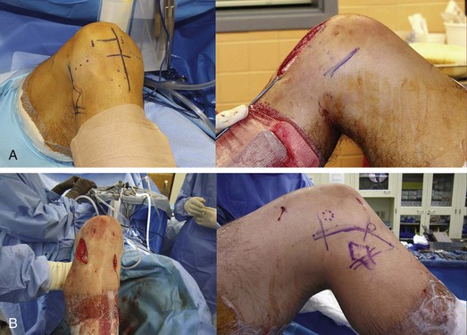
Chapter 86 The diagnosis and management of the multiple ligament–injured knee presents a clinical challenge to even the most experienced orthopedic surgeon. We consider multiligamentous knee injury to be a spectrum that can range from a cruciate plus a collateral ligament injury to a bicruciate (dislocated knee) injury with or without collateral involvement. It is important to remember that the severity of injury varies widely and that every knee is different. Certain ligaments can be partially injured and may heal with nonoperative management (medial collateral ligament [MCL]; posterior cruciate ligament [PCL]). The most commonly encountered multiligamentous injuries at our institution are combined anterior cruciate ligament (ACL)–MCL and PCL–posterolateral corner (PLC) injuries, followed by bicruciate ruptures with an associated collateral ligament tear. Treatment strategies should follow basic clinical guidelines, which may need to be adjusted on an individual basis.1 These injuries typically have one of three presentations: immediately after a traumatic event in the emergency room, in the clinical setting after acute evaluation and stabilization, and in the setting of chronic instability. Assessment begins with a thorough history and physical examination to establish the mechanism of injury; to characterize the functional, employment, and athletic status of the patient; and to identify concomitant injuries and prior knee pathology. Initial evaluation must be performed expeditiously to identify indications for emergent intervention and minimize potentially limb-threatening complications including compartment syndrome, popliteal artery lacerations, and peroneal nerve injuries. Dislocation should be considered in knees with gross instability of two or more ligaments because more than 50% reduce spontaneously before evaluation.2 The physical examination of the knee begins with assessing for abrasions, lacerations, previous surgical scars, and the presence of significant intra-articular or lower extremity swelling. A careful neurovascular examination to detect pulse asymmetry and distal motor or sensory deficits is performed, and concern for arterial injury should prompt urgent evaluation with noninvasive vascular studies (ankle-brachial indexes [ABIs]), computed tomography (CT) angiography, or popliteal arterial angiography.3 Characterization of the pattern of ligamentous instability is performed with an evaluation of patellar stability and Lachman, anterior and posterior drawer, and varus and valgus testing at full knee extension and 30 degrees of flexion. Additional rotatory testing including a dial test and evaluation of posteromedial and posterolateral rotatory instability is performed as indicated. It is important to evaluate clinical laxity with the tibiofemoral joint initially reduced, because abnormal positioning of the joint may lead to overestimation or underestimation of tibial translations, especially during posterior drawer or rotatory testing. Comparison with the contralateral extremity allows detection of subtle clinical laxities, although swelling, pain, and guarding may interfere with the ability to accurately identify and grade ligament injuries. We grade all injuries with the International Knee Documentation Committee (IKDC) standard clinical laxity scale (grade 1, 3 to 5 mm; grade 2, 5 to 10 mm; grade 3, more than 10 mm). By definition, a partial ligamentous injury is categorized as grade 1+ or 2+, and a complete tear as grade 3+.4 We obtain standard radiographs of both knees in all patients to evaluate for fracture, associated arthritis, and joint reduction. The presence of subtle findings including avulsion fracture, joint subluxation, and patellar height is also assessed. Magnetic resonance imaging (MRI) assists in the grading of ligamentous injuries and identification of pathology of the menisci, chondral surfaces, and related structures.5 The MRI findings are reviewed with the direct input of our musculoskeletal radiology colleagues before an individualized treatment plan is established. Acute surgical indications include vascular injuries requiring intervention, open or irreducible knee dislocation, and compartment syndrome. We previously reported on performance of ligamentous repair or reconstruction within 1 to 3 weeks of injury,4,6 although we now prefer delaying surgery to allow for resolution of the acute inflammatory phase of injury (i.e., swelling), interval healing of previous surgical interventions (e.g., vascular repair, fasciotomy), restoration of knee range of motion (ROM), and the return of quadriceps motor control. Relative indications for acute surgical intervention (within 14 days) in our hands include grade 3 PLC avulsions and displaced longitudinal meniscal tears; anatomic repair can be performed before the onset of significant scarring or retraction. Primary cruciate ligament reconstruction can be deferred for 6 to 8 weeks with frequent clinical evaluation to assess the pattern of ligamentous instability and detection of factors including deep vein thrombosis (DVT) or wound complications. A detailed discussion of the diagnosis, the treatment options and alternatives, and the risks and complications of surgical intervention is performed before surgical consent is obtained and the procedure is begun. The surface anatomy is identified and the planned surgical incisions are marked. Important landmarks include the inferior patellar pole, tibial tubercle, Gerdy tubercle, fibular head, and medial and lateral joint lines. The soft spot at the fibular neck is palpated and marked to correspond to the expected location of the peroneal nerve. The anterolateral portal is placed 2 to 3 mm lateral to the edge of the patellar tendon at the level of the inferior pole of the patella, and the anteromedial portal is placed 1 cm medial to the patellar tendon at the same level. A careful assessment of the presence of patella alta or baja is performed to allow consistent placement of the arthroscopic portals with respect to the joint line. A superolateral outflow portal is placed 1 cm proximal to the superior pole of the patella between the vastus lateralis and the iliotibial (IT) band. If a posteromedial portal is required, the site is determined during arthroscopy with a spinal needle (Fig. 86-1). The surgical incisions are injected with 0.25% bupivacaine and 1 : 100,000 epinephrine for hemostasis. Our standard anterior surgical incision for tibial tunnel placement and hamstring autograft harvest is longitudinally centered between the anterior and posterior borders of the anteromedial tibial cortex approximately 3 cm distal to the joint line. An anterior longitudinal incision from the midpatella to the distal aspect of the tibial tubercle, overlying the medial border of the patellar tendon, is used for patellar ligament autograft harvest, and a 6-cm longitudinal incision centered over the proximal patella and quadriceps tendon is used for quadriceps tendon autograft harvest. Lateral procedures are performed through a 12-cm curved incision centered between the biceps femoris tendon and IT band proximally and the fibular head and Gerdy tubercle distally. Medial ligament injuries are addressed through a proximal extension of the anteromedial tibial incision that is centered over the junction of the middle and posterior third of the medial femoral condyle and extends toward the distal edge of the vastus medialis obliquus (VMO). We attempt to preserve the largest skin bridge possible (minimum 7 cm) between incisions to minimize wound healing complications. Midline incisions are avoided.7
Multiligament Knee Reconstruction
The Pittsburgh Approach
Preoperative Considerations
Surgical Technique
Surgical Landmarks, Incisions, and Portals
Related posts:
 Knot-Tying and Suture-Passing Techniques
Arthroscopic Remplissage for Management of Engaging and Deep Hill-Sachs Lesions
Open Repair of Multidirectional Instability
Arthroscopic Meniscus Repair: Inside-Out Technique
Tendon Augmentation Devices in Rotator Cuff Repair
Arthroscopic Lateral Retinacular Release and Lateral Retinacular Lengthening
Knot-Tying and Suture-Passing Techniques
Arthroscopic Remplissage for Management of Engaging and Deep Hill-Sachs Lesions
Open Repair of Multidirectional Instability
Arthroscopic Meniscus Repair: Inside-Out Technique
Tendon Augmentation Devices in Rotator Cuff Repair
Arthroscopic Lateral Retinacular Release and Lateral Retinacular Lengthening
![]()
Stay updated, free articles. Join our Telegram channel

Full access? Get Clinical Tree


Musculoskeletal Key
Fastest Musculoskeletal Insight Engine