Morton’s Neuroma


Morton’s Neuroma


Stephen J. Miller

Aprajita Nakra


Metatarsalgia or generalized pain in the plantar forefoot area has many causes. A common source of such exasperating discomfort is a peripheral nerve affliction that may involve one or more common digital plantar nerves as they pass through the intermetatarsal spaces.

The first anatomic description of this localized nerve enlargement was clearly illustrated by the Italian anatomist di Civinini in 1835 (1). However, the neuralgic symptoms of this disorder were first accurately described by the English chiropodist, Durlacher, in 1845 (2). He characterized it as attacking “the plantar nerve … between the third and fourth metatarsal bones.”

Ironically, this entity was named after T.G. Morton of Philadelphia, who believed that the pain developed about the fourth metatarsophalangeal articulation because of nerve irritation from its lateral osseous neighbor (3). He should not be confused with T.S.K. Morton, who reported six cases of “Morton’s metatarsalgia” in 1893 (4), or with D.J. Morton, who described the functional anatomic entity, Morton’s syndrome, in 1935 as a cause of metatarsalgia (5).


DEFINITION, ANATOMY, AND INCIDENCE

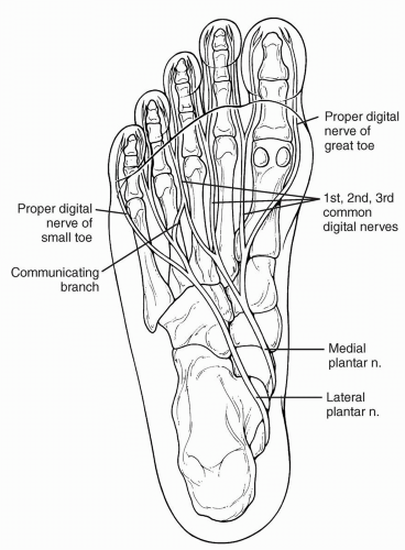
Morton’s neuroma is a misnomer used to describe this painful pedal neuropathy. The lesion is a benign enlargement of the third common digital branch of the medial plantar nerve located between, and often distal to, the third and fourth metatarsal heads. The region is supplied by a communicating branch from the lateral plantar nerve as well (6, 7, 8, 9) (Fig. 1).

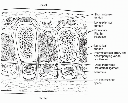
Classically, the nerve passes beneath the deep transverse intermetatarsal ligament on its plantar aspect. The only other structures passing in this immediate area are the third plantar metatarsal artery, with its accompanying vein or veins, and the tendon slip from the third lumbrical muscle that inserts into the extensor hood apparatus on the medial aspect of the fourth toe. This is also the area in which pacinian corpuscles are normally found in the subcutaneous tissues (10). The neurofibrous lesion is separated from the sole by the fat pad, plantar fascia, and connective tissue compartments. The superficial transverse metatarsal ligament is situated superior to the nerve when the region is viewed from the plantar aspect (Fig. 2).

The incidence of Morton’s neuroma in the general population has not yet been determined. Youngswick observed that of 4,000 patients presenting to his clinic with foot complaints, 9.3% were diagnosed with a neuroma (11).

In addition to the third intermetatarsal space, an intermetatarsal neuroma may also occur in other locations, usually within the second intermetatarsal space but rarely in the first or fourth. Out of 113 histologically confirmed neuromas, Bartolomei and Wertheimer found 64% in the third intermetatarsal space, 29% in the second, 6% in the fourth, and 1% in the first (12). Addante et al. evaluated 152 neuroma resections and found that 66% involved the third intermetatarsal space, 18% the second, 4% the fourth, and 2.6% the first (13). Mann and Reynolds found the neuromas evenly distributed between the second and third intermetatarsal spaces (14), whereas Bennett et al. found 68% in the third interspace and 32% in the second (15). Friscia et al. evaluated 366 patients with neuromas and found that 91% of these lesions involved the third intermetatarsal space (16).

The lesions can occur bilaterally as well (0% to 12%) (12,14,17). Less frequently (1% to 4%), neuromas can be found in two intermetatarsal spaces in one foot and rarely in three intermetatarsal spaces in the same foot (12,14,17, 18).

Histologic evidence strongly supports the concept that Morton’s neuroma syndrome is an entrapment neuropathy, resulting from compressive forces, especially against the deep transverse intermetatarsal ligament (19, 20, 21, 22, 23, 24, 25, 26). Incriminating factors include densely packed whorls of collagen, Renaut’s bodies, and thickened endoneural capillaries all formed within the subperineural space of interdigital nerves (25). Also seen are local demyelination and telescoping of myelin in opposite directions on either side of the lesion (26). Confusion has resulted from the many names applied to this lesion, which may be better described as a perineural fibroma (Table 1).


FIG. 1. Anatomy of the plantar nerves and location of classic Morton’s neuroma beneath the deep transverse intermetatarsal ligament. Note the contribution from segments of the medial and lateral plantar nerves.


FIG. 2. Cross section through the forefoot at the level of the metatarsophalangeal joints. The metatarsal artery lies above the neuroma with its accompanying venae comitantes. The lumbrical tendon lies lateral to the neuroma.


TABLE 1. Terminology used to describe Morton’s neuroma


Term


Authors and year


Neuralgic affection … plantar nerve


Durlacher, 1845


A peculiar and painful affection of the fourth metatarsophalangeal joint


Morton, 1876


Luxation podalgia


Dana, 1885


Anterior metatarsalgia


Pollosson, 1889


Metatarsal neuralgia


Bradford, 1891


Morton’s metatarsalgia


Morton, 1893


Morton’s painful affection of the foot


Hoadley, 1893


Morton’s disease


Tubby, 1896


Plantar neuralgia


Jones and Robert, 1897


Morton’s painful disease


Stern, 1904


Morton’s neuralgia


Cush, 1937


Neuritis of the fourth digital nerve


Bettx, 1940


Morton’s toe


McElvenny, 1943


Plantar neuromas


Bickel and Dockerty, 1947


Morton’s toe syndrome


Kite, 1947


Plantar digital neuritis


Nissen, 1948


Lateral plantar neuritis


Hiss, 1949


Metatarsal neuroma


Sandel, 1958


Plantar metatarsal neuritis


Pincus, 1962


Interdigital neuroma


Hauser, 1971


Morton-Durlacher neuralgia


Silverman, 1976


Intermetatarsal neuroma


Goldman, 1979


Thomas Morton disease


Gauthier, 1979


Morton’s neuritis


Milgram, 1980


Plantar interdigital neuroma


Viladot, 1982


Pseudoneuroma


Alexander, Johnson, and Parr, 1987


DIAGNOSIS


Signs and Symptoms

Most symptomatic intermetatarsal neuromas are found in women (12,14,15) and are commonly diagnosed between the fourth and the sixth decades, although these lesions may sometimes be found in teenagers. The patient may be overweight (12). In milder or early cases, the patient may describe a sensation of walking on a wrinkle in the stocking or a lump in the shoe. If the neuroma is large enough, the adjacent toes may be forced to spread apart on weight bearing. Because Morton’s neuroma causes a distinct set of symptoms, the diagnosis can most often be made from the patient’s history (Table 2).


Pain

The pain from this syndrome has been described as sharp, dull, or throbbing, but it classically manifests as a paroxysmal burning sensation “like walking on a hot pebble” or “having a hot poker thrust between the toes.” It is most often localized to the region of the third and fourth plantar metatarsal heads and may radiate distally into adjacent toes, transversely to adjacent metatarsal heads, or proximally up the leg to the knee and, in rare instances, as high as the hip. Bizarre symptoms such as metatarsalgia during orgasm or defecation have been reported, with relief on excision of the neuroma (27).

Even though the patient may describe numbness in the third and fourth toes, a sensory deficit is seldom present. At times, the digits may be hyperesthetic. Patients may describe a cramping sensation in the arch, forefoot, or toes; however, there is no evidence of muscle cramping (28).

The pain is greatly aggravated by walking in shoes and is relieved somewhat by rest, although in acute stages, lancinating or aching pains may occur during periods of inactivity. Sometimes the patient finds irritation even from bed sheets intolerable. A characteristic and virtually pathognomonic indication of a neuroma is the overwhelming desire to remove the shoe, rub the forefoot, and flex the toes. This usually provides dramatic, although transient, relief of pain.

Symptoms are present for a few weeks to several years. However, if the pain is acute, the patient usually seeks help within a few weeks. In many instances, acute pain appears after an incident of trauma which may or may not seem mild. Examples would be stepping on a rock, twisting an ankle, striking the edge of a curb, jamming the foot into the floorboard
in a motor vehicle accident, or simply changing into a new pair of shoes or doing an extraordinary amount of walking. The use of narrow or tight-fitting shoes can both instigate and aggravate symptoms.


TABLE 2. Symptoms of unoperated Morton’s neuroma as reported by patients from two studies


Study A percentage (n = 65)


Study B percentage (n = 115)


Pain increased with walking


91


Relief by rest


89


Plantar pain


77


Pain in forefoot


76


Relief by removing shoe


70


60


Pain radiating to toes


62


Burning pain


54


Pain between toes


43


Numbness into toes or foot


40


23


Aching or sharp pain


40


Inability to wear fashionable shoes


39


Pain up foot or leg


34


11


Cramping sensation


34


Pain at rest


19


History of associated injuries


15


Pain at night


10


Pain in entire foot


9


A, From Mann RA, Reynolds JC. Interdigital neuroma: a critical clinical analysis. Foot Ankle 1983;3:238. B, From Bennett GL, Graham CE, Mauldin DM. Morton’s interdigital neuroma: a comprehensive treatment protocol. Foot Ankle Int 1995;16:762.


Palpation

Tenderness and sometimes reproduction of pain can be elicited by squeezing each intermetatarsal space in a dorsoplantar direction at or distal to the metatarsophalangeal joints. On occasion, this maneuver deliver a palpable mass dorsally or plantarly, especially during the application of simultaneous transverse compression of the first and fifth metatarsal heads in the plantar sulcus when the toes are hyperextended. This maneuver also causes the patient a varying amount of pain.

Hyperextending the patient’s toes with one hand and rolling the opposite thumb transversely across the metatarsal hands on the sulcus side may reveal a palpably thickened longitudinal mass (Bratkowski’s sign), which is tender when Morton’s neuroma is present (29).

Mulder’s sign is positive when lateral compression of the forefoot combined with plantar and dorsal pressure produces a silent, palpable, and sometimes painful click in the affected intermetatarsal space as the neuromatous lesion is squeezed plantarly between the adjacent metatarsal heads (30,31). This clicking may also be caused by an adventitial bursa that forms in close association with the neuroma between the metatarsal heads (32).

Local tenderness should be differentiated from arthritic pain by direct palpation of the adjacent metatarsophalangeal joints. Further palpation of the nearby metatarsal shafts helps to rule out a stress fracture.


Differential Diagnosis

Many other disorders have symptoms that may resemble Morton’s neuroma. Some of these are listed in Table 3. Differentiation can usually be made by means of a thorough history and physical examination, although when an element of doubt remains in the face of persistent symptoms, diagnostic testing may be implemented. Even though Morton’s neuroma is a benign lesion, local malignant sarcomas have caused similar presenting symptoms in rare instances (33,34). In addition, other benign neoplasms have been reported to cause “Morton’s metatarsalgia” (35,36).


TABLE 3. Differential diagnosis of Morton’s neuroma


Metatarsal stress fracture


Rheumatoid arthritis


Autoimmune arthritides


Osteochondritis dissecans (Freiberg’s)


Localized vasculitis


Ischemia


Tarsal tunnel syndrome


Nerve root compression syndromes


Peripheral neuritis


Peripheral neuropathy (e.g., diabetic)


Intermetatarsal bursitis


Metatarsophalangeal joint capsulitis


Metatarsophalangeal joint predislocation syndrome


Transfer metatarsalgia


Diagnostic Tests


Radiographs

Weight-bearing radiographs may be taken to help rule out other pathologic conditions and also to evaluate for rotation, enlargement, or juxtaposition of the metatarsal heads that may contribute to the origin of the neuroma (37, 38, 39). Grace et al., in their study of 48 feet with pathologic confirmation of neuroma and 100 asymptomatic patients, found no statistical relationship between radiologic findings or metatarsus proximus and digital divergence (“Sullivan’s sign”) and the physical occurrence of neuromas (40,41). Instead, an increased intermetatarsal angle of the affected interspace was noted in the neuroma-afflicted group. The neuroma itself is not visible on x-ray films or xeroradiography.


Sensory Nerve Conduction Tests

Electrodiagnostic techniques for evaluating Morton’s neuroma are not highly accurate, primarily because of the difficulty in isolating a single interdigital nerve with an electrode (24). However, in one study, the diagnosis was confirmed by electrophysiologic testing of five patients. Positive results were characterized by an abnormal-dip phenomenon, a relatively normal conduction velocity, and normal duration of the sensory compound nerve action potential. These findings are the hallmarks of a neuropathy with a predominant axonal degeneration (42). Therefore, such testing may be reserved for further assessment of patients with metatarsalgia who lack the classic physical signs of Morton’s neuroma (22,43).


Magnetic Resonance Imaging

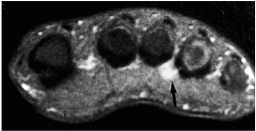
Cross-sectional magnetic resonance imaging (MRI) through the forefoot has differentiated a Morton’s neuroma mass from the surrounding bone and soft tissues (44). The neuroma appears isointense relative to muscle on T1-weighted images and homogenously or inhomogenously hypointense relative to fat tissue on T2-weighted images. In other words, it is best seen on T1-weighted images as a well-demarcated mass of lower signal intensity (45) (Fig. 3). Studies have shown that T1-weighted axial and coronal images with an axial fast spin-echo T2-weighted sequence detect neuromas more consistently than an enhanced T1-weighted fat-suppressed sequence (46,47). Although the neuroma is often invisible on T2-weighted images, on occasion the presence of fluid-filled bursae will lead to a high signal intensity proximal to the neuroma (48,49).


FIG. 3. Magnetic resonance image of an intermetatarsal neuroma. Coronal T2-weighted image of a neuroma in the third interspace. (Courtesy of George Vito, D.P.M.)

However, MRI has been shown to produce false-negative images, so the accuracy of the scans is not ensured (50). For this reason, as well as for cost considerations, MRI for the diagnosis of Morton’s neuroma may be best considered in patients with recurrent neuroma, atypical symptoms, or coexisting neurologic conditions (51), or it may be used when a neuroma is suspected in an adjacent interspace.


Ultrasonograhy

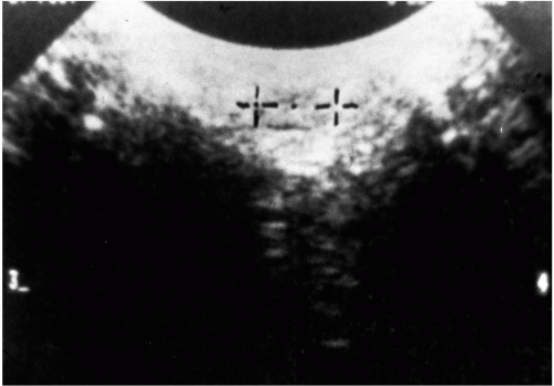
High-resolution ultrasound appears to be the most promising of the objective imaging techniques for visualizing Morton’s neuroma (52). Studies of ultrasound evaluation compared with surgical and pathologic confirmation have demonstrated a diagnostic accuracy of 95% to 98% (18,53, 54, 55). However, one limitation is that ultrasonography is highly dependent on the operator and the machine. An ultrasonographer with inadequate experience or the use of a low-resolution transducer can produce false-negative test results. The typical sonographic appearance of a neuroma is that of an ovoid, hypoechoic mass oriented parallel to the long axis of the metatarsals, best seen on coronal section through the forefoot (52,55) (Fig. 4). Normal interdigital nerve diameter in that area is about 2 mm. Initial data suggest that the lesion becomes symptomatic when it reaches 5 mm or more in diameter. This test is especially helpful for determining the presence of neuromas in adjacent intermetatarsal spaces preoperatively. It can also be of assistance in diagnosing postsurgical recurrence of intermetatarsal neuroma (56).


FIG. 4. Sonographic image of Morton’s intermetatarsal neuroma. Note the hypoechoic mass oriented parallel to the long axis of the metatarsals. (Courtesy of George Vito, D.P.M.)


Histopathology

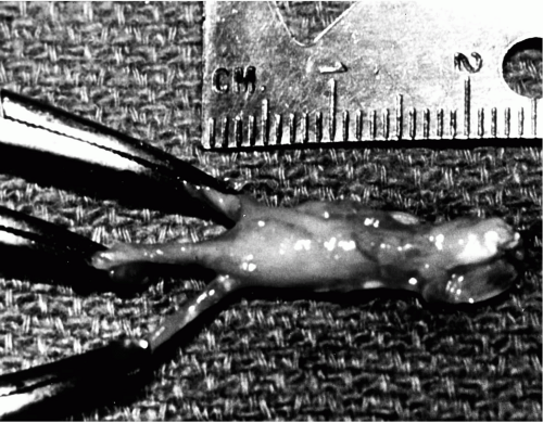
On gross examination, a resected specimen of Morton’s neuroma has a shiny, glistening surface that is white to buff yellow. It is generally fusiform and should have two or more easily identifiable digital branches with a distinct bifurcation. Either branch may have a swelling along its course as well. Usually, the nodule can be dissected easily from the surrounding fibroadipose tissue. However, it can be firmly attached to the intermetatarsal bursa or enwrapped in the bursa itself (Fig. 5). In some instances, the bursa may also envelop the lumbrical tendon. The findings of several investigators indicate that the neuroma does not have to be particularly large, nor does it have to be present for a substantial amount of time, to cause pain and for the nerve to undergo significant pathologic changes (38,57,58).

The histopathology is central to the diagnosis and origin of Morton’s neuroma. No histologic examinations of Morton’s neuroma were published until that of Betts in 1940 (59). Since then, there have been many investigations into the histopathology of this lesion (Table 4). Actually, Morton’s neuroma is neither a true neuroma nor a neoplasm. The term neuroma refers only to tumorous nodules that are formed by neural proliferation and dense fibrosis involving both axons and sheath (Schwann’s cells) (60).

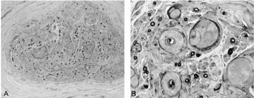
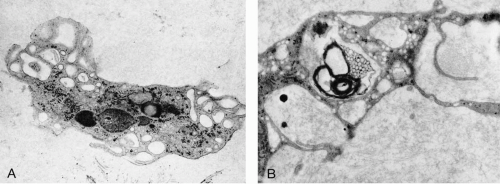
The histopathologic investigations of numerous authors support the observations that Morton’s neuroma consists of degenerative alterations of one or more intermetatarsal nerves (Fig. 6). This process is characterized by the following: endoneural and neural edema (early stages); perineural, epineural, and endoneural fibrosis and hypertrophy (late stages); Renaut’s body formation, usually in a subperineural location; hyalinization of the walls of endoneural blood vessels; subintimal and perivascular fibrosis that may lead to occlusion of local blood vessels (resembling a healed vasculitis); mucinous changes endoneurally and perineurally; and demyelination with axonal loss (60, 61, 62, 63, 64, 65, 66, 67, 68, 69, 70, 71, 72, 73, 74). Goldman noted perineural epithelial cells lying freely in the endoneurium and assumed that they played a phagocytic role (64) (Fig. 7).


FIG. 5. Gross specimen of resected Morton’s neuroma. Bifurcate digital branches and the fusiform enlargement are clearly visible.


TABLE 4. Histopathology of Morton’s neuroma: a chronology


Authors and year


Material


Findings


Betts, 1940


19 cases


The first to make a histologic examination; found increased fibrous tissue elements


Cottrell, 1940


Found progressive thickening of the walls of the endoneural vessels and the perineurium with aging in normal nerves


McElvenny, 1943


12 specimens, 11 patients


Neurofibromas or angioneurofibromas; Renaut’s bodies


Baker and Kuhn, 1944


14 specimens, 11 patients


Resembled amputation neuroma: reactive hyperplasia


King, 1946


5 cases


“Sclerosing neuroma”


Bickel and Dockerty, 1947


18 cases


Interstitial edema and irregular demyelination


Nissen, 1948


27 cases


Local vascular degeneration: an ischemic lesion


Winkler, Felner, and Kimmelstiel, 1948


20 cases


Essentially a degenerative process


Ringertz and Unander-Scharin, 1950


18 specimens, 21 controls


Endoneural edema and demyelination are the characteristic features of Morton’s neuroma


Scotti, 1957


17 cases


“Endarteritis-fibrosis complex’


Viladot and Moragas, 1966


7 cases


Similar degenerative changes; no sign of inflammation or thrombotic injury


Meachin and Abberton, 1971


23 specimens, 19 patients, 23 controls


Nonspecific degenerative reactive changes


Reed and Bliss, 1973


118 cases


“Regressive and productive intermetatarsal elastofibrositis;” elastin tissue proliferation in the fibroadipose tissue surrounding Morton’s neuroma


May, 1976


31 specimens


“Traumatic neuritis” with proliferation of connective tissue elements: neurofibroma


Lassmann, 1976


105 patients


Thickened perineurium, thickened walls of endoneural vessels, endoneural edema and sclerosis, axonal degeneration without wallerian degeneration


Tate and Rusin, 1978


50 specimens


Marked fibrosis and collagenous infiltration; demyelination; axonal degeneration


Goldman, 1979, 1980


133 cases


Demyelination diagnostic of intermetatarsal neuroma; observed distal wallerian degeneration as evidence of axonal degeneration and shift to left in axon diameter population; pathologic process not sparing any components of the peripheral nerve; detailed degenerative activity plus some axon regeneration; Renaut’s bodies


Lassmann, 1979


133 cases


Endoneural vascular fibrosis


Mann and Reynolds, 1983


65 specimens, 11 recurrent neuromas


Similar degenerative changes; classic pathologic findings of neuromas a traumatic neuroma in recurrent lesions


Graham and Graham, 1984


24 neuromas


Venous congestion, entrapment changes, nerve enlargement distal to the transverse metatarsal ligament


Guiloff, Scadding, and Klenerman, 1984


16 patients


Similar degenerative changes: Renaut’s bodies common


Shereff and Grande, 1991


10 specimens


Edema of the endoneurium, fibrosis beneath the perineurium, axonal degeneration and necrosis characteristic of nerve impingement


Bourke, Owen and Machet, 1994


15 patients


“Except for possible demyelination, the so-called typical histologic changes of fibrosis and swelling are similar to that seen as presumed degenerative disease in elderly patients”


FIG. 6. A: Light micrograph. Note the endoneural and perineural fibrosis, the large hyalinzed nodules in the endoneurium, and the dropping out of myelimated fibers (800× magnification). B: Light micrograph. Note the vessels with thickened walls, the hyalinized nodules, and the endoneural fibrosis (2,000× magnification). (Courtesy of Flair Goldman, D.P.M.)

In advanced cases of Morton’s neuroma, Renaut’s bodies (hyaline granules) have been observed in the endoneural tissue (25,61,64). These degenerative structures, first described in 1881 (75), are a nonspecific response to nerve trauma, particularly to compression (76, 77, 78).


FIG. 7. A: Electron micrograph. Note the giant vacuolated endoneural fibroblast (20,000× magnification). B: Electron micrograph. Note the degenerating myelin engulfed by the perineural epithelial cell. Also note multiple micropinocytotic vesicles (25,000× magnification). (Courtesy of Flair Goldman, D.P.M.)

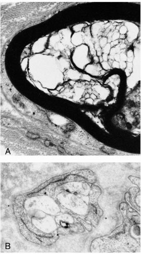
Finally, wallerian degeneration, or fatty degeneration of a nerve fiber caused by loss of nutrition, has been observed in Morton’s neuroma. In addition, evidence of regeneration of axons may in part explain the symptoms associated with the neuroma (24,42,79) (Fig. 8).

Several of the degenerative changes seen histologically in the plantar intermetatarsal nerves are the same in both control specimens and in patients with symptomatic pathologically confirmed Morton’s neuroma. More characteristic
of Morton’s neuroma are demyelination, endoneural edema, and a greater degree of subperineural fibrosis (74,80). Although endoneural edema can be associated with inflammation, only one study observed a leukocytic infiltration into the nerve, and that was only in half of their specimens (n = 10) (73).


FIG. 8. A: Electron micrograph. Note the vesicular degeneration of myelin (18,200× magnification). B: Electron micrograph. Note the cluster of regenerating axons (34,000× magnification). (Courtesy of Flair Goldman, D.P.M.)

In summary, Morton’s neuroma is a progressive, degenerative, and at times regenerative process in which early and late pathologic changes maybe found. Although the intermetatarsal nerves and blood vessels undergo many degenerative changes, endoneural edema, excess fibrosis, and demyelination are diagnostic of the disorder. Therefore, perineural fibroma may be a better term for this lesion. It appears from histopathologic studies that Morton’s neuroma is the result of both crushing and stretching trauma to the interdigital nerves, particularly in the third intermetatarsal space. Goldman was more specific and suggested that the epineurium responds to mechanical compression, whereas the perineurium responds to stretch (64).


ETIOLOGY AND BIOMECHANICS

Many theories have been proposed for the etiology of Morton’s neuroma (Table 5). Histologic findings leave little doubt that the syndrome is indeed a mechanical entrapment neuropathy (23, 24, 25, 26,73). The deep transverse intermetatarsal ligament appears to be the anatomic structure most likely to be responsible for this process.

Less clear is the exact source of these mechanical entrapment forces. A common observation is that most intermetatarsal neuromas occur in the pronated foot (12,38,81,82), where there are not only excessive stretch forces imposed on the interdigital nerves but also compressive and shearing forces from the adjacent hypermobile metatarsal heads (83, 84, 85). The nerve is also subjected to increased trauma by the motion occurring between the medial and lateral columns of the foot, which are divided between the third and fourth metatarsals.

In addition, as the medial and lateral plantar nerves dive plantarly under the arch, a stretch is placed on these nerves during prolonged midstance pronation as the foot is everted, abducted, and dorsiflexed. Tension is increased as these nerves travel around the flexor digitorum brevis sling and are drawn up tightly against the plantar and anterior edge of the unyielding deep transverse intermetatarsal ligament (65). This may help to tether the nerves in both the third and the second intermetatarsal spaces. Further tension and compression on the nerve occurs across this ligament when the toes hyperextend at the metatarsophalangeal joint (23,57,59,61,65,86, 87, 88). Thus, occupations requiring repetitive toe hyperextension can result in the development of an intermetatarsal neuroma, regardless of foot type.

Finally, pointed-toe or narrow shoes can further add compressive forces that favor the production of intermetatarsal neuromas in any foot type (61,65). High-heeled shoes not only throw weight forward onto the ball of the foot and jam the toes into the narrow front of the shoe, but also they force the toes into hyperextension and encourage nerve compression.


Intermetatarsal Bursa

At the level of adjacent metatarsophalangeal joints, an intermetatarsal bursa lies in close proximity to Morton’s neuromas (12,38,58,62,66,67,89, 90, 91, 92, 93, 94, 95). These features have been confirmed on MRI scans (49). Inflammation of these structures creates swelling and compression on the adjacent nerve and may explain some of the acute symptoms associated with neuromas, especially when the patient has a prompt response to corticosteroid injections.

Only gold members can continue reading. Log In or Register to continue

Stay updated, free articles. Join our Telegram channel

Jul 26, 2016 | Posted by in MUSCULOSKELETAL MEDICINE | Comments Off on Morton’s Neuroma

Full access? Get Clinical Tree

Get Clinical Tree app for offline access