Juvenile Hallux Abducto Valgus Deformity
Luke D. Cicchinelli
Aprajita Nakra
Juvenile hallux abducto valgus is a commonly encountered complex deformity. This condition has also been referred to as adolescent bunion, metatarsus primus varus, and metatarsus primus adductus. Specific criteria for defining juvenile or adolescent hallux abducto valgus deformity are lacking, although some investigators have classified the condition as occurring in a person aged 20 years or less because of the relatively plastic nature of the components of the deformity (1). Other investigators have referred to the deformity as manifesting during the formative years of life, roughly from 11 to 19 years of age (2, 3, 4, 5).
The origin of hallux abducto valgus is multifactorial. In 1895, Boniface categorized the various etiologies into four groups: mechanical, muscular, diathetic, and anatomic (6). Root et al. described the primary etiologic factors for development of hallux abducto valgus as hypermobility of the first ray, rheumatic inflammatory disease, neuromuscular diseases, and postsurgical malfunction (7). Schwitalle et al. devised a classification that was more specific for children, including congenital, neurogenic, and idiopathic types (8). Congenital hallux valgus is almost always associated with other deformities such as polydactyly, cleft foot, and tumors, especially enchondroma. Some congenital cases may represent a limb bud deficiency or an actual developmental anomaly (1). Neurogenic hallux valgus is the result of spastic or paralytic palsy with a muscular imbalance that typically affects the foot as a whole. As such, the clinical and radiographic evaluation may reveal some of the pathologic entities affecting the overall alignment of the first metatarsophalangeal joint (9). However, in other patients, the cause is less certain.
Hereditary factors are perhaps the most commonly cited cause of juvenile hallux abducto valgus deformity. A positive family history for the condition has been noted in 68% (10) to 80% (11) of patients. The factor predisposing to the evolution of a hallux valgus may be transmitted by an autosomal dominant trait showing incomplete penetrance. Full penetrance may result in a much earlier onset and a more severe deformity.
Although tight or poorly fitted shoes are often blamed by patients for the presence of hallux abducto valgus deformity, the role of shoes in the creation of this condition is likely overstated. However, the incidence of juvenile hallux valgus has been shown to be higher in the shod population than among those who walk barefoot (12,13). Poorly fitted shoes have been proposed as a contributing factor in the progression of the juvenile hallux valgus deformity (14), although the association of tight shoes with juvenile hallux valgus has not been examined (15). Shoes should be viewed as more of an aggravating factor once the condition is evident than a primary cause.
Juvenile hallux valgus is usually a manifestation of abnormal biomechanical function of the lower extremity that is related to anatomic variations in structures and acquired changes resulting from external forces. With the exception of congenital anomalies or inflammatory diseases, the progression of hallux abducto valgus in the juvenile patient is usually related to the severity of biomechanical disorder (16). According to Piggott, a congruous metatarsophalangeal joint is stable and does not progress to significant hallux abducto valgus deformity (5). However, once the joint has begun to sublux, it is at significant risk for progressive deformity.
Numerous anatomic and biomechanical conditions have been proposed as leading to the development of juvenile hallux valgus (17). This diversity is probably a result of the different emphasis placed on each entity by different investigators, and it also reflects an inconsistent interpretation of the relationship between potential causes and effects (8). Furthermore, many conditions may be responsible for the development of juvenile hallux abducto valgus, and the origin may be multifactorial.
Truslow described an oblique position of the first metatarsal bone as the initial deforming influence in the creation of metatarsus primus varus (18). This divergence was interpreted as an anatomic variation and not an acquired deformity. Lapidus believed that the atavistic cuneiform, similar
to the prehensile great toe of higher primates, accounted for the increased medial deviation of the first metatarsal (19). He believed that an inherent instability in the medial column of the foot predisposed it to the formation and propagation of a bunion deformity. Other anatomic variants that have been purported to predispose a young patient to hallux valgus include a round first metatarsal base, long and short first metatarsal, oblique positioning of the first metatarsocuneiform joint, and the presence of a lateral exostosis or an os intermetatarsia at the base of the first metatarsal (16,20). However, definitive scientific evidence to confirm the influence of these factors in the development of juvenile hallux valgus is still lacking.
to the prehensile great toe of higher primates, accounted for the increased medial deviation of the first metatarsal (19). He believed that an inherent instability in the medial column of the foot predisposed it to the formation and propagation of a bunion deformity. Other anatomic variants that have been purported to predispose a young patient to hallux valgus include a round first metatarsal base, long and short first metatarsal, oblique positioning of the first metatarsocuneiform joint, and the presence of a lateral exostosis or an os intermetatarsia at the base of the first metatarsal (16,20). However, definitive scientific evidence to confirm the influence of these factors in the development of juvenile hallux valgus is still lacking.
In staging the progression of hallux abducto valgus, some authors considered that the abduction of the hallux preceded the development of metatarsus primus adductus (7,21,22). Piggott found that subluxation of the first metatarsophalangeal joint occurred with intermetatarsal angles as small as 7 degrees (5). Kalen and Brecher also showed that metatarsus primus varus was nearly absent in adolescents with hallux abducto valgus deformity, yet the hallux was almost always abducted (23). The opposite view, that an increased intermetatarsal angle is the initial abnormality associated with hallux abducto valgus, has been explained by authors such as Truslow and Lapidus, as described previously.
Many investigators agree that any factor that causes abnormal subtalar joint pronation and a subsequent hypermobile first ray enhances the progression of juvenile bunions (24). Although they are seldom the reason for seeking professional care, metatarsus adductus, pes valgus deformity, ankle equinus, and suprastructural deformities such as medial femoral or tibial torsion may directly influence the bunion deformity (25,26). Yu et al. recommended that the hallux valgus deformity should be regarded as a sign or symptom of another underlying deformity rather than as an isolated condition (27).
Pontious et al. proposed a classification system based on the belief in two distinct types of patients with juvenile hallux abducto valgus deformity (28). The first group (type I) consists of a patient with significant metatarsus adductus, more severe deforming forces (equinus and flatfoot), a strong familial history, and an earlier onset. The second group (type II) is composed of patients with moderate deformity, a more rectus foot type, controllable deforming forces, or later onset with slower progression of deformity (28).
CLINICAL FEATURES
The typical patient with juvenile hallux abducto valgus presents between the ages of 10 and 16 years. Pain is not as common a complaint as in the adult, but the primary concern may be the appearance of the deformity. However, girls may describe an inability to wear a certain style of shoes, discomfort from activity-specific shoes, and increased concerns regarding self-image and perception of their bodies. Adolescent boys may be less concerned with appearance, but they frequently have questions regarding discomfort in sport shoes. There may be a family history of the condition, and therefore the patient or parents may express concerns regarding the progression of the deformity, a desire to explore treatment options that may control or prevent further development of the deformity, or a need to understand surgical options and timing. The patient’s ability to participate in physical activities may help to determine the level of disability in those patients reticent to complain. A family history of hallux abducto valgus deformity may assist in determining the likelihood of further progression.
Clinical evaluation of the affected side reveals the amount of transverse and sagittal plane deviation. Juvenile hallux valgus differs significantly from the adult bunion in that the deviation of the toe is less pronounced, the medial eminence is smaller, bursal thickening is rare, and a rotation deformity of the hallux is present only in patients who have a more pronounced condition. Degenerative changes at the metatarsophalangeal joint are minimal, and the phalangeal and metatarsal epiphyses may have yet to close (29).
The presence of digital deformities or plantar callosities may also help in determining the overall nature of the condition. The range of motion of the first metatarsophalangeal joint may be assessed in both the resting position and under manual reduction. Lack of ability to reduce the hallux passively is referred to as “a track bound joint” and historically was believed to indicate deviation of the affected articular cartilage, but it more likely represents displacement of the sesamoid apparatus and contracture of the soft tissues (26,28). However, in patients with juvenile hallux abducto valgus deformity, a higher degree of proximal articular set angle deviation has been observed (28).
Examination of the alignment of the foot and the motions therein may be helpful in identifying potential influences in the formation of juvenile hallux valgus deformity. Ankle equinus can have a profound effect on midfoot stability by unlocking the midtarsal joint with resultant distal hypermobility and juvenile hallux valgus. Pes valgus deformity and metatarsus adductus can potentially compromise the repair of juvenile hallux valgus and may need to be considered in the surgical approach to the patient.
Progression of the deformity may lead to secondary problems such as digital contractures and plantar callosities. Painful degenerative hallux valgus in an adult is often the result of untreated deformities in the growing adolescent. Appropriate and timely treatment of juvenile hallux valgus is helpful in avoiding the more pronounced and permanent ligamentous, articular, and osseous changes that ensue over time.
The treatment protocol for juvenile hallux valgus remains controversial. Many factors must be taken into consideration (27), including the following:
The age of the patient and the status of the growth plate (open or closed)
The flexibility of the deformity
The onset and progression of the deformity
The coexisting etiologic factors
The presence of a coexisting deformity
Family history
The degree of symptoms
The patient’s and parents’ expectations
RADIOGRAPHIC EVALUATION
Radiographs are an important part of the evaluation of a patient with juvenile hallux abducto valgus. In most instances, this evaluation includes the assessment of weight-bearing dorsal plantar and lateral views. Sesamoid axial and oblique views may also be of benefit in some instances. Neutral-position lateral and dorsal plantar views have been recommended in some instances to help reveal compensated deformity such as metatarsus adductus (25). However, this is seldom necessary. The relationships that appear to be of primary importance are the intermetatarsal angle, the metatarsus adductus and hallux abductus angles, and the sesamoid position. Other features that may be evaluated are the amount of hallux abductus interphalangeus, the overall shape of the first metatarsal head, the presence of any accessory bones between the first and second metatarsal bases, and the length of the first metatarsal. A relatively low intermetatarsal angle or a long first metatarsal may belie an underlying metatarsus adductus deformity (27) (Figs. 1 and 2). Proximally, other features to observe are the growth plate and any apparent obliquity of the first metatarsocuneiform joint.
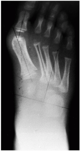 FIG. 1. Although the intermetatarsal angle is low, early bunion deformity is appreciated in association with a substantial metatarsus adductus deformity. |
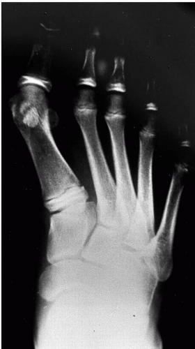 FIG. 2. Note the appearance of a long first metatarsal in the presence of metatarsus adductus as a result of the transverse plane adduction of the forefoot on the midfoot. |
Of particular note is the lateral displacement of the sesamoid bones. Deviation of these structures is evidence of a mechanical advantage of the lateral soft tissue structures and flexor hallucis longus. If the condition is allowed to persist, these soft tissue structures will increase the likelihood of further progression of the deformity (27).
Attempts to assess the proximal articular set angle radiographically are unreliable, and this relationship is best assessed intraoperatively (28). Historically, the shape of the first metatarsal head was believed to be important in determining the transverse plane stability of the metatarsophalangeal joint. A round metatarsal head was purportedly associated with a greater amount of joint instability and a greater tendency toward hallux abducto valgus, whereas a square head was considered more likely to resist abductory forces at the first metatarsophalangeal joint. However, screening of 6,000 school children between the ages of 9 and 10 years for hallux abducto valgus deformity revealed only a weak relationship between the shape of the metatarsal head and the degree of hallux abducto valgus (30).
Assessment of the rearfoot and midfoot joints may reveal positional or structural abnormalities that may influence the forefoot and the formation of juvenile hallux valgus deformity. This is particularly pertinent in patients with suprastructural deformities or collapsing pes valgus conditions. Yu et al. proposed that juvenile hallux valgus deformity may
be classified radiographically as either static or dynamic (27). The dynamic form results primarily from hypermobility of the forefoot secondary to biomechanical abnormalities, with the first metatarsophalangeal joint the primary site of deformity. In these cases, the extent of lateral deviation of the great toe is more likely to create a retrograde buckling force on the metatarsal head and therefore to increase the splaying between the first and second metatarsals. In addition, the eccentric loading of the metatarsophalangeal joint may cause lateral articular cartilage deviation and abduction.
be classified radiographically as either static or dynamic (27). The dynamic form results primarily from hypermobility of the forefoot secondary to biomechanical abnormalities, with the first metatarsophalangeal joint the primary site of deformity. In these cases, the extent of lateral deviation of the great toe is more likely to create a retrograde buckling force on the metatarsal head and therefore to increase the splaying between the first and second metatarsals. In addition, the eccentric loading of the metatarsophalangeal joint may cause lateral articular cartilage deviation and abduction.
The static form of juvenile hallux abducto valgus primarily presents as a deformity of the metatarsal itself or of the metatarsal cuneiform joint. The hallmark of the dynamic deformity is abduction of the hallux and an increased intermetatarsal angle. However, patients with the static deformity present with or without abduction of the great toe, yet with a high intermetatarsal angle. A rhomboidal shape of the metatarsal may result from an abnormally wide and hypoplastic lateral epiphyseal growth plate. Deviation may also arise from the articular surface of the medial cuneiform (27). Investigators have suggested that an increase in obliquity of the distal articular facet of the medial cuneiform indicates a hyperadduction of the first ray (26).
ASSOCIATED DEFORMITIES
Deformities associated with juvenile hallux abducto valgus may be those that contribute to the origin of the condition or those that are produced as byproducts of the early onset of hallux valgus. As previously discussed, predisposing conditions such as torsional deformities, pes valgus deformity, and ankle equinus may affect the long-term success of correction of juvenile hallux valgus. A significant contributing factor to juvenile hallux valgus is metatarsus adductus. The association of metatarsus adductus with juvenile or adolescent hallux valgus deformity has been recognized by several authors (17,28,31,32). Greater levels of metatarsus adductus have been associated with greater amounts of hallux abductus in adult patients (32). A statistically significant association has been identified between increasing metatarsus adductus angle and hallux abducto valgus in adolescent patients undergoing surgery (17). Pontious et al. also noted a 75% incidence of metatarsus adductus in adolescent patients with hallux abducto valgus (28). Furthermore, significantly higher rates of recurrence of deformity were noted in juvenile and adolescent patients because of an unappreciated metatarsus adductus deformity. Metatarsus adductus has also been noted to be more prevalent in adult patients with hallux abductus (31).
Related posts:

Stay updated, free articles. Join our Telegram channel

Full access? Get Clinical Tree


