Metastatic Disease About the Knee: Evaluation and Surgical Treatment

Chapter 152 Metastatic Disease About the Knee


Evaluation and Surgical Treatment


According to the latest cancer statistics, at least 1,368,030 new cancer cases are diagnosed annually in the United States.12 Those cancers with a propensity for bone metastases rank among the most commonly diagnosed types of new cancers. Both breast and prostate cancer rank first in numbers of new cases for women and men, respectively. Lung cancer ranks second for men and women, kidney cancer ranks seventh among men, and thyroid cancer ranks eighth among women.


Metastatic disease to bone occurs less commonly about the knee than it does in more proximal femoral sites, but the distal femur is not an uncommon site of metastatic lesions or pathologic fractures.8 Metastatic disease is particularly rare distal to the knee joint, just as it is distal to the elbow in the upper extremity. Metastatic disease to the tibia has been estimated to account for only 4% of pathologic fractures, although lesions in this location are probably more frequent.10,13 Metastatic involvement in the patella is rare but has been reported.14,17 In part, the less frequent involvement by metastatic disease in these sites has presented a challenge to its orthopedic management because of the paucity of literature available on which to base guidelines.


In addition, the relatively distal location in the extremity presents its own challenges for diagnosis and treatment. The distal femur and proximal tibia are comprised predominantly of cancellous metaphyseal bone, so lesions may become large before they are evident radiographically. The occurrence of metastatic disease to bone or synovium following total knee replacement is a rare occurrence but should be kept in mind as a potential source of pain after total knee replacement.1,4,6,20


Fixation is also a potential problem. Supracondylar femur and tibial plateau fractures in the older population most commonly affected by metastatic disease frequently occur in osteoporotic bone, making fixation difficult, even when there is no tumor involvement. The presence of tumor increases the level of difficulty. Currently available fixation devices designed specifically for the distal femur do not allow the surgeon to accomplish the goal of protecting the entire femur, including the intertrochanteric region and femoral neck, from coexistent or subsequent development of other lesions. Plate and screw devices designed for the proximal tibia carry the same limitation below the knee, whereas intramedullary devices for the proximal tibia may be technically challenging.


Hence, metastatic disease about the knee is a topic of great interest to the orthopedic oncologist and general orthopedic surgeon alike. Comparatively little is known and much research is needed. This chapter will describe the general treatment principles of metastatic disease to bone, evaluation of the patient with suspected metastatic disease, prediction of pathologic fracture, and surgical management.


Treatment Principles


Operative management of metastatic lesions to the distal femur or proximal tibia should follow the same principles of treatment as for metastatic lesions to any site. The most important principle is that a patient’s recovery time from surgery should not outlast their expected survival. Even patients with a 4- to 6-week life expectancy are likely to benefit from improved quality of life if the operative intervention improves their overall function. Although the recovery time following an intramedullary rodding or plate fixation is relatively short, poor fixation, which requires a prolonged period of restricted weight-bearing to allow healing, does not benefit the patient. Cement supplementation or prosthetic replacements that obviate the need for bone healing and allow full weight bearing immediately decreases the recovery time and does benefit the patient. Hard and fast rules regarding minimum life expectancy to warrant operative intervention are less important than an individualized assessment of the patient in conjunction with the medical oncologist and family members. In general, a patient should be expected to survive the hospitalization, from 30 to 90 days postoperatively. A moribund, immediately preterminal patient is not considered a good operative candidate.


The corollary to the minimum survival rule is that the operation should result in a stable reconstruction that allows immediate weight bearing and durability for the patient’s shortened life span. The improved function and pain relief provided by an immediately stable construct will translate into improved quality of life over the period of limited survival. Although a cemented endoprosthetic reconstruction will usually accomplish this goal, the same may often be achieved by internal fixation. In a fracture situation, supplementing internal fixation with bone cement will often allow immediate weight-bearing, but fixation durability relies on fracture healing. If the patient survives long enough and the fracture fails to heal, the construct will likely fail. The healing of pathologic fractures is slowed by local disease progression and by radiotherapy, but is also closely related to the underlying disease process and expected survival. Patients with metastatic lung cancer, for example, rarely survive longer than 3 to 6 months, so their fractures rarely heal. Some fractures in patients with breast carcinoma metastases will heal, given appropriate treatment, because many will live longer than 6 months. Even the best reported healing rate for pathologic fractures, which occurred for multiple myeloma, was only 67%.7


The role of polymethylmethacrylate (PMMA) as an adjunct to the treatment of pathologic fractures is well established, both for internal fixation devices and for endoprostheses. As a supplement to internal fixation devices, bone cement improves pain relief and ambulation for lower extremity fixation devices more than that of internal fixation alone. This is particularly true in metaphyseal regions, such as those about the knee. In contrast, when metastatic lesions occur in diaphyseal locations, a number of series have demonstrated the success of intramedullary rodding without adjuvant bone cement. Even in diaphyseal locations, however, cement adds stability to the construct, so its use in addition to intramedullary rodding remains controversial. As the means of fixation for endoprostheses, bone cement obviates the need for healing by bony ingrowth into porous-coated implants which can be hampered not only by the underlying disease process but also by radiotherapy.


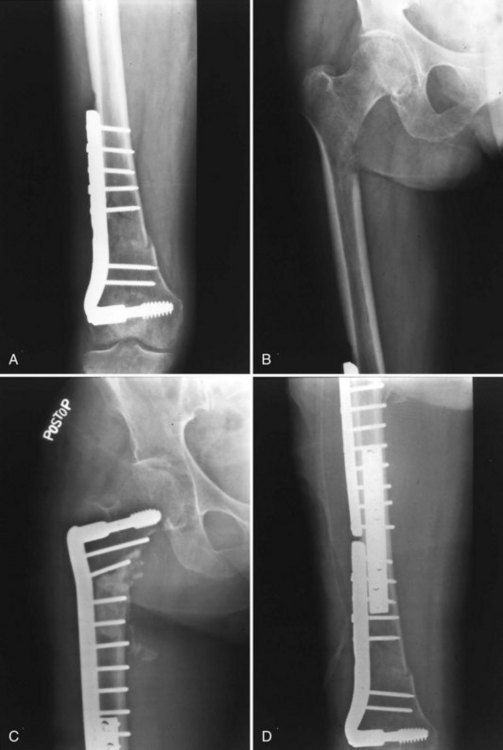
One of the most important but often neglected axioms for orthopedic treatment of metastatic disease is the protection of the entire bone proximal and distal to the lesion (Fig. 152-1). This is especially important to address preexisting bone lesions that may progress to weaken the bone or cause fracture, but it is also recommended prophylactically to address new sites that may arise as the disease progresses. With fixation of the distal femur with a retrograde intramedullary nail or long-stemmed femoral total knee component, the intertrochanteric region and/or femoral neck remain unprotected and are frequent sites of metastases. Failure to consider proximal femoral impending pathologic fractures may result in pathologic fracture proximal to an internal fixation or prosthetic device. Prior to operative intervention, radiographs of the entire affected bone should be reviewed.


Finally, radiotherapy should be used postoperatively to protect the entire instrumented region of the bone. When intramedullary reaming is done, the entire bone should receive irradiation. Failure to irradiate postoperatively leads to a higher rate of implant failure and lower functional benefit overall.18 Consultation should be obtained with a radiation oncologist preoperatively or in the early postoperative course for this purpose. When incisions are laterally placed, they can often be avoided by anteroposterior-posteroanterior (AP-PA) radiation treatment, but incisions within the actual radiation field should usually be allowed to heal completely before initiating radiotherapy.


Evaluation


Before treatment may be undertaken, the correct diagnosis must be established. The diagnosis of metastatic disease as the cause for a specific new bone lesion in any location should never be assumed unless the patient already has biopsy proven metastatic disease. The danger lies in incorrectly treating a primary bone sarcoma as a metastatic lesion, with typically disastrous results for the patient and physician. In the age group of those older than 40 years, sarcomas are rare compared with metastatic disease, multiple myeloma, and lymphoma, but they do occur. Furthermore, solitary metastatic lesions from renal cell or thyroid carcinoma may be treated by resection for cure rather than internal fixation, so this scenario should also be given due consideration. Even in a patient with a history of a malignancy with a propensity for bone involvement, the first bone metastasis usually should be proven by biopsy before proceeding with operative intervention. Beyond the consideration of mistreating a sarcoma, some lesions, such as those from metastatic renal carcinoma, myeloma, and thyroid carcinoma, are notorious for being highly vascular. Consideration should be given to preoperative embolization for these vascular lesions.


Primary malignancies with a propensity for bone involvement include breast, prostate, lung, kidney, and thyroid carcinoma, although almost all primary sites have been reported to involve bone. Most breast and prostate bone metastases will present in patients with an established diagnosis of the corresponding primary, whereas the most common sources for a bone metastasis without an established primary are lung and renal carcinomas.


For patients without an established diagnosis, a comprehensive history should seek any history of prior biopsies or tumor excisions, however remote (Table 152-1). Physical examination should also be comprehensive, including evaluation of the breast or prostate, abdomen, and thyroid and may be best performed by an internist or oncologist. Lymphadenopathy should also be sought. Serologic examination, including serum and urine protein electrophoresis for multiple myeloma and prostate-specific antigen in men, should be done along with checking the serum calcium level to identify hypercalcemia. Radiographic evaluation, including chest radiography and mammography, when appropriate, should be done initially, followed by chest computed tomography (CT), and abdominal-pelvic evaluation by CT or ultrasound. This approach will reveal the diagnosis in approximately 85% of patients.16


Table 152-1 Key Elements in Evaluation of Patient With Metastatic Disease of Unknown Primary


Key Element Features of Metastatic Disease to Evaluate
History Carcinomas, biopsies; purpose of hysterectomy, TURP
Physical examination Breast or prostate examination, thyroid, abdominal masses, lymphadenopathy
Laboratory evaluation CBC, SPEP-UPEP, PSA, LDH, calcium levels
Radiographic evaluation Chest radiograph, radiographs of entire affected bone
Total skeleton 99Tc bone scan
CT scanning of chest, abdomen, pelvis

CBC, Complete blood count; LDH, lactate dehydrogenase; PSA, prostate-specific antigen; SPEP, serum protein electrophoresis; 99Tc, technetium-99; TURP, transurethral resection of prostate; UPEP, urine protein electrophoresis.


Submitting intramedullary reamings as biopsy material during placement of a retrograde nail for a distal femoral lesion is not appropriate when the diagnosis is in doubt. The same can be said for reamings obtained during placement of a tibial intramedullary nail for a proximal tibial lesion. If the patient is subsequently found to have a sarcoma, the entire knee joint, as well as the reamed portion of the femur or tibia, respectively, would have to be considered to be contaminated. A straight, longitudinal, laterally placed extracapsular incision is appropriate for lesions of the distal femur. However, when the distal femoral cortex is breached medially and there is soft tissue extension in that direction, a medial biopsy is preferred. The proximal tibial is more commonly biopsied anteromedially. When possible, the defect in the cortex in the distal femur or proximal tibia should be made through the thin metaphyseal bone to minimize the additional weakening that occurs when more structurally sound diaphyseal cortex is breached. When biopsy of a small metaphyseal or epiphyseal lesion establishes the diagnosis of metastatic disease, curettage and cementation of the region, with or without reinforcement pins, followed by postoperative irradiation, may suffice as treatment.


Although the surgical treatment principles for multiple myeloma are essentially the same as those for metastatic carcinoma, the diagnosis of multiple myeloma can usually be made based on the finding of a monoclonal spike on the serum protein electrophoresis or Bence-Jones proteins in the urine. Hence, biopsy is less frequently needed once the serologic diagnosis of multiple myeloma has been established.

Stay updated, free articles. Join our Telegram channel

Aug 26, 2016 | Posted by in ORTHOPEDIC | Comments Off on Metastatic Disease About the Knee: Evaluation and Surgical Treatment

Full access? Get Clinical Tree

Get Clinical Tree app for offline access