Fig. 46.1
Plain radiograph of the proximal humerus. Purely lytic lesion, destroying the cancellous bone. This represents a completely lytic osseous metastasis in women with a follicular carcinoma of the thyroid
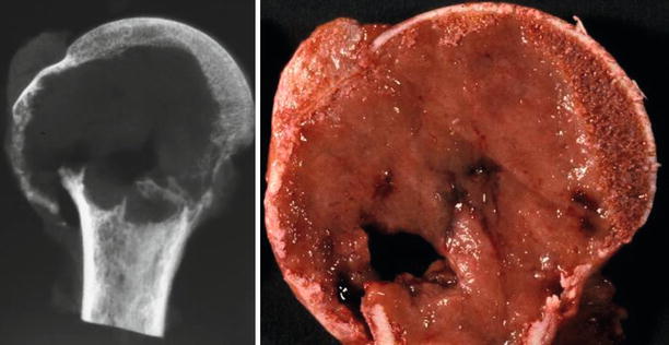
Fig. 46.2
Plain radiograph of the surgical specimen: The lesion is entirely lytic with pathologic fracture. The gross image shows a solid homogeneous parenchymatous brown lesion, with focal hemorrhage
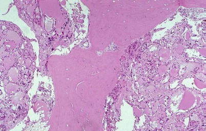
Fig. 46.3
Follicular adenocarcinoma of the thyroid infiltrating the cancellous bone. Follicular structures containing pink eosinophilic colloid material are present
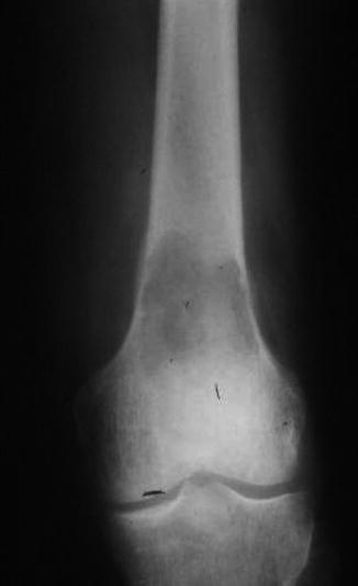
Fig. 46.4
Plain radiograph shows a large lucent lesion on the distal metaphysis of the femur
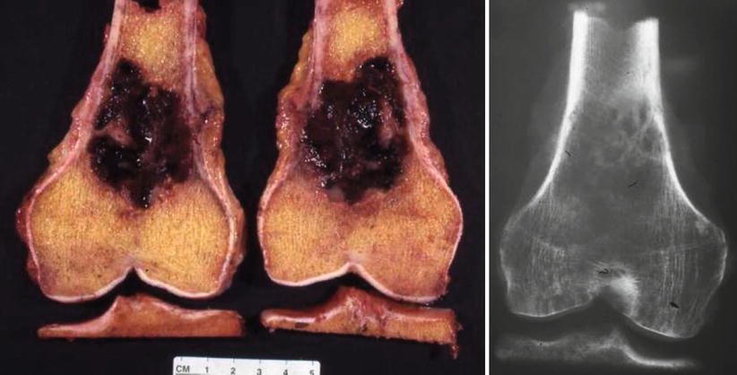
Fig. 46.5
Gross image shows a hemorrhagic lytic destructive lesion along with radiograph of the surgical specimen. The expansile red tumor destroys the cancellous bone and focally involves the cortical bone
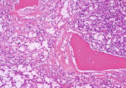
Fig. 46.6


The histology shows metastatic renal cell carcinoma, clear cell type. There is preexisting residual cancellous bone embedded in a mainly clear and focally eosinophilic malignant cell proliferation
Related posts:


Stay updated, free articles. Join our Telegram channel
Full access? Get Clinical Tree
