Fig. 51.1
Metaphyseal fibrous defect (MFD) of the distal femur. (a, b) Anteroposterior and lateral X-ray showing a large radiolucent lesion, with well-defined sclerotic border. (c) Axial CT scan showing the defect with posterior disruption of the cortex
Fig. 51.2
(a, b) Anteroposterior and lateral X-ray showing radiolucent, eccentrically lesion in the distal metaphysis of the femur, juxtaposed to the endosteal surface of one cortex
Fig. 51.3
(a, b) MFD. Roentgenographic anteroposterior and oblique view. Lytic, eccentric elongated lesion in the distal femur; the long axis of the lesion is parallel to the host bone
Fig. 51.4
(a, b) MFD in the posterior aspect of the femur. Anteroposterior and lateral radiographs showing a large lesion with the characteristic pattern
Fig. 51.5
X-ray anteroposterior view. (a) Male, 11 years old, with a small fibrous defect in the distal metaphysis of the femur. (b) Same lesion, 3 years later
Fig. 51.6
(a, b) Radiographic and computed tomographic scan image. Long evolution of MFD with sclerosis of the lesion
Fig. 51.7
(a, b) Radiographic images. Metaphyseal fibrous defect complicated with pathologic fracture
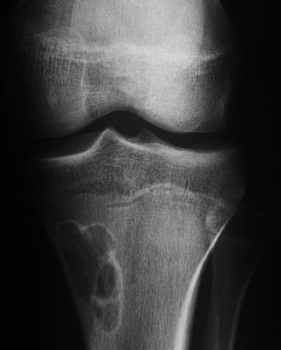
Fig. 51.8
MFD in the upper metaphysis of the tibia. Anteroposterior radiograph shows radiolucent, eccentric lesion with well-defined, sclerotic inner border
Fig. 51.9
(a, b) MFD of the tibia. Anteroposterior and lateral radiographs showing a lesion with the characteristic pattern
Fig. 51.10
(a, b) Anteroposterior and lateral X-ray, sclerotic old lesion in the proximal metaphysis of the tibia. (c) Axial CT image of the lesion clearly shows the sclerosis