Meniscal Transplantation without Bone Plugs
Fotios P. Tjoumakaris
Anthony M. Buoncristiani
Christopher D. Harner
The meniscus plays a vital role in the successful performance and well-being of the knee. Loss of the meniscus, in part or in total, significantly alters joint functions and predisposes the articular cartilage to degenerative change. Meniscal transplantation has been developed in an attempt to alter the natural history of the postmeniscectomy knee. The procedure is performed in hopes of forestalling progressive joint deterioration by theoretically restoring part of the meniscal function.
Surgical techniques for meniscal transplantation have evolved over the last decade from mainly open arthrotomies to arthroscopically assisted techniques, with or without the use of bone plugs (or blocks) at the meniscal horn attachments. Proper patient selection as well as realistic patient goals and expectations are vital to avoid unnecessary failures. Results from long-term outcome studies should help to further refine meniscal allograft techniques as well as its indications and improve our understanding of where this procedure belongs in the treatment algorithm of the postmeniscectomy knee.
INDICATIONS/CONTRAINDICATIONS
The ideal candidate for a meniscal allograft transplant has joint line pain secondary to meniscal deficiency and fits the following criteria: (a) documented evidence of a previous subtotal or total meniscectomy or its biomechanical equivalent; (b) ligamentously stable or concurrently performed ligament reconstruction; (c) early grade I or II articular cartilage changes as determined by the use of a 45 degrees posteroanterior (PA) flexion weight-bearing radiograph; (d) no evidence of malalignment as determined on standing long cassette views; and (e) no evidence of articular incongruity on diagnostic arthroscopy. All patients are informed during preoperative counseling that the ultimate decision to proceed with the transplant is made in the operating room. Contraindications to transplantation include grade III or IV changes in the involved compartment, ligamentous instability, malalignment that would place significant stress on the allograft, and a patient with unrealistic expectations regarding outcome.
PREOPERATIVE PLANNING
Physical Examination
Candidates being considered for meniscal transplant surgery require a comprehensive examination of both lower extremities. Height and weight should be recorded as an indication of body habitus as well as the standing alignment of the lower extremities. The patient’s ability to squat and any limitations in movement and associated discomfort are noted. It is important to observe the gait of the patient for any abnormalities. Both knees are examined for the presence of an effusion.
The patella must be examined and a thorough ligamentous examination performed with special attention placed on the anterior cruciate ligament (ACL) status. This is done through the Lachman test, pivot shift, and anterior drawer in neutral, internal, and external rotation. The posterior cruciate ligament (PCL) is evaluated with the posterior drawer and posterior sag with the hip and knee flexed to 90 degrees. Posterolateral rotatory instability is ruled out through the dial test, reverse pivot shift, and the posterolateral drawer. The collaterals are tested with varus and valgus stress at 0 and 30 degrees of knee flexion. It is important to elicit the presence or absence of joint line tenderness over the involved and the uninvolved compartments as well as the presence of hyperflexion signs.
Imaging
We routinely obtain four views of the knee in meniscal transplant candidates. This includes a PA 45degree flexion weight-bearing view, Merchant, lateral, and long cassette from the hips to the ankles to evaluate the mechanical axis. From these studies the alignment and joint space of the involved knee is calculated and compared with the opposite asymptomatic knee. Patients who have a mechanical axis deviation toward the involved compartment require a staged or combined osteotomy in addition to transplantation. We routinely obtain a magnetic resonance image (MRI) of the knee to assess the quality and amount of remaining meniscal tissue. In patients whose pain is nonspecific, a three-phase 99Tc bone scan is obtained. Increased uptake in the compartment indicates early degenerative changes.
Before the definitive procedure, the amount of remaining meniscus, the quality of the articular surface, and any associated ligamentous injuries must be verified so they may be addressed at the time of surgery. If necessary, this can involve an examination under anesthesia and a diagnostic arthroscopy staged before transplantation. The plain radiographs are also used to determine the appropriate size for the allograft tissue as required for the tissue bank.
SURGERY
Positioning, Draping, and Operating Room Organization
The patient is positioned supine on a regular operating room table with a sandbag taped to the table at the foot of the bed to hold the leg at 90 degrees of knee flexion. A standard side post is used at the level of the greater trochanter to provide a fulcrum for valgus stress and to provide lateral support with the knee at 90 degrees of flexion. A tourniquet is not routinely used for the procedure. The extremity is prepped from the tourniquet distally with alcohol and betadine and draped with a nonimpervious stockinette, extremity drape, and a half sheet to cover the post. A bolster is placed between the lateral thigh and the post (Fig. 10-1).
The operating room should be organized to provide a controlled environment where traffic and the potential for contamination are kept to a minimum. It is necessary to have a separate table for graft preparation that has an oscillating saw, drill, and instruments.
Specific Approaches
All incision sites as well as the joint are marked before prepping and draping, prepped out with a betadine stick, and injected with 1% lidocaine with epinephrine. A standard arthroscopy is performed using an anterolateral-viewing portal just adjacent to the patellar tendon and above the joint line and an anteromedial working portal 1 cm medial to the medial edge of the patellar tendon. Two main approaches are used in both the medial and lateral transplants: the anteromedial or lateral and the posteromedial or lateral approaches. The anterior approach involves an extension of the arthroscopy skin incision superiorly along the patellar tendon. A medial or lateral parapatellar arthrotomy is
made. Often it is necessary to remove a small amount of the fat pad to visualize the joint. The anterior horn of the remaining meniscus should be well visualized. Three No. 2 braided nonabsorbable sutures are passed around the remaining anterior horn, leaving the needles attached for later fixation to the allograft.
made. Often it is necessary to remove a small amount of the fat pad to visualize the joint. The anterior horn of the remaining meniscus should be well visualized. Three No. 2 braided nonabsorbable sutures are passed around the remaining anterior horn, leaving the needles attached for later fixation to the allograft.
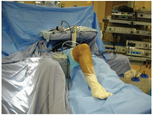 FIGURE 10-1 The extremity is prepped, draped, and positioned so that it can rest at 90 degrees of flexion. |
The posteromedial approach involves a skin incision 3 cm long just anterior to the posterior margin of the medial femoral condyle. One third of the incision should be above the joint line. Careful dissection is used to divide the subcutaneous fat, and care is taken to look for and protect the infrapatellar branch of the saphenous nerve, which typically crosses just above the joint line. The sartorial fascia (layer 1) is then divided in line with the skin incision and a posterior capsulotomy is made between the posterior border of the medial collateral ligament and the posterior oblique ligament (layer 2). Three No. 2 nonabsorbable sutures are passed around the posterior oblique ligament for later closure. The posterolateral approach involves a 3-cm skin incision just posterior to the lateral collateral ligament, two thirds below and one third above the joint line. The interval between the iliotibial band and the biceps femoris is developed using a combination of blunt and sharp dissection. The plane between the lateral head of the gastrocnemius and the posterolateral capsule is developed and a popliteal retractor is placed.
Details of Procedure
A standard arthroscopy is initially performed to evaluate the condition of the articular surface as well as the amount of remaining meniscal tissue. The graft can be thawed after this information is obtained and it is determined that the patient is a suitable candidate. The remaining edge of the meniscus is trimmed using a square-tipped biter to provide an edge to which the transplant is sutured. This is easiest to perform arthroscopically in a lateral meniscal transplant and open in the medial transplant. Care is taken to preserve a portion of the remaining meniscus for fixation to the allograft. The graft is then prepared on a separate table. We prefer to begin preparing the graft before performing the arthrotomies and trough preparation because there have been cases in which the incorrect graft has been sent. The graft preparation, trough formation, and arthrotomies can then be performed simultaneously.
Lateral Meniscal Transplant
Related posts:
 Patella and/or Extensor Mechanism Allograft Reconstruction
 Acute Quadriceps Tendon Repair
 Anatomic Anterior Cruciate Ligament Double-Bundle Reconstruction
 Double-Bundle Posterior Cruciate Ligament Reconstruction
 High Tibial Osteotomy in Knees with Associated Chronic Ligament Deficiencies
 Opening Wedge Osteotomy—Proximal Tibia and Distal Femur
Stay updated, free articles. Join our Telegram channel

Full access? Get Clinical Tree


