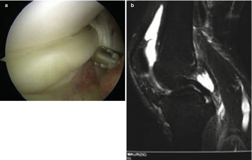
Fig. 37.1
Vertical or longitudinal tears. (a) Arthroscopic view. (b) Preoperative magnetic resonance imaging

Fig. 37.2
Bucket-handle tears. (a) Arthroscopic view. (b) Preoperative magnetic resonance imaging

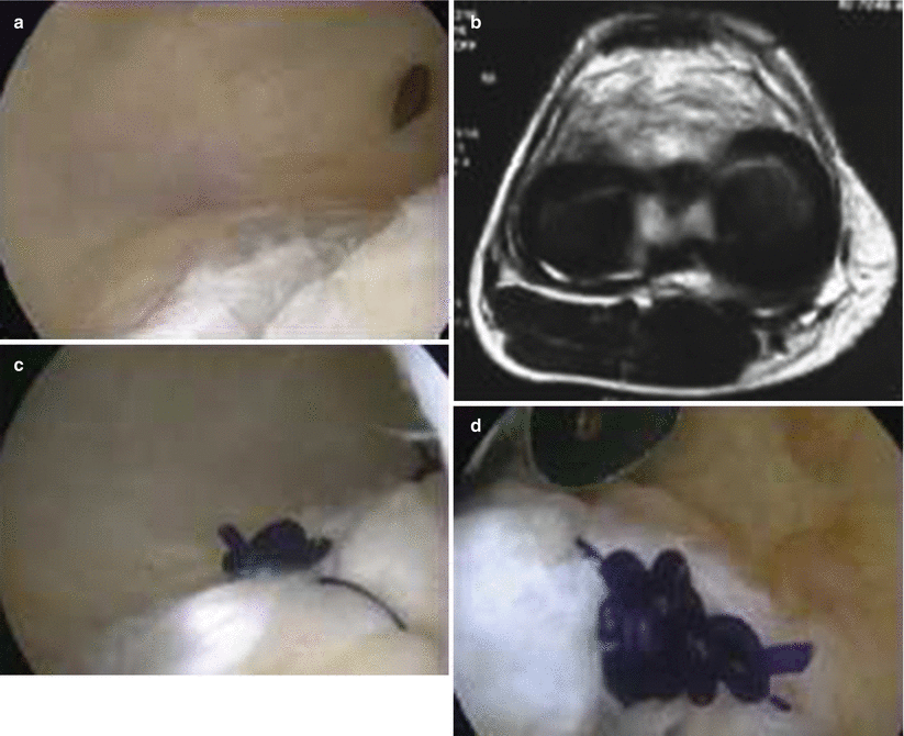
Fig. 37.3
“Ramp lesion” or “hidden lesion” of the posterior horn of the medial meniscus (MM). (a) Arthroscopic view. (b) Preoperative magnetic resonance imaging. (c, d) Final suture with a PDS suture. Suture through the posteromedial portal with a hook
37.4 Clinical and Diagnostic Examination
Meniscal tears may present with joint locking, limitation to extension, joint effusion and possible associated lesions of the collateral ligaments and/or ACL [41, 55, 56]. Prior to surgery, a presumptive diagnosis and a differential diagnosis can be formulated on the basis of a careful history, thorough clinical examination, and appropriate imaging investigations.
Generally, a meniscal tear will present with:
1.
Acute joint pain localised to the medial joint line.
2.
Joint effusion. If the tear involves the inner areas of the meniscus, the effusion generally develops gradually over a few hours. In peripheral lesions, the effusion develops much more rapidly (within minutes), and because the peripheral region is vascular, the effusion may contain blood.
3.
Joint locking is a common symptom after a meniscal injury. Locking usually occurs at 20–45° of extension. It is due to a displaced meniscal fragment that becomes trapped between the femoral condyle and the tibial plateau [57].
4.
A sensation of giving way may occur. In these circumstances, it is necessary to distinguish a meniscal lesion from joint instability due to other causes such as ACL and/or quadriceps femoris insufficiency. During the clinical examination, the uninvolved leg should be used as a reference for comparison of the qualitative and quantitative results of the involved leg. The examination should include inspection, palpation, range of motion (ROM), gait pattern, girth measurements and tests to assess the integrity of the menisci and other structures of the knee joint [58].
37.4.1 Tests
Several different specific tests can be applied to assess meniscal involvement. A positive result on any one test cannot by itself establish the presence of a meniscal lesion, but together with other objective findings, it can help in the differential diagnosis.
Diagnostic accuracy is improved by considering the results of three tests in combination. In general, all clinical tests tend to be less reliable in the presence of a concurrent ligamentous lesion and are less accurate in patients with degenerative lesions than in young patients with acute injuries [59]. Below are some tests that can be performed.
37.4.1.1 McMurray Test [60]
This test is positive in central tears or tears of the posterior horn. With the patient supine, the examiner holds the heel with one hand, and with the other, he/she supports the lower part of the knee, trying to extend the knee completely while rotating the tibia first internally and then externally. Pain during forced flexion suggests a lesion of the posterior meniscal horns (these meniscal portions slide back when the knee is flexed and are compressed between the proximal tibial surface and the femoral condyles); pain during forced extension suggests a lesion of the anterior meniscal horns (during extension, the menisci tend to slide forward and the anterior horns are compressed between the tibial and femoral condyles). Pain at 90° of flexion indicates a lesion of the meniscal body. Specificity is 81 %, and sensitivity is 44 %.
37.4.1.2 Apley Test [61]
The patient lies in the prone position, with knee flexed to 90° and thigh anchored to the table with the examiner’s knee. The examiner distracts the patient’s knee while rotating the tibia inwards and outwards. The presence of pain during this manoeuvre indicates involvement of the ligaments. The examiner then compresses the knee (grinding test) while rotating the tibia inwards (for the lateral meniscus) and outwards (for the medial meniscus). Pain during this manoeuvre indicates a meniscal tear. Specificity is 86 %, and sensitivity is 42 %.
37.4.1.3 Finocchietto Sign [62]
Sensation of a painful palpable click during the anterior drawer test is caused by entrapment of the meniscus between the femur and tibia during knee flexion and extension.
37.4.1.4 Childress Test [63]
The patient squats with knees completely flexed and attempts a “duckwalk”. Limitation of movement indicates a possible meniscal lesion. However, pain in this position may indicate a meniscal lesion or involvement of the femoro-patellar articulation [63]. The Childress test has proved to be more accurate than other tests for the detection of meniscal lesions with associated ACL tears, with a sensitivity of 68 % and an accuracy of 66 %. More specific tests are the Steinmann I sign and the Apley test.
Other tests for meniscal lesions are the O’Donoghue test, the Payr sign and the Steinmann I sign.
37.4.2 Imaging Investigations
Anteroposterior (AP) and lateral knee radiography in young patients with acute trauma [64] may be combined with a Rosenberg projection [65] and a skyline view in subjects older than 40 years of age with suspected degenerative lesion. These imaging studies are required for detecting osteophytes or degenerative chondral changes through assessment of the joint space. They are also useful for detecting malalignment [66].
Magnetic resonance imaging (MRI) is the most powerful and accurate modality for the diagnosis of meniscal lesions. It provides additional information about the state of the ligaments and cartilage [67]. It depicts many of the essential features of meniscal tears needed for making treatment decisions: location, shape, length and depth [68, 69]. As for diagnostic accuracy, it has 86–96 % sensitivity in the detection of medial meniscus tears, with a specificity of 84–94 % [70, 71]. In reality, if a pathological signal is detected on one section only, the accuracy of the diagnosis of a medial meniscal tear is 55 %. The accuracy increases if the pathological signal is seen in at least two consecutive images [71]. MRI is also accurate in the detection of repairable meniscal lesions, and it has good sensitivity for determining irreparable lesions. One study has shown that 89 % (103 of 116) of meniscal lesions were classified equally at MRI and subsequent arthroscopy with regard to reparability. However, the same study demonstrated that MRI had variable accuracy in predicting the configuration of the meniscal lesion later identified with arthroscopy [72]. MRI diagnosis is, however, associated with a risk of false-negative or false-positive results. False-negative results are more common with lesions located in the outer half of the posterior horn of the medial meniscus or in the inner third of the meniscal body. These are small lesions detected clinically by palpation of the joint line; they are stable and may be treated conservatively [73]. False-positive results are more frequent and concern the posterior horn of the medial meniscus. They are caused by chondrocalcinosis in the meniscus or healed meniscal lesions, which often continue to show high signal intensity. In the presence of chondrocalcinosis, MRI sensitivity in the medial meniscus is 89 %, specificity is 72 % and accuracy is 81 %. One of the causes of the diagnostic difficulties lies in the differences in descriptive terminology and interpretation used by orthopaedic surgeons and radiologists with regard to the lesion being examined [74, 75].
37.5 Treatment Strategy
Stable lesions <1 cm in length and not causing any significant mechanical symptoms can be treated with plain observation [76]. Surgery becomes necessary when the lesions give rise to mechanical symptoms. Lesions >1 cm can be distinguished into reparable and irreparable, depending on location. Only 20 % of meniscal tears are suitable for repair. The factors influencing success include the age of the lesion, the location, the type of lesion, the patient’s age and the possible presence of associated lesions [77, 78].
37.5.1 Total Meniscectomy
In the past, meniscal lesions were treated with total meniscectomy. This occurred because the true functions of the meniscus were unknown, and in his 1940 paper “The Semilunar Cartilages”, McMurray wrote that “The cause of failure of meniscal removal lies in the failure to remove the entire affected cartilage” [79]. In 1948, Fairbanks published what is now a classical paper, in which he described the characteristic radiographic changes produced by meniscectomy. Whereas today it is clear that these changes reflect the disruption of the tibio-femoral joint due to loss of the meniscal protective function, Fairbanks observed that “It seems likely that narrowing of the joint space will predispose to degenerative changes, but a connection between these appearances and later osteoarthritis is not yet established and is too indefinite to justify clinical deductions” [50]. During most of the twentieth century, therefore, orthopaedic surgeons carried out a large number of total meniscectomies with good short-term outcomes [80]. However, looking at the results over longer follow-up periods, the negative outcomes became increasingly apparent. For example, Tapper and Hoover from the Mayo Clinic reported on the results of a retrospective study of 213 patients followed up from 10 to 30 years after their meniscectomy, in which only 68 % had good or excellent results and only 38 % were asymptomatic. Moreover, they found that those with a bucket-handle tear but with intact peripheral meniscal rim had higher rates of satisfaction [81].
In the light of these results, there is an increased awareness of the importance of meniscal function [82], and the current motto of orthopaedic surgeons is “Save the meniscus”. Recently, R. Verdonk stated that we have to “keep the meniscus… vestigial soft tissue structure in a self-maintaining transmission system” [83].
37.5.2 Partial Meniscectomy
The development of arthroscopic techniques and related surgical instruments has facilitated the performance of partial meniscectomy, which has become a pillar of the treatment of symptomatic lesions. The short-term results of partial meniscectomies are very satisfactory. Jareguito et al. have reported that in 90 % of cases, they obtained good or satisfactory results at 2-year follow-up and that 85 % of these patients returned to the desired activity level. However, at 8-year follow-up, the results remained good or excellent in 62 % of cases, and only 48 % maintained the pre-injury level of activity [84]. All in all, the differences in the functional scores between total and partial meniscectomy decrease over longer follow-up periods, on average after 7–8 years. Lee et al., in a biomechanical study, demonstrated that also partial meniscectomy influences the transmission of articular loads. They concluded that the goal is to preserve the greatest amount of meniscus possible [85]. Thus, we have biomechanical and clinical evidence suggesting the superiority of partial meniscectomy, despite its limitations of not being able to maintain an adequate biomechanical behaviour and avoid degenerative changes.
Medial meniscectomy is performed with the patient under spinal anaesthesia or with local anaesthesia and sedation depending on the surgeon’s preference and anaesthesiological concerns. Generally, a tourniquet is applied to the upper thigh, although some surgeons prefer to carry out the arthroscopy without it. The limb may be placed in a leg holder or on a flat table with a lateral post: both devices allow a valgus force to be applied in order to correctly visualise the internal compartment. The classical arthroscopic access points are used (antero-medial and antero-lateral); the use of the supero-medial portal for the outflow cannula is optional. Using an arthroscopic probe, the lesion is carefully assessed: a flap, a radial lesion, a dislocated bucket handle, and degenerate appearance of a complex lesion are all indications for partial meniscectomy. In the case of a flap tear, the probe is used to accurately visualise the site of attachment to then go on to resect it and remove the free meniscal fragment. Bucket-handle tears can be treated with a variety of techniques. The one we prefer consists in first resecting the anterior attachment base and then, after having caught the meniscal fragment with an appropriate grasper through an accessory antero-lateral portal (just below the standard portal), resecting the posterior attachment from the medial portal while applying traction and torsion. If visualisation of the area of the posterior attachment base proves difficult, a varus force with the semiflexed knee can be applied to facilitate the passage of the instrumentation: this will help to open up the space between the medial femoral condyle and the posterior cruciate ligament. Finally, the contour is smoothed with a shaver. If the procedure involves the posterior horn of the medial meniscus, a valgus force with extended and internally rotated knee may be required. In all cases, before ending the procedure, the stability of the meniscal remnant needs to be assessed. In patients with excessively tight MCL, it may be justified to release it to ensure correct visualisation of the medial compartment: this can be done by performing multiple needle punctures subcutaneously.
37.5.2.1 Complications
2.
Recurrent joint effusion. This is due to the conflict between the femoral condyle and tibial plateau in the absence of the meniscus.
3.
Synovial fistula (take care to suture the posteromedial portal).
4.
Embolic events. Data from phlebographic studies report an incidence of deep vein thrombosis (DVT) similar to that observed in other surgical procedures at moderate-to-high risk (18 % total DVT and 5 % proximal DVT) even though other, ultrasonography-based, studies have reported lower incidence rates. Application of the tourniquet seems to be an additional risk factor, but, on the other hand, it allows for shorter procedure times. In a study conducted on low-risk patients, reviparin prophylaxis (at a daily dose of 1,750 IU, suitable for moderate risk) for an average of 8 days was associated with a reduction of DVT from 4.1 to 0.85 % [88].
5.
Iatrogenic damage to cartilage. A recent study found that treatment of lesions of the medial posterior horn caused mild damage to the femoral condyles in 28 % of cases and moderate-to-severe damage in 6 % of cases [89].
6.
Ligamentous lesions. An uncommon complication: Small reported two cases of MCL stretching in a series of 1,184 arthroscopies [90].
7.
Breakage of surgical instruments. This is a real, though uncommon, complication (0.3 %) which has decreased over the years with the introduction of new surgical instruments and growing surgeons’ experience [91].
8.
Vascular damage. Mostly pseudoaneurysms of the popliteal artery or arteriovenous fistulas, with an incidence of 0.05 % [92].
9.
Nerve damage. Although definitely more common during meniscal repair, this complication has also been reported in meniscectomies, with a rate of 0.6 %. In most cases, the infrapatellar branch of the saphenous nerve is injured during creation of the arthroscopic portals [93].
10.
Osteonecrosis. This complication was initially observed in cases of arthroscopic meniscectomy: osteonecrosis of the postoperative knee (ONPK). In fact, more recently it has also been observed following other arthroscopic procedures (chondroplasty, ACL reconstruction) [94–97]. It predominantly affects the medial femoral condyle (82 %), followed by the lateral femoral condyle, the lateral tibial plateau and finally the medial tibial plateau. In 65 % of cases, there is pre-existing chondral damage [95, 98, 99]. Possible causes are:
An aggressive postoperative rehabilitation may contribute to the development of this condition. Rapid return to weight-bearing activities and exercise (before the bone remodelling stimulated by the changes in load distribution) induces insufficiency fractures [100].
An increase in cartilage permeability to arthroscopic fluid (due to cartilage lesions or iatrogenic damage during the procedure) may lead to increased interosseous pressure, with oedema and necrosis [101].
Direct injury from thermal effects (laser) or photoacoustic trauma (gas bubbles = shock wave), during radiofrequency use. The inflammatory response leads to bone oedema, locally increased intraosseous pressure and osteonecrosis [102, 103].
Even the presence of a medial meniscus lesion has been suggested as a potential aetiological factor. In particular, a tear of the posterior meniscal root has been implicated (incidence of 2.8 % among 1,500 knees examined). In 80 % of knees with this diagnosis, an osteonecrosis manifested clinically. The suggested pathogenic mechanism is a change in the loads transmitted to the femoral condyle due to loss of the dissipation function of the condyles themselves as a result of increased compartmental pressure and therefore osteolysis and osteonecrosis [104].
37.5.3 Meniscal Repair
Prompted by the studies of Arnozcky and Warren [5] on meniscal vascularity and healing potential, and also thanks to the introduction of new arthroscopic techniques, meniscal repair has become more widespread. The indications can be summarised as follows:
Patient-related factors: instability, locking, effusion, grinding and pain
Factors deriving from the physical examination: joint-line tenderness, effusion and limitation of motion
Specific tests for meniscal tears
MRI (tear of the red-red or red-white zone)
Arthroscopy: reducible tear
The contraindications are degenerate or poorly vascular meniscus tissue, patient’s age, patient’s limited compliance with the postoperative physiotherapy protocol, untreated knee instability, osteoarthritis, chronic lesions, longitudinal tears shorter than 1 cm, or radial tears. Several different techniques are available for meniscal repair:
1.
Scott and co-workers [105] and Cannon [106] developed the inside-out repair technique. This involves the use of double-lumen, zone-specific cannulae [107] through which needles preloaded with suture material are passed. In the inside-out repair of posterior horn tears, a counter-incision is made to prevent neurovascular complications. In the repair of medial meniscus tears, the incision must be posterior and parallel to the MCL and anterior to the medial gastrocnemius. The knee must be flexed to 5–15°. The technique can be used for tears of the posterior horn and meniscal body.
2.
Warren [108] was the first to describe the outside-in repair technique, which is relatively inexpensive (use of an 18-gauge needle + monofilament) and simple. Its main indication is repair of the middle third of the meniscus and of the anterior horn. Possible complications are iatrogenic lesions to the meniscus due to multiple attempts at suture needle placement or neurovascular lesions.
3.
Morgan introduced the all-inside technique utilising arthroscopic knots [109]. This is indicated in longitudinal tears of the posterior horn and ramp lesions. It is a difficult technique that requires creation of a posteromedial access through which a special hook or pigtail needle is passed to perform a vertical PDS suture of the posterior horn. This technique has seen the greatest efforts of manufacturers of orthopaedic materials to provide increasingly precise and easy-to-use devices to make the technique more accessible. Surgeons have at their disposal solid implants (polylactic acid anchors) [110] – in actual fact used increasingly less often because of the possible postoperative complications – or anchoring systems (partially bioabsorbable filaments mounted on bioabsorbable anchors) [90].
The characteristics and location of the tear and the operator’s experience can influence the type of technique adopted, even though all-inside techniques are being increasingly used since they are relatively easy to apply and do not require accessory surgical accesses. However, several studies have raised the problem of the failure of these methods even though the results are all in all similar to those achieved with the inside-out technique. The possible complications of repair techniques for tears of the medial meniscus are:
1.
2.
Involvement of the popliteal artery with the T-Fix system (Smith & Nephew Endoscopy) if no depth penetration limiter is utilised [112].
3.
Risk of penetrating the sartorius tendon or deep fascia of MCL, which may slow functional recovery because of postoperative pain [113].
4.
Get Clinical Tree app for offline access

Failure and migration of the solid implants with cartilage lesions and/or synovitis [114]. Technique: debride the fibrous tissue with a rasp or shaver. In some cases, needling at the level of the meniscal joint line may be done to stimulate bleeding of the vascular plexus. Debridement of the posterior region of the medial meniscus may no doubt prove difficult, and a possible solution is to create an accessory posteromedial portal. Once the meniscus has been prepared, the various repair techniques can be performed:
(a)


In all-inside procedures, the sutures are generally inserted via the ipsilateral portal for posterior horn tears and via the contralateral portal for meniscal body tears.
Stay updated, free articles. Join our Telegram channel

Full access? Get Clinical Tree


