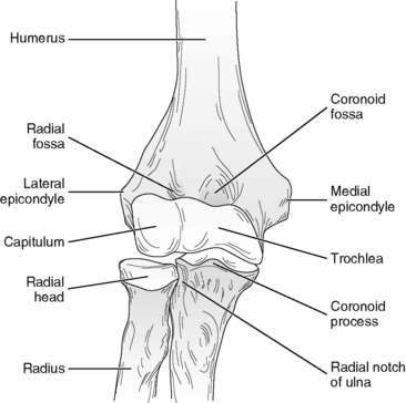
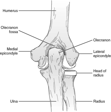
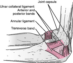
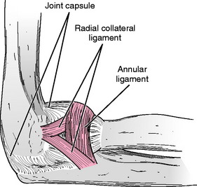
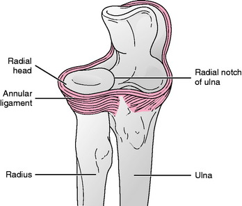
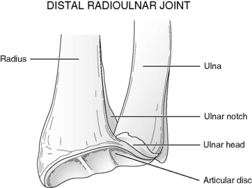
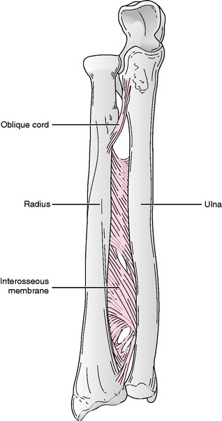
Chapter 4 Within the elbow joint capsule are three articulations, two that make up the elbow joint complex and one that is part of the forearm complex. The humeroradial and humeroulnar joints make up the joint complex known as the elbow (Figs. 4-1 and 4-2). The humeroradial joint consists of the articulation between the convex capitulum of the distal humerus and the slightly concave proximal surface of the radial head. The articulation between the somewhat hourglass-shaped trochlea of the humerus and the concave, semilunar-shaped trochlear notch of the ulna forms the humeroulnar joint. Both joints are located within a single joint capsule that also is shared by the proximal radioulnar joint.2 Ligamentous reinforcement of the elbow joint occurs primarily on the medial and lateral sides of the joint via the ulnar (Fig. 4-3) and radial (Fig. 4-4) collateral ligaments, respectively. These ligaments resist valgus and varus stresses to the joint throughout the full range of elbow motion.18,26,21 Additional stability of the elbow joint is provided by the high degree of bony congruency between the articular surfaces that make up the joint. Although the elbow joint traditionally has been classified as a hinge joint, the hinge component occurs at the humeroulnar articulation, and the humeroradial joint is classified as a plane joint.2 Motions available at the elbow are flexion and extension, which occur in a plane oriented slightly oblique to the sagittal plane, owing to the angulation of the trochlea of the humerus.10 The axis of rotation for flexion and extension of the elbow is centered on the trochlea, except at the extremes of flexion and extension, where the axis moves anteriorly and posteriorly, respectively.13 During the movements of elbow flexion and extension, the concave surface of the trochlear notch of the ulna glides along the convex trochlea of the humerus. Simultaneously, at the humeroradial joint, the concave head of the radius glides along the convex capitulum of the ulna. Both radial and ulnar articular surfaces glide anteriorly as the elbow flexes and posteriorly as it extends. At the extremes of flexion and extension, rolling motions of the ulna and radius replace the gliding motion.13,28 Elbow flexion range of motion (ROM) is limited by soft tissue approximation between the structures of the anterior arm and the forearm, particularly during active flexion of the joint when contact between contracting flexors of the arm and forearm stops the motion. The range of elbow flexion tends to be greater when the joint is moved passively because there is less interference by contracting muscle bulk. Elbow extension ROM is limited by contact of the olecranon process of the ulna with the olecranon fossa of the humerus.10 Information regarding normal ROM for the elbow is located in Appendix B. Gray’s Anatomy2 describes three articulations that interconnect the bones of the forearm: the proximal and distal radioulnar joints and the middle radioulnar union. The proximal radioulnar joint is located anatomically within the capsule of the elbow joint and consists of the articulation between the rim of the radial head and the fibro-osseous ring formed by the annular ligament and the radial notch of the ulna (Fig. 4-5). The distal radioulnar joint is located anatomically at the wrist, although inside a separate joint capsule. This joint is formed by the articulation between the concave ulnar notch of the radius and the convex head of the ulna (Fig. 4-6).8 A third articulation between the radius and ulna, the middle radioulnar union, has been classified as a syndesmosis, although this articulation is not classified as a joint at all by the Nomina Anatomica.30 The middle radioulnar union consists of the shafts of the radius and ulna held firmly together by the interosseous membrane and by the oblique cord, a small ligament that attaches from the ulnar tuberosity to just distal to the radial tuberosity (Fig. 4-7).17 Ligamentous reinforcement of the proximal radioulnar joint occurs via two ligaments. The annular ligament is attached to the anterior and posterior margins of the radial notch of the ulna and encircles the radial head, holding it firmly against the radial notch (see Figs. 4-3 through 4-5).16 A second ligament, the quadrate ligament, runs from the inferior aspect of the radial notch to the neck of the radius, reinforces the joint capsule, and has been attributed with stabilization of the proximal radioulnar joint during the extremes of pronation and supination.29 The distal radioulnar joint is reinforced by a triangular articular disc that is positioned on the distal end of the ulna. This disc binds the distal ulna and radius together and is the primary reinforcement for the joint. The dorsal and palmar radioulnar ligaments assist in stabilization of the distal radioulnar joint.11 During pronation and supination of the forearm, motion occurs at the proximal and distal radioulnar joints simultaneously. At the proximal joint, the convex radial head spins within the ring formed by the radial notch of the ulna and the annular ligament. The radial head spins anteriorly during pronation and posteriorly during supination. Distally, the concave ulnar notch of the radius rolls and slides anteriorly on the ulnar head during pronation and posteriorly during supination.21 Supination of the forearm is limited by tension in ligamentous structures (anterior radioulnar ligament and oblique cord).25 Limitation of forearm pronation occurs as the result of contact between the bones of the forearm (radius crossing over ulna) and tension in the medial collateral ligament of the elbow and the dorsal radioulnar ligament of the distal radioulnar joint.7,21 Information regarding normal ranges of motion for forearm supination and pronation is located in Appendix B. Most functional activities require a fairly large amount of elbow flexion ROM (Figs. 4-8 to 4-10). A recent study by van Andel and colleagues31 reported that all functional tasks examined in their study required a minimum of 85 degrees of elbow flexion. These results were similar to those reported by Vasen et al,32 who used a motion-restricting brace to determine the functional ROM of the elbow. Of 50 subjects examined, 49 were able to perform all 12 functional activities included in the study, with elbow motion limited to a range of 75 degrees to 120 degrees of flexion. Numerous other investigators have attempted to quantify the amount of elbow and forearm motion required to perform various functional activities.3,6,14,15,19,20,22–24 A summary of elbow and forearm range of motion related to various functional activities is provided in Table 4-1. Most of the studies from which data were derived were performed in healthy adults, although some data were obtained from elderly and pediatric subjects. Essentials of the study populations and the instrumentation used are included in the table. Caution should be used in extrapolating these data to the general population because sample sizes for all studies were small. For more in-depth information on each study, the reader is referred to the reference list at the end of this chapter. Elbow flexion and extension may be measured with the patient in the upright (standing or sitting), supine, or side-lying position. Because of greater stability provided to the humerus, the supine position is preferred for measurement of ROM. The American Academy of Orthopaedic Surgeons5 recommends that the patient be in the upright position with the shoulder flexed to 90 degrees when measurements of elbow flexion and extension are taken. In patients with tightness of the long head of the triceps, such positioning may limit flexion of the elbow. Therefore, motions of the elbow joint should be measured with the shoulder maintained in the anatomical position.
MEASUREMENT of RANGE of MOTION of the ELBOW and FOREARM
ELBOW JOINT
OSTEOKINEMATICS
ARTHROKINEMATICS
LIMITATIONS OF MOTION
FOREARM JOINTS
ARTHROKINEMATICS
LIMITATIONS OF MOTION
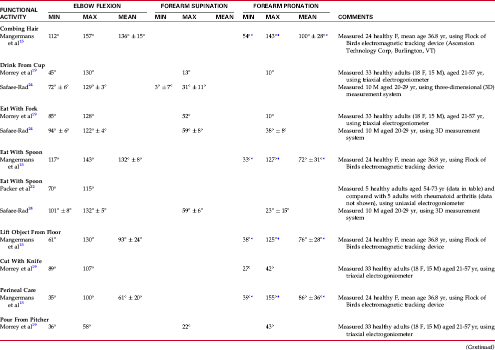
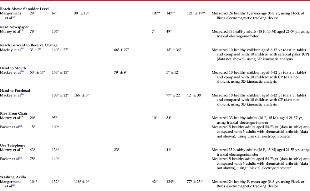
RANGE OF MOTION AND FUNCTIONAL ACTIVITY
TECHNIQUES OF MEASUREMENT
Related posts:
 RELIABILITY and VALIDITY of MEASUREMENTS of RANGE of MOTION and MUSCLE LENGTH TESTING of the UPPER EXTREMITY
MEASUREMENT of RANGE of MOTION and MUSCLE LENGTH: CLINICAL RELEVANCE
MEASUREMENT of RANGE of MOTION of the KNEE
MEASUREMENT of RANGE of MOTION of the HIP
MEASUREMENT of RANGE of MOTION of the ANKLE and FOOT
PEDIATRIC RANGE of MOTION
RELIABILITY and VALIDITY of MEASUREMENTS of RANGE of MOTION and MUSCLE LENGTH TESTING of the UPPER EXTREMITY
MEASUREMENT of RANGE of MOTION and MUSCLE LENGTH: CLINICAL RELEVANCE
MEASUREMENT of RANGE of MOTION of the KNEE
MEASUREMENT of RANGE of MOTION of the HIP
MEASUREMENT of RANGE of MOTION of the ANKLE and FOOT
PEDIATRIC RANGE of MOTION
![]()
Stay updated, free articles. Join our Telegram channel

Full access? Get Clinical Tree


Musculoskeletal Key
Fastest Musculoskeletal Insight Engine