Management of Arthrofibrosis of the Knee

Chapter 90


Management of Arthrofibrosis of the Knee


Arthrofibrosis is the proliferation of fibrotic tissue within and surrounding a joint. It results in decreased range of motion (ROM), pain, decreased function, and subsequent strength loss secondary to disuse. In the knee joint, arthrofibrosis is a potential complication after fracture treatment or intra-articular surgery, including arthroscopy, anterior cruciate ligament (ACL) reconstruction, and total knee arthroplasty. Arthrofibrosis is a difficult condition to treat, and precautionary measures should be taken to prevent this potential postoperative complication.


Arthrofibrosis of the knee results in the loss of extension, and in some patients loss of knee flexion also occurs. Loss of knee extension is usually more symptomatic compared with loss of flexion.1


Arthrofibrosis is most effectively treated via a multidisciplinary approach, with the physician, rehabilitation staff, and patient working closely together. The treatment should be viewed as a process that includes intensive rehabilitation, followed by arthroscopic scar resection when needed and postoperative rehabilitation. In addition, patients often require a lot of emotional support and encouragement throughout this process and must be well informed and actively involved in their own care.


Preoperative Considerations


Physical Examination


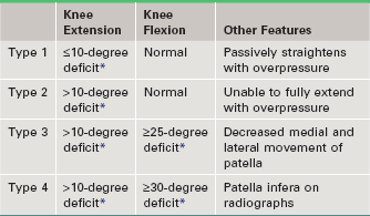
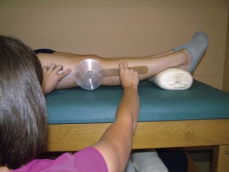
Normal ROM of the knee may include some degree of hyperextension and varies from person to person. Therefore ROM of the uninvolved knee must be assessed first to gain a comparison point, and any difference between the uninvolved and involved knee ROM is noted as a deficit. Hyperextension of the knee is often overlooked, but is a normal finding in most knees. DeCarlo and Sell2 found that 95% of males and 96% of females have some degree of knee hyperextension; the mean for males was 5 degrees, and the mean for females was 6 degrees. Knee extension must be assessed with the patient supine and both heels propped on a bolster, allowing the knees to fall into hyperextension (Fig. 90-1). Unless extension is measured this way and compared with that of the opposite, normal knee, a slight loss of knee extension could easily be overlooked. The amount of knee extension and flexion loss is noted and can be used to classify the severity of arthrofibrosis to assist with treatment planning (Table 90-1).3


Physical examination should also include an assessment of patellar mobility, quadriceps muscle control, observed muscle atrophy, and patella height. Decreased patellar mobility is observed in patients with type 3 or 4 arthrofibrosis, and patients with type 4 arthrofibrosis will have a shortened patellar tendon.


Imaging


Bilateral standing posteroanterior, lateral, and Merchant view radiographs should be obtained. It is important to measure patellar height and compare it between the involved and uninvolved knees to detect any side-to-side difference. The length of the patellar tendon can be measured on the lateral radiograph from the inferior pole of the patella to the tibial tubercle. The normal values for this measurement can vary considerably, making a side-to-side comparison necessary for detection of tendon contraction. Patients with type 4 arthrofibrosis will demonstrate patella infera from a contracted patellar tendon.


A Merchant view radiograph,4 in which both patellae are viewed, can be used to detect disuse osteopenia in the involved patella. This is a sign that the patient has been significantly favoring the involved leg by standing with the weight shifted onto the uninvolved leg and avoiding use during other activities of daily living.


Magnetic resonance imaging (MRI) is useful in determining the extent of scar formation and proliferation within the knee and surrounding tissues.


Surgical Planning


The optimal timing for surgical intervention will vary considerably among patients with arthrofibrosis. It is important for patients and clinicians to understand that treatment of arthrofibrosis is a process that is not isolated to surgical intervention alone. Appropriate and directed preoperative and postoperative rehabilitation is a crucial aspect of caring for patients with this condition.


The decision of when to proceed with surgery is based not on timeframes, but rather on the progress made with rehabilitation. Patients with type 1 arthrofibrosis may be able to regain symmetrical knee extension with rehabilitation alone, and surgical intervention may not be necessary. When needed, surgical intervention for arthrofibrosis is most successful when performed after the patient has worked diligently to improve knee ROM but has reached a plateau and can no longer make improvements with rehabilitation alone. In addition, the patient must be mentally prepared for surgery and have a full understanding of the plan of care, the postoperative rehabilitation required, and the prognosis of this condition.


Rehabilitation


Preoperative rehabilitation is divided into two distinct phases: the primary focus of the first phase is to maximize knee extension, and the goal of the second phase is to maximize knee flexion without any loss of knee extension. In our experience, working on knee extension and flexion at the same time is counterproductive, and patients become frustrated with the lack of progress. Similarly, working on lower extremity strength is also delayed until maximal or full ROM has been achieved.


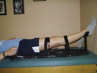
Owing to the difficulty in regaining knee extension in patients with arthrofibrosis, we routinely prescribe the use of a passive knee extension device (Kneebourne Therapeutic, Noblesville, IN) in this patient population (Fig. 90-2). This device provides a patient-controlled, sustained stretch to the knee to help patients regain symmetrical knee extension, including hyperextension. The patient can use this device independently at home three to five times per day for 10 to 15 minutes per session. Patients also perform towel stretch (Fig. 90-3), prone hang, and heel prop exercises to improve knee extension. In addition, patients work to improve quadriceps muscle control with the goal of regaining the ability to perform an active heel lift (Fig. 90-4) and achieve active hyperextension.


Stay updated, free articles. Join our Telegram channel

Sep 11, 2016 | Posted by in SPORT MEDICINE | Comments Off on Management of Arthrofibrosis of the Knee

Full access? Get Clinical Tree

Get Clinical Tree app for offline access