Lumbar Pedicle Subtraction Osteotomy


Lumbar Pedicle Subtraction Osteotomy


Sigurd H. Berven

Praveen Mummaneni


INDICATIONS/CONTRAINDICATIONS

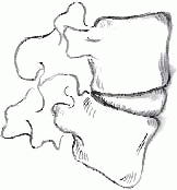
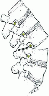
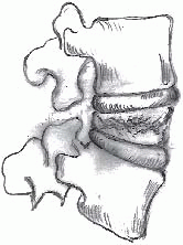
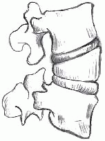
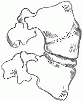
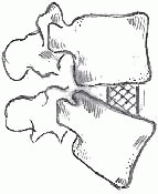
Posterior-based osteotomies contraction (PSO) in the lumbar spine encompass a spectrum of techniques that are intended to realign the spine through resection of posterior elements with variable resection of the anterior column of the spine. The spectrum of posterior-based osteotomies is listed in Table 20-1. The spectrum of osteotomies is a continuum, and the osteotomy techniques share steps including resection of the facet joints, decancellation, and partial or complete corpectomy from a posterior approach. The spectrum of osteotomies begins with a resection of the facet joints alone and realignment of the spine with an axis of rotation at the anterior longitudinal ligament in a mobile spine (type 1) or anterior column osteoclasis through a rigid spine (type 2). Decancellation of the vertebral body through the pedicle permits a controlled fracture of the anterior column and may be useful in realigning the spine in the coronal and sagittal plane, as described by Heinig in the “eggshell osteotomy” (type 3). The lumbar pedicle subtraction osteotomy is a wedge resection of the vertebral body from below the pedicle to the anterior cortex of the vertebra, and the wedge resection may be entirely intraosseous with a hinge on the superior third of the vertebral body (type 5) or at the supraadjacent disc (type 6). Finally, a posterior resection of one or more vertebra and discs may be useful in treating more complex deformity including translation of the trunk. The purpose of this chapter is to describe the technique of the lumbar pedicle subtraction osteotomy.

The lumbar pedicle subtraction osteotomy (transpedicular vertebral wedge resection) is a useful technique for the operative correction of both sagittal and coronal plane deformity (4). The clinical impact of deformity is determined and predicted primarily by the sagittal plane (12,18). The primary indication for the technique is correction of sagittal plane deformity in the patient who has had a previous circumferential fusion of the spine. The transpedicular wedge resection technique was first described by Thomasen (27) in 1989 for the management of fixed sagittal plane deformity in ankylosing spondylitis. Common etiologies of fixed sagittal plane deformity include ankylosing spondylitis, or iatrogenic causes of fixed sagittal plane deformity including flatback syndrome (8) and kyphotic decompensation syndrome (10). More generally, lumbar spine kyphosis with variable degrees of rigidity may be the result of degenerative change, congenital anomaly, trauma, neoplastic disease with pathologic fracture, or infection. The rigidity of lumbar kyphosis and the apex of the deformity are important determinants of the surgical approach. In a spine with a mobile anterior column, or a flexible deformity, posterior-based osteotomies with facet resection and deformity correction through the disc space or osteoclasis of the anterior column may be useful. In a spine with an apex of deformity at L4 or L5, a combined anterior and posterior approach to the spine may be useful in recreating lordosis at the lumbosacral segments and correcting a rigid coronal obliquity of a fractional curve from L4 to S1. The lumbar pedicle subtraction osteotomy may be used at any level of the lumbar spine and is most useful for rigid deformity, which requires resection of bone including a portion of the anterior column for mobilization and realignment. The technique may also be used above the level of the conus including the cervical and thoracic spine.


TABLE 20-1 Spectrum of Posterior-Based Osteotomies


Type


Description


Diagram


Reference


1


Resection of posterior elements from mid-pars above to pedicle below with realignment of the spine through hinging through a mobile disc anteriorly


image


Ponte


2


Resection of posterior elements from mid-pars above to pedicle below with realignment of the spine through hinging through the anterior column of the spine which is ankylosed. The opening involves osteoclasis rather than movement through a mobile intervertebral disc


image


Smith-Peterson


3


Posterior-based transpedicular decancellation of the vertebral body with realignment through controlled fracture of the anterior column


image


Heinig


4


Posterior-based intraosseous wedge resection of the vertebral body with realignment through osteoclasis of the proximal third of the anterior vertebral body


image


Thomasen


5


Posterior-based wedge resection with extension of the osteotomy into the supraadjacent disc and realignment hinging on the anterior column at the intervertebral space


image


Modified Thomasen


6


Posterior-based vertebral column resection including one or more vertebra with adjacent discs


image


Suk


The lumbar pedicle subtraction osteotomy fits best in the spectrum or posterior-based osteotomies as an option for patients with a sagittal deformity that is correctable with an apical resection of approximately 30 degrees and coronal imbalance less than 8 cm (1). Although the lumbar pedicle subtraction osteotomy may be used in the patient with open disc spaces and mobility of the anterior column of the spine, the authors prefer to use a combined anterior and posterior approach (5), or posterior-based osteotomies as described by Ponte for the patient with a mobile anterior column as these techniques permit a more gradual and harmonious realignment of the spine through the creation of trapezoidal intervertebral disc spaces (11). If osteoclasis is possible for the anterior column, the Smith-Peterson osteotomy may be useful to create segmental realignment (23). In the patient with a severe lumbar kyphosis that requires more than 40 degrees of correction, or trunk translation and coronal imbalance greater than 8 cm, a vertebral column resection may prove more advantageous (6,26).


PREOPERATIVE PREPARATION


Patient Assessment

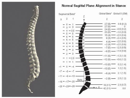
The patient with a fixed sagittal plane imbalance presents with an inability to maintain horizontal gaze, fatigue to the thighs and hips, and back pain that is often intractable. Normal sagittal alignment of the spine follows a vertical axis from the center of C2, in front of T7, behind L3, and to the posterior margin of the sacrum (Fig. 20-1). Forward displacement of C7 relative to the pelvis may be due to regional malalignment in the lumbar, thoracic, and cervical spine. Extraaxial causes of global sagittal malalignment may commonly be caused by hip flexion contracture, especially in older patients, patients with neuromuscular scoliosis, and patients with ankylosing spondylitis. A differentiation of the anatomic source of sagittal plane deformity may be gleaned from a thorough physical examination. On standing, the patient’s deformity is best appreciated with the knees fully extended. On sitting, if sagittal balance corrects or if the trunk appears with good balance relative to the pelvis, then a hip flexion contracture may be the cause of sagittal plane malalignment. A hip flexion contracture can be demonstrated using the Thomas test. If the patient remains with forward displacement of the occiput relative to the pelvis on sitting, then the patient should be evaluated in the supine position. In the patient with deformity localized to the lumbar spine, the patient will be able to lie with shoulders on the table in the supine position with flexion of the knees and extension of the hips. In contrast, if the patient remains with head and upper thoracic spine elevated from the table in the supine position, fixed deformity in the cervical and/or thoracic spine is likely.


FIGURE 20-1 Normal sagittal alignment of the spine.


The patient with fixed sagittal imbalance will present with a characteristic stance and gait pattern. Patients extend at the hips and flex at the knees in order to maintain sagittal balance. This compensatory mechanism leads to significant fatigue of the quadriceps and limited standing and walking tolerance. Patients typically walk with short steps and may require a walker for support in cases of more severe imbalance. The patient with a wide-based gait or ataxia should be evaluated for myelopathy and possible spinal cord pathology. Hip extension may be quantified by measuring the pelvic tilt radiographically. On clinical exam, extension of the hips is identifiable by inspection of the position of the anterior superior iliac spine compared with the posterior superior iliac spine and by an apparent flattening of the buttocks. The cervical spine may also be affected by global deformity in the thoracic and lumbar spine, and neck pain is a common clinical complaint of patients with sagittal deformity due to hyperextension of the cervical spine to maintain horizontal gaze.


Radiographic Evaluation

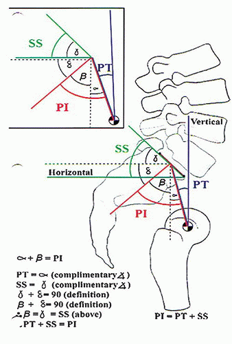
Standing 14 × 36 inch radiographs in the posteroanterior and lateral projections are most useful for an assessment of the global and regional alignment of the spine. The lateral radiograph represents the spinal deformity most accurately with the knees fully extended and the shoulders at 30- to 60-degree forward flexion (28). Placing the proximal interphalangeal joints in the clavicular fossa may result in a more accurate radiographic assessment of sagittal alignment and less radiographically apparent sagittal malalignment than having the arms in 60 degrees of forward flexion (13). Inclusion of the proximal femur in the lateral view permits an assessment of the femoropelvic contribution to sagittal deformity and measurement of pelvic tilt and pelvic incidence (Fig. 20-2). Normal lumbar lordosis is closely correlated to the pelvic incidence. Realignment of the spine should aim to match lumbar lordosis to within 10 degrees of pelvic incidence (1). Patients with a high pelvic incidence may require significantly more than 40 degrees of lumbar lordosis for optimal sagittal alignment of the spine. Even small losses of segmental lordosis in the lower lumbar spine may translate to large changes in global alignment between C7 and the sacrum. Flexion and extension radiographs, including imaging over a bolster, are useful to determine the flexibility of the deformity above and below the planned osteotomy.


FIGURE 20-2 Measurement of lumbopelvic parameters. PT, Pelvic tilt; PI, Pelvic incidence; SS, Sacral scope.

Only gold members can continue reading. Log In or Register to continue

Stay updated, free articles. Join our Telegram channel

Jun 14, 2016 | Posted by in ORTHOPEDIC | Comments Off on Lumbar Pedicle Subtraction Osteotomy

Full access? Get Clinical Tree

Get Clinical Tree app for offline access