Fig. 40.1
Leiomyosarcoma of the metaphysis and diaphysis of a femur. Specimen photography and radiography. The lesion involved metallic implants from a previous surgical procedure. (a) Whitish-pink firm and elastic lesion occupying the marrow space in a mostly longitudinal fashion, permeating the cortex and involving adjacent soft tissue. (b) Absence of mineral deposits in tumor tissue
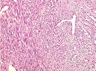
Fig. 40.2
Low-power microscopic view. Long and interwoven bundles of spindle cells
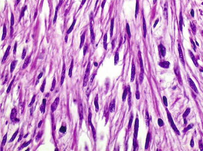
Fig. 40.3
High-power microscopic view. Spindle cells with hyperchromatic nuclei, sometimes showing blunt ends. Atypical mitoses are frequent
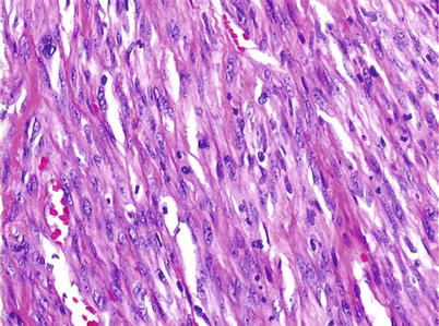
Fig. 40.4
Medium-power microscopic view of a leiomyosarcoma of a higher grade than the previous figure. Immunohistochemistry may be needed to properly identify the neoplasia
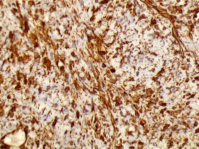
Fig. 40.5


Immunohistochemistry – SMA positive in neoplastic cells
Related posts:


Stay updated, free articles. Join our Telegram channel
Full access? Get Clinical Tree
