Lateral Approach to the Valgus Total Knee
James B. Stiehl
HISTORICAL PERSPECTIVE
Surgical techniques evolve to create easy, reproducible methods that simplify a procedure and reduce complications. These objectives were the guidelines of Dr. Peter A. Keblish, the originator of the lateral retinacular or valgus approach to the knee in total knee arthroplasty (TKA). The surgical problem in this case was how to do an adequate and complete release of contacted lateral ligaments often combined with articular deformity and lateral femoral condyle hypoplasia in valgus-deformed knees (Figs. 2-1 and 2-2). This approach was further necessitated by the fact that early extension ligament balancing was needed for the “tibia cut first method” adopted around the world for mobile-bearing TKA. Following the principles of Insall, the ligaments had to be completely balanced before the femoral bone cuts could be initiated; otherwise, potential flexion space instability could arise if balancing was altered after trials had been implanted. The most efficient method of balancing the lateral ligaments was to go directly at them through a lateral skin and fascial incision. This approach is desirable for any valgus-deformed knee but is optimal in cases of severe valgus deformity and patellar dislocation when extensile lateral releases are mandated. The experienced surgeon should use the approach on simpler cases such that the difficult case is not magnified by unfamiliarity with the approach.
ANATOMICAL CONSIDERATIONS
The direct lateral approach (1, 2, 3) is a technique that offers many advantages in correction of fixed valgus deformity, as well as other challenges that confront the knee surgeon, primarily, patella alignment and soft tissue considerations. It is less commonly used (and understood) than the standard medial approaches but has been shown to improve stability and patella results in fixed valgus TKA (4, 5, 6, 7). Fixed contractures in valgus TKA require sequential releases that include the lateral capsule, iliotibial band (I-TB), vastus lateralis tendon, and very occasionally, the lateral collateral ligament (LCL), the popliteus, lateral gastrocnemius, and inner aspect of the fibular head (preserving and lengthening the LCL). These releases are best addressed by the direct access using the lateral approach.
Fixed valgus deformity is usually associated with lateral femoral condyle hypoplasia, femoraltibial malrotation, resorption of the lateral femoral condyle and tibial plateau, and a relatively large medial condyle. Lateral structures are tight, and the patella is frequently deformed and subluxed over the deformed lateral condyle. The valgus knee deformity is most prevalent in females (9:1), and rheumatoid arthritis is more common. Prosthetic cover and joint seal can be a problem because the skin and soft tissue are often deficient. Excessive undermining, increased tension, or lack of a soft tissue layer between the skin and the prosthesis can lead to skin necrosis, a potentially devastating complication of TKA.
The fascia lata extension envelops the quadriceps with attachment to the posterior aspect of the femur. The distal lateral confluence becomes the iliotibial band with distinct insertion into Gerdy’s
tubercle and the lateral tibial plateau. Transverse and oblique fibers extend to the patellar mechanism (lateral retinaculum), and longitudinal fibers attach to bone via Sharpey’s fibers and extend to the myofascia of the anterior compartment. The I-TB and the lateral retinaculum are deforming factors in the fixed valgus knee. The I-TB attachment to the upper tibia produces a valgus moment with external rotation, and the oblique and transverse extensions produce a lateral (subluxing) moment to the patella. The extra-articular (superficial) layer also includes the lateral hamstring, the fabellofibular ligament, the lateral head of the gastrocnemius, and the popliteus. These structures may be contracted secondary to long-standing valgus. Bony deformity may further increase concave side contractures.
tubercle and the lateral tibial plateau. Transverse and oblique fibers extend to the patellar mechanism (lateral retinaculum), and longitudinal fibers attach to bone via Sharpey’s fibers and extend to the myofascia of the anterior compartment. The I-TB and the lateral retinaculum are deforming factors in the fixed valgus knee. The I-TB attachment to the upper tibia produces a valgus moment with external rotation, and the oblique and transverse extensions produce a lateral (subluxing) moment to the patella. The extra-articular (superficial) layer also includes the lateral hamstring, the fabellofibular ligament, the lateral head of the gastrocnemius, and the popliteus. These structures may be contracted secondary to long-standing valgus. Bony deformity may further increase concave side contractures.
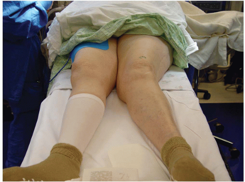 FIGURE 2-1 Severe valgus deformity in a 74-year-old female. |
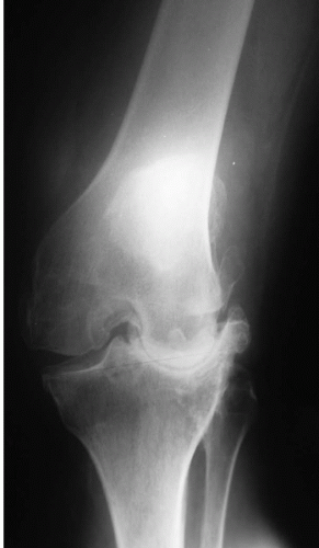 FIGURE 2-2 Valgus deformity noted with lateral femoral hypoplasia and severe erosion of the lateral tibial plateau with a mechanical axis alignment of 23 degrees valgus. |
The lateral superficial layer differs from the (compliant) medial oblique retinaculum of the vastus medialis in that the lateral retinaculum is relatively noncompliant. This noncompliant lateral fascial extension to the patellar mechanism, coupled with contractures of the deeper layer, becomes a major determinant of the soft tissue deformity in the valgus knee.
The popliteus tendon, LCL, fabellofibular ligament, arcuate ligament, and capsule form the posterolateral complex. Anteriorly, the vastus lateralis inserts at the proximal patellar facet. The tendon of the vastus lateralis is usually of substantial thickness and joins the lateral aspect of the central quadriceps (rectus tendon). This structure is covered by a capsular and/or synovial layer in the joint. The muscles, by definition, have an extra-articular origin. The LCL differs from the medial collateral ligament in that its distal insertion is at the fibular head. The deep anterior and posterior lateral soft tissue layers are usually contracted to different degrees, depending on factors such as the underlying pathology, longevity of the deformity, and bony pathology. Management of superficial and deep layer contractures in valgus TKA must be understood and represents a key to correction of tibial rotation, centralization of the patella, and achieving proper flexion/extension gap balancing. In either case, the direct lateral approach enhances exposure and provides other advantages that will be discussed in the Technique section.
INDICATIONS
The standard medial parapatellar and subvastus/midvastus variations are the most commonly used approaches in TKA (8). There is a general consensus that sequential releases should be performed
from the medial side prior to or after prosthetic insertion in fixed valgus (5, 6, 7, 8, 9, 10). However, the medial approach in valgus TKA fails to address the pathologic anatomy directly, and releases may be overdone because exposure is limited after patella relocation and trial testing. Patella maltracking is more common (10), and there is increased potential for inaccurate flexion-extension gap balancing and less than optimum femoral-tibial stability. Other technical disadvantages include (a) increased external rotation of the tibia, (b) access to the posterolateral corner is more difficult, (c) an extensive lateral release is still required, (d) joint seal and prosthetic soft tissue coverage is difficult, (e) vascularity to the quadriceps patella tendon mechanism and lateral skin (beneath the extensive lateral release) is decreased (12), (f) it does not allow for optimal correction of the external rotation contracture of the tibia, and (g) it may encourage over-release of deep soft tissues.
from the medial side prior to or after prosthetic insertion in fixed valgus (5, 6, 7, 8, 9, 10). However, the medial approach in valgus TKA fails to address the pathologic anatomy directly, and releases may be overdone because exposure is limited after patella relocation and trial testing. Patella maltracking is more common (10), and there is increased potential for inaccurate flexion-extension gap balancing and less than optimum femoral-tibial stability. Other technical disadvantages include (a) increased external rotation of the tibia, (b) access to the posterolateral corner is more difficult, (c) an extensive lateral release is still required, (d) joint seal and prosthetic soft tissue coverage is difficult, (e) vascularity to the quadriceps patella tendon mechanism and lateral skin (beneath the extensive lateral release) is decreased (12), (f) it does not allow for optimal correction of the external rotation contracture of the tibia, and (g) it may encourage over-release of deep soft tissues.
The lateral approach in valgus deformity, by contrast, addresses the pathologic anatomy in a rational and sequential manner (17,18). The approach (a) is direct, (b) accomplishes the extensive “lateral release” with the exposure, (c) decreases skin undermining, (d) internally rotates the tibia with improved access to the pathologic posterolateral corner, (e) allows for better titration of sequential releases based on flexion-extension gap balance requirements, (f) preserves vascularity because the medial side is untouched, (g) allows for planned soft tissue gap and prosthetic coverage, (h) centralizes the quadriceps patella tendon mechanism, which optimizes patella tracking, (i) improves femoral-tibial alignment stability, and (j) rehabilitation is unimpeded because the medial quadriceps remains intact.
CONTRAINDICATIONS
There are few contraindications to this approach, although it may not be advisable in the presence of a nearby scar if the distance between the incision and prior scar is less than 6 to 8 mm, because the viability of the intervening skin bridge could be compromised. Additionally, given the more limited access and exposure of the medial side of the knee compared to medial approaches, this surgical approach is relatively contraindicated in the presence of a fixed varus deformity, because it is difficult to release medial side contractures through the lateral approach.
TECHNIQUE
The surgical technique of the direct lateral approach differs substantially from the standard medial parapatellar approach (14,17,18). The surgeon is less familiar with the lateral side of the knee; orientation is reversed, and more careful handling of the soft tissues is required. The recommended skin incision in the virgin knee follows the Q-angle and is slightly lateral to the patella, lateral border of the patellar tendon, and the tibial tubercle. Long incisions are preferred, especially in short, large legs (Fig. 2-3). It is important to avoid unnecessary undermining. In previously operated knees, the existing incision should be incorporated and extended proximally and distally. If multiple incisions are present, select the most direct or latest.
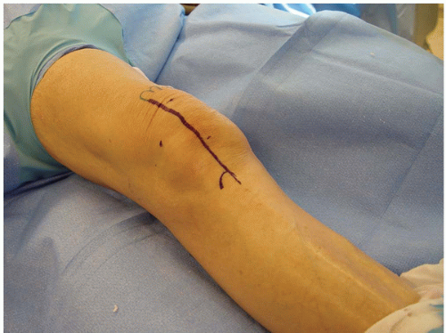 FIGURE 2-3 Skin incision follows the Q-angle and is slightly lateral to the midline. |
Six major steps of the direct lateral approach are as follows:
• | Step 1: | I-TB release/lengthening |
• | Step 2: | Lateral arthrotomy, coronal plane Z-plasty |
• | Step 3: | Patella dislocation, joint exposure |
• | Step 4: | Deep concave side releases (options) |
Tibial sleeve release, osteoperiosteal | ||
Distal LCL lengthening | ||
Proximal LCL lengthening | ||
Sleeve or osteoperiosteal | ||
Sliding lateral condyle osteotomy | ||
• | Step 5: | Instrumentation/prosthetic insertion |
• | Step 6: | Soft tissue closure deep to superficial layer |
I-TB Release/Lengthening
Classically, the I-TB is exposed proximally by separating the inner fascial sleeve from the vastus lateralis muscle. The band is released from the posterior femur and “finger stripped” to the posterolateral corner. A varus stress at the knee joint will “bowstring” the tight fascial bands, allowing for a multiple puncture “pie-crusting” lengthening under visual and digital control. The release is performed approximately 10 cm proximal to the joint line. The peroneal nerve can be palpated or explored, but this is seldom required and not recommended except in very severe cases. A simpler method preferred by the author is to expose the distal insertion of the I-TB into Gerdy’s tubercle and either sharply incise or use a periosteal elevator to elevate the distal fascial insertion (Fig. 2-4). The simple I-TB release with subsequent lateral capsular release will allow the anatomical correction of all but the most severe deformities (>90%).
Lateral Retinacular Incision (Superficial Layer)
Related posts:
 Minimally Invasive Total Knee Arthroplasty
 Cruciate-Retaining Total Knee Arthroplasty
 Simultaneous Total Knee Arthroplasty and Femoral Osteotomy
 Impaction Bone Grafting for Large Bone Defects
 Implanting the Revision Total Knee Arthroplasty
 Soft Tissue Balancing Technique for Posterior Stabilized Total Knee Arthroplasty
Stay updated, free articles. Join our Telegram channel

Full access? Get Clinical Tree


