Fig. 3.1
Monopolar: electrical current is conducted into the tissue to the grounding pad. Bipolar: current is conducted away from the tip to the return electrode on the probe shaft
Table 3.1
Tissue impedance
Tissue/substance | Impedance (Ω) |
|---|---|
Saline | 90–120 |
Ligaments | 100–140 |
Cartilage | 350–500 |
Bone | 1,100 |
Monopolar and bipolar devices both require a conductive milieu such as normal saline or lactated Ringer’s. The tips of the RF probes are available in many shapes and sizes to accommodate the anatomy of the problem being treated.
While the following discussion focuses on the use of the Ho:YAG laser, radiofrequency (RF) devices can be used in place of the laser [6]. The only exception to this generalization is the ablation of bone, which is more readily accomplished with the laser. The RF probes can be used through the same portals described for the laser probes. Radiofrequency devices can ablate and shrink tissue. However, one must be careful while using RF wands to not overheat the joint or the structures adjacent to the joint. Adequate inflow/outflow is essential while using the RF devices. Prolonged use of the RF probes without an adequate heat sink (fluid flow) can lead to diffuse thermal injury of the joint surfaces and adjacent peri-articular structures. The RF energy penetrates the tissue to a depth of 4 or more mm as compared to the 0.5 mm penetration of the laser. There is therefore a greater potential for injury to adjacent extraarticular structures (nerves) when using the RF probes.
Laser/RF-Assisted TFCC Debridement
Andrew Palmer [7] devised a classification scheme for TFCC tears that divides TFCC tears into traumatic tears, type I and degenerative tears, type II [7]. While both types of tears can be treated arthroscopically, types I-A, II-C, and II-D lend themselves to laser-assisted debridement (Fig. 3.2).
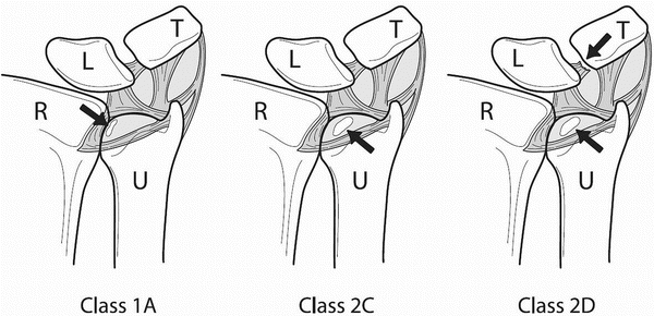
Fig. 3.2
Palmer classification of TFCC tears [Adapted with permission from Palmer AK. Triangular fibrocartilage complex lesions: a classification. J Hand Surg Am 1989;14:594–606. With permission from Elsevier]
Before starting arthroscopic treatment of TFCC tears, the ulnar variance must be evaluated. This is done by taking an X-ray with the shoulder abducted to 90° and the elbow flexed to 90° with the hand flat on the X-ray cassette (the “90 × 90” view of Palmer) [8]. Triangular fibrocartilage debridement in face of an ulnar-plus variance is doomed to fail, as the simple debridement of the TFCC is insufficient to decompress the ulnar side of the wrist. In such cases of ulnar abutment syndrome, an ulnar shortening is needed. The results of TFCC debridement in patients with an ulnar-zero variance can be good, but there lingers the possibility of having to perform an ulnar shortening later. It is recommended that this possibility be discussed with the patient preoperatively. In contrast to the patient with an ulnar plus variance, the patient who presents with an ulnar minus variance is very likely to respond to simple debridement of the central portion of the triangular fibrocartilage [9, 10].
The technique of laser-assisted triangular fibrocartilage debridement is similar to that of mechanical debridement of the triangular fibrocartilage with the exception that the arthroscope can be left in the 3-4 portal while the laser is kept in the 4-5 portal. The laser is set to 1.4–1.6 J at a frequency of 15 pulses per second. With the help of a side-firing 70-degree laser tip, the triangular fibrocartilage can be very rapidly and precisely debrided. The 70-degree laser tip permits ablation not only of the radial and palmar portions of the TFCC tear, but also the ulnar and dorsal components. There is no need to bring the laser probe in through the 3-4 portal. During the debridement, care must be taken not to injure the ulnar head. This is avoided by firing the laser tangentially to the head of the ulna or passing the probe beneath the triangular fibrocartilage and firing distally. This latter technique presents minimal danger to the lunate or triquetrum, in that the fluid used to expand the joint acts as a heat sink and absorbs the laser energy as it emerges from beneath the triangular fibrocartilage. The central portion of the TFCC is debrided back to stable edges, taking care not to injure the dorsal and palmar radioulnar ligaments.
The arthroscopic treatment of the ulnar abutment syndrome is facilitated by using the Ho:YAG laser [11]. Hyaline cartilage is very efficiently removed with the laser at higher energy settings (2.0 J at 20 pulses per second). Not only are the ulnar head hyaline cartilage and subchondral bone rapidly removed, but, in contrast to burring, they are removed without producing much debris. Once the cancellous bone of the ulnar head is exposed, however, the bur becomes the most effective tool to complete the ulnar shortening. This is because it becomes very time consuming to focus the laser beam on each trabecula. During the ulnar head resection, care must be taken not to injure the sigmoid notch with either the laser or the bur. Also, care must be taken not to detach the insertion of the triangular fibrocartilage from the fovea at the base of the ulnar styloid. The successful arthroscopic ulnar shortening relies on teamwork. The assistant brings the surfaces of the ulnar head to be resected to the laser being held by the operating surgeon. By progressively supinating and pronating the forearm, an appropriate amount of ulnar head is excised. The goal is to resect sufficient ulna to produce a 2 mm negative ulnar variance. The amount of ulna resected must be verified with intraoperative fluoroscopy. Occasionally, complete visualization of the ulnar head requires that the scope be placed in the 4-5 portal with the laser entering the distal radioulnar joint through the distal radioulnar joint portal. This portal is established just proximal to the 4-5 portal and TFCC.
An effort is made to leave a smooth surface on the remaining distal ulna. The trabeculae of the distal ulna always produce a somewhat rough distal ulna at the completion of the procedure. These irregularities, however, disappear during the months following the surgery (Figs. 3.3 and 3.4). Large irregularities must be avoided, as they can catch on the proximal surface of the residual TFCC during supination and pronation.
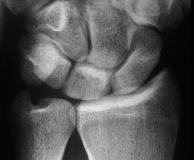
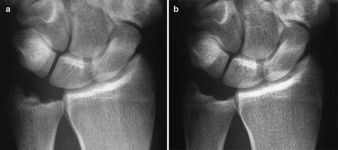
Fig. 3.3
Ulnar abutment
Fig. 3.4
(a) Early and (b) late post-laser-assisted arthroscopic ulnar shortening demonstrating smoothing of resection site with time. a is 6 weeks postoperative and b is 6 months postoperative
The postoperative regimen after TFCC debridement, with or without ulnar shortening, includes providing the patient with a wrist splint to be worn as needed, as well as a home therapy program consisting of active and passive range of motion exercises. The sutures (wounds are closed using subcuticular sutures of 4-0 Prolene) are removed at 2 weeks. Strengthening exercises can be started at 6 weeks if needed. Premature resumption of heavy lifting or repetitive activities will lead to radiocarpal synovitis. Some patients feel so good after as little as 2 weeks that the surgeon must temper the patient’s desire to return to full activity. In the case of an ulnar shortening, the recovery can be as long as 6 months, as suggested by Feldon [12]. However, the majority of patients will be improved long before 6 months.
Other Indications for Laser/RF-Assisted Wrist Arthroscopy
Synovectomy
Synovectomy is probably the most frequently performed laser-assisted procedure. This procedure is often needed to permit complete joint visualization, particularly of the lunotriquetral and ulnocarpal joints. The laser, set at 1.2–1.5 J and 15 pulses per second, vaporizes the inflamed synovium and scar tissue quickly and with minimal bleeding due to the hemostatic effect of the laser. The hemoglobin in the inflamed synovium absorbs the laser energy better than the adjacent capsule, thus providing an extra level of safety for the capsule. Scar tissue and synovitis in the radiocarpal, ulnocarpal, and midcarpal joints can be rapidly debrided. When performing a dorsal wrist synovectomy, or for that matter anytime the laser is being used, care should be taken to avoid aiming the laser at the arthroscope, as the laser energy will destroy the scope.
Partial Interosseous Ligament Tears
Partial tears of the scapholunate and lunotriquetral ligaments can be nicely treated with the laser set at 0.2–1.0 J and 15 pulses per second. The ablation of these tears can be done very precisely without scuffing or injuring the adjacent intact articular cartilage.
Chondromalacia
Chondromalacia has been treated with the laser and RF devices. There is, however, evidence that at least in regard to the RF devices, significant injury to the underlying healthy cartilage can occur even when exercising caution and using low power settings [13, 14]. Based on this information, it is difficult to recommend RF treatment of chondromalacia. Chondromalacic fronds can, however, be gingerly vaporized with the laser set at 0.2–0.8 J and 15 pulses per second. The laser beam must be oriented tangentially to the joint surface so that only the fronds of frayed cartilage are treated. Great care must be taken to not injure the underlying healthy cartilage. Because the laser radiation can be directed selectively toward the chondromalacic fronds, sparing the underlying cartilage, it is safer in this situation than are RF devices. However, great care must be exercised. It should be kept in mind that the long-term effectiveness of debridement of chondromalacic fronds has not been established, and the potential for significant injury to healthy cartilage cannot be ignored even with the laser.
Bone Resection
We have seen that the Ho:YAG laser can be used to resect the distal ulna. Similarly, the laser can be used to perform radial styloidectomies, osteophytectomies, and complete resection of the ulnar head. The principles outlined in the section describing the laser-assisted arthroscopic Feldon procedure apply to these procedures as well. The articular cartilage and subchondral bone are vaporized with the laser, while the cancellous bone is removed with a bur.
Radial styloidectomy is performed with the arthroscope in the 4-5 portal and the laser and bur entering through the 1-2 and 3-4 portals. (The 1-2 portal is approached with caution, as the radial artery and branches of the superficial radial nerve course through this area. Only blunt dissection should be used in establishing the 1-2 portal.) A clear junction usually exists between the area of the radial styloid to be debrided (exposed subchondral bone) and the adjacent healthy cartilage. If this is not the case, a K-wire can be placed under both fluoroscopic and arthroscopic control through the radial styloid at the ulnar limit of the proposed bone resection. This provides an intra-articular landmark. The amount of styloid resected should be just enough to solve the problem being addressed, taking care to leave the attachments of the radioscaphocapitate and long radiolunate ligaments intact. Postoperative care after this procedure is similar to that described for a TFCC debridement.
Laser-assisted arthroscopic Darrach procedures and matched ulnar resections are logical extensions of the laser-assisted arthroscopic Feldon procedure. The technique used for these procedures is essentially the same as that used for the laser-assisted arthroscopic Feldon procedure. One would anticipate less morbidity with this technique, though no published series are currently available. The use of the laser to treat grade IV chondromalacia has been successful in our hands in a limited number of cases. Two approaches are used, depending on the clinical presentation. If the joint surfaces involved cannot be unloaded (i.e., the proximal lunate), the laser is used to ablate the detached cartilage and subchondral plate. The laser debridement is extended to expose a healthy cartilage/bone interface. This “crater” margin is “freshened” with the bur. The subchondral bone is burred back to bleeding, cancellous bone. This last step is needed, as laser cauterization slows the fibrous tissue ingrowth necessary for the success of chondroplasty in this setting. Early range of motion is essential. The use of continuous passive motion has proven to be important.
Related posts:

Stay updated, free articles. Join our Telegram channel

Full access? Get Clinical Tree


