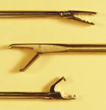
Chapter 3 Clinical and Surgical Pearls and Pitfalls • Create accessory portals under direct vision using a spinal needle to assess angle of entry to be able to successfully pass suture through tissue. • An attempt should be made to place the portal in an adequate position to allow for a reasonable amount of swelling. For example, placement of the lateral portal too close to the lateral aspect of the acromion may lead to difficulty performing adequate acromioplasty and bursectomy. • Maximize visualization before starting the reparative procedure. Remove obstructing bursa or soft tissue. • Perform a complete survey of the joint and periarticular structures before beginning the repair. Focusing on only the known pathology may lead to missed diagnoses. • Make a plan. Make sure all potential equipment and devices are available before the case. • Work quickly on secondary procedures such as acromioplasty to minimize unnecessary soft tissue extravasation. • Obtain adequate hemostasis. Failure to do so may ultimately lead to longer operative times with difficulty visualizing the structures. The anesthesia team should maintain blood pressure less than 100 mm Hg systolic to maximize visualization of work in the subacromial space via the arthroscopy. • Prevent tangling of sutures when multiple anchors are used. We recommend that sutures from each anchor be placed in different portals to prevent entanglement. Other options include keeping the sutures “outside” of a cannula interposed between the cannula and soft tissue, or using a small stab incision to serve as a suture repository while other suture limbs are being passed. • Different repairs require different suture passers. Gaining comfort with multiple suture-passing techniques allows for better tissue fixation and a quicker operative procedure. • A clear cannula in the working portal allows visualization and prevents soft tissue from interfering with the knot as it slides to the tissue. • Only one set of suture should be retrieved into the working portal used for tying the knot. • Use the portal that is best directed over the anchor to allow better suture sliding. Be sure to check that the suture slides easily before attempting to tie a sliding knot. • If the suture does not slide, a nonsliding knot with reversed half hitches is necessary for maximum fixation to be obtained. • Before the knot is tied, a knot pusher may be passed down the post suture to untwist the suture.1 • For nonsliding knots, we tie at least six half hitches, alternating the post and reversing the throws with each (underhand and overhand). • Advanced arthroscopists may choose to forego the use of a cannula. If this method is chosen, we recommend that a ring forceps be placed around both suture limbs inside the working space and retrieved together to avoid soft tissue interposition. • Select the suture limb that will function as the “post” that allows for best tissue approximation and compression. In a mattress suture configuration, the post can be either limb. In the simple suture configuration, pick the post away from articular cartilage. This suture limb is usually on the tissue side, allowing for maximal compression of the tissue against bone and also directing the knot away from the joint, thus avoiding articular injury from the resultant knot.1 • Place a clamp to the end of the suture post limb before tying a knot. This prevents the knot pusher from sliding off the post and provides resistance as the knot is tightened. • Visualize the knot as it slides to the tissue to ensure that the tissue is compressed to the desired location. • Maintain tension on the post limb as the knot is seated to avoid loosening. • “Past pointing” is a technique by which the knot pusher is used to tension the knot by switching the tension to the loop limb and pushing past the knot with the post limb of suture (see Fig. 3-7). This technique allows the knot to fully seat, which increases the knot security provided by the knot’s internal friction. • After an initial sliding knot is tied, reversed half hitches on alternating posts should be thrown and seated with the knot pusher, using past pointing to prevent the knot from coming loose or backing out. • Be patient. Allow extra time on all arthroscopic cases in the beginning. • Practice your knot-tying skills. The time to practice is before the case when you are not under pressure. When practicing, use bigger string or rope to view the knot configuration. Dry and wet laboratories are extremely helpful and should be used for training when possible. • Management of suture requires careful attention. You must practice and visualize your knot tying. Make it as easy and automatic as tying your shoes. The advent of the suture anchor has dramatically expanded the options for tissue repair. Numerous suture anchor designs are available; anchors come in multiple sizes, allowing maximum fixation strength of tissue to bone.2 Anchors may be made of metal, absorbable material, or plastic and should allow for sutures to slide easily through the eyelet. The anchor, when inserted into the bone, allows suture to be passed through soft tissue and affixed to the desired anatomic location in a predictable fashion. Multiple sutures may be preloaded into the anchor, allowing for multiple points of soft tissue fixation and decreased load on each suture knot.3 Although the choice of the best anchor for each surgical procedure is beyond the scope of this chapter, a basic understanding of anchor types is advised. Arthroscopic cannulas allow for suture passage through tissues, avoiding incorporation of unwanted soft tissue in the repair constructs.4 Sutures and instruments that are not passed through a cannula can be trapped in soft tissues, causing significant difficulty in knot tying, which may result in increased operative times, less secure fixation, and generalized frustration for the surgeon. Cannulas also allow the surgeon to keep sutures organized, prevent suture entanglement, provide easy access to the joint, and facilitate visualization. The ideal cannula size to allow for passage of typical arthroscopic instruments is 8.5 to 10 mm. Cannulas are important tools that play an integral part in the surgical plan. After insertion of the anchor, specialized instruments will be necessary to assist with management of sutures to facilitate a secure repair.4 Suture retrievers are the workhorse of any arthroscopic procedure (Fig. 3-1). These devices can be locking or ratcheting, and function to grasp the suture. Some devices will securely hold the suture, whereas others secure the suture but allow it to slide in the jaws (suture retrieval forceps, or “loopie”). Suture retrieval forceps can facilitate removal of suture from the joint by allowing it to slide as it is extracted. This prevents the suture limb from sliding through the anchor unintentionally. Another option for suture management is the crochet hook instrument.4 This device allows the surgeon to place the suture at various places within the joint for retrieval and passage. Some crochet hooks have a modification that allows for sutures to be pushed with the tip in addition to being pulled with the hook (“push-me, pull-me”).
Knot-Tying and Suture-Passing Techniques
Instrumentation
Suture Anchors
Cannulas
Arthroscopic Instruments
Related posts:
 Patient Positioning, Portal Placement, Normal Arthroscopic Anatomy, and Diagnostic Arthroscopy
 Osteochondral Autograft for Cartilage Lesions of the Knee
 Arthroscopic Rotator Cuff Repair: Double-Row Techniques
 Surgical Treatment of Posterolateral Instability of the Elbow
 Primary Repair of Osteochondritis Dissecans in the Knee
 Arthroscopic Meniscus Repair: All-Inside Technique
![]()
Stay updated, free articles. Join our Telegram channel

Full access? Get Clinical Tree


Knot-Tying and Suture-Passing Techniques
 Video
Video