Fig. 49.1
JBC in the femoral neck. X-ray shows a well-delimited oval osteolytic radiolucent small lesion outlined by a sclerotic rim
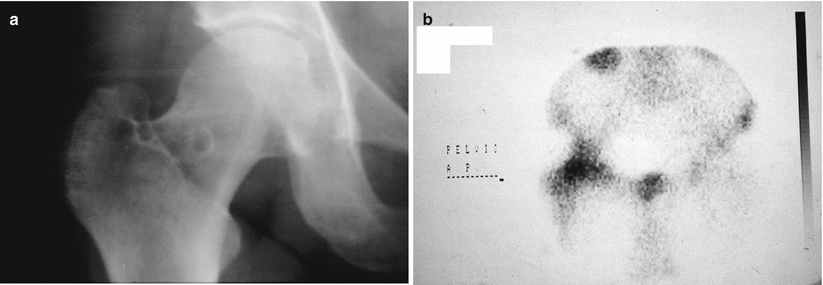
Fig. 49.2
(a, b) JBC arising in the femoral neck. Bone scan occasionally shows a hot spot
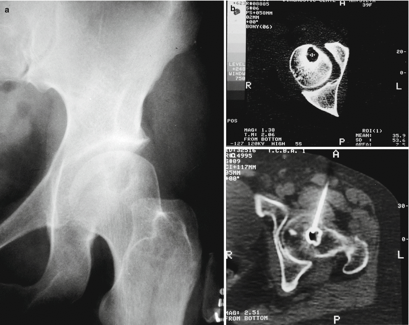
Fig. 49.3
(a) JBC located in the femoral head. (b) CT scan evidencing a lytic lesion with a peripheral sclerotic rim. (c) The diagnosis was made by core needle biopsy

Fig. 49.4
(a) X-ray and (b) CT show a large JBC arising in the femoral head
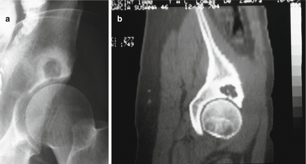
Fig. 49.5
(a) Roentgenogram and (b) CT scan of a JBC arising in the acetabulum, not related to joint osteoarthritic pathology
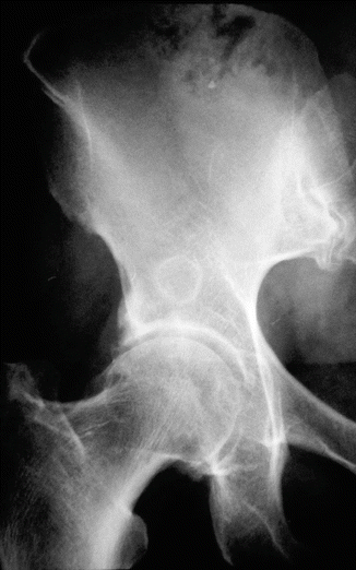
Fig. 49.6
Typical JBC located in the acetabulum
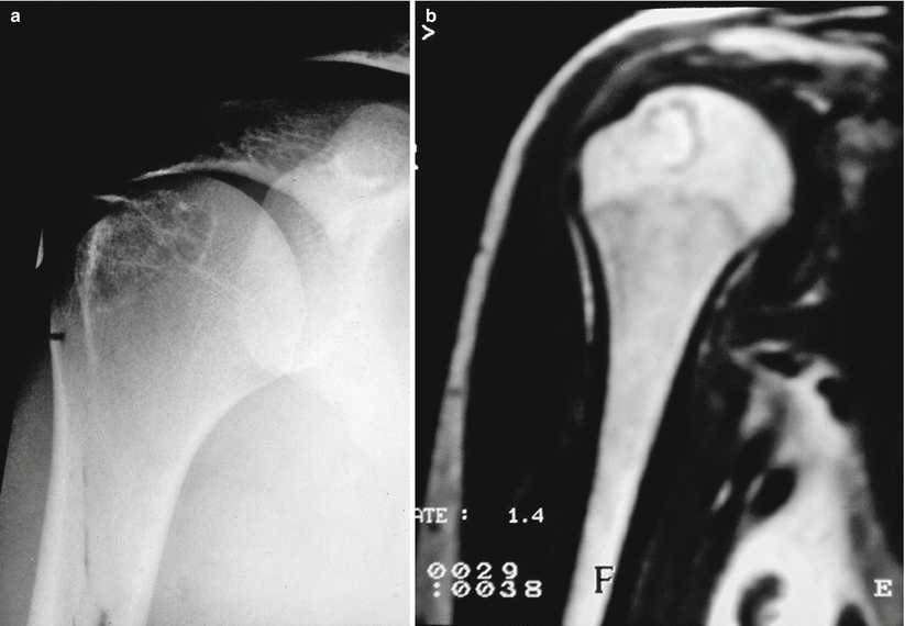
Fig. 49.7
(a) Humerus epiphyseal location of a JBC. (b) MRI confirms the fluid content of the lesion, low signal in the peripheral sclerotic margin and edema in the peripheral bone marrow
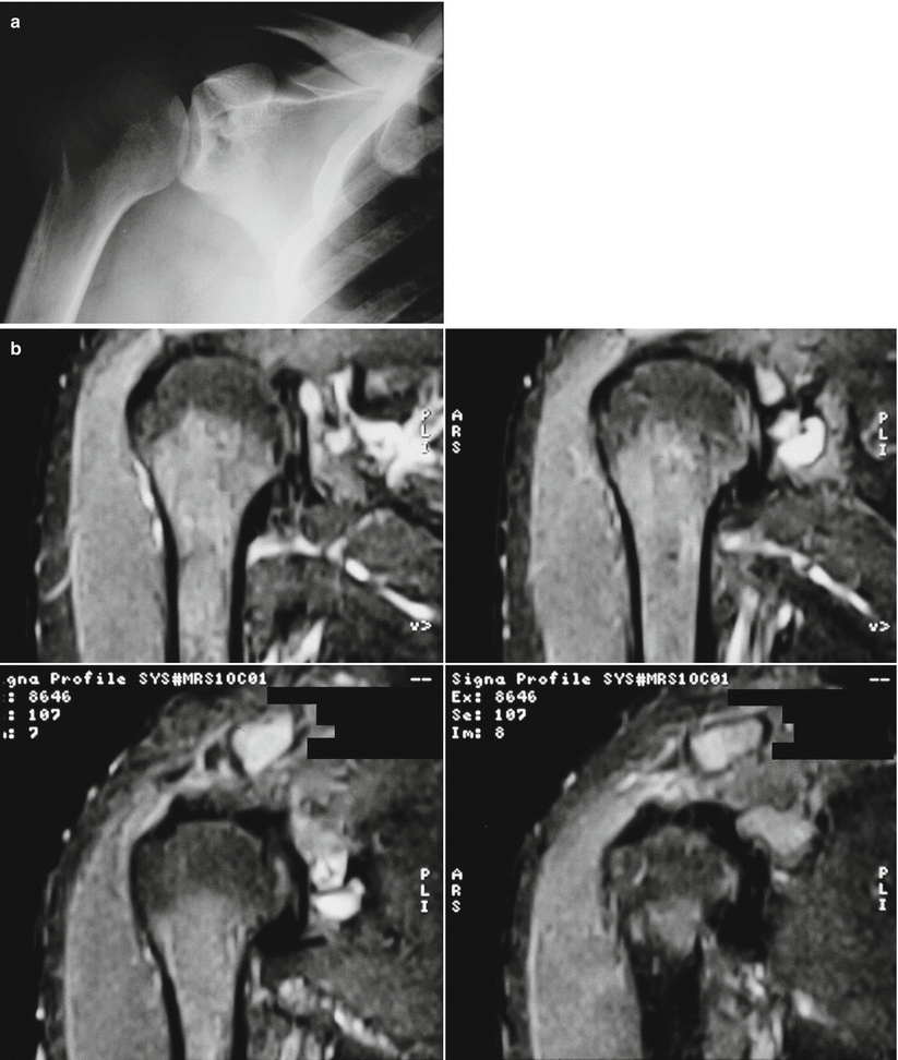
Fig. 49.8
(a) X-ray and (b) MRI showing a JBC in the glenoid of the scapula
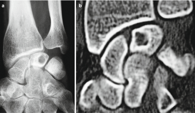
Fig. 49.9
(a) X-ray and (b) CT showing a typical JBC arising in the lunate bone
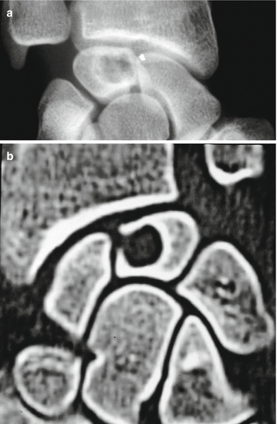
Fig. 49.10


(a) Roentgenogram of a JBC in a lunate bone. (b) CT shows the communication between intraosseous cyst and the adjacent joint
Stay updated, free articles. Join our Telegram channel

Full access? Get Clinical Tree


