Juvenile Idiopathic Arthritis


Juvenile Idiopathic Arthritis


Pamela F. Weiss


OVERVIEW OF PEDIATRIC RHEUMATIC DISEASE ENCOUNTERED BY THE PEDIATRIC ORTHOPAEDIC SURGEON

Joint pain is a common childhood complaint. Each year, as many as 1% of all children will be evaluated by a physician for joint pain (1). Approximately 15% of healthy children reported on a health questionnaire that they had episodes of musculoskeletal pain (2). Further, healthy children in day care centers have approximately one painful episode every 3 hours, arising from play, disciplining, or interaction with peers (3). The orthopaedic surgeon is often the first specialist to encounter the child with joint, limb, or back pain. In a study of subspecialty referrals of juvenile arthritis, most children with pauciarticular juvenile rheumatoid arthritis (JRA) (62%) were referred to orthopaedic surgeons prior to referral to pediatric rheumatology care (4). Among children who are evaluated by a physician for pain in the joints, only 1 in 100 will eventually be diagnosed as having arthritis, but among those who present to an orthopaedist, the frequency of arthritis is surely higher. Accordingly, it is important that the orthopaedic surgeon be able to identify the most likely cause of the pain and either initiate treatment or refer the patient to an appropriate medical specialist.

The purpose of this chapter is to provide the orthopaedic surgeon with an in-depth understanding of the presentation, differential diagnosis, and management of children with arthritis. With this framework, the orthopaedic specialist should be able to identify children with juvenile arthritis and to differentiate arthritis from benign pains of childhood, psychogenic pain syndromes, benign musculoskeletal back pain, infection, malignancy, or other systemic autoimmune diseases (lupus, dermatomyositis, and vasculitis). Infectious, malignant, congenital, mechanical, or traumatic causes of arthralgias and arthritis are presented in order to contrast the symptoms with those of juvenile arthritis; detailed presentations on these conditions can be found elsewhere in this text.


CLASSIFICATION OF JUVENILE ARTHRITIS

Juvenile arthritis is a term for persistent arthritis lasting >6 weeks of unclear etiology. A diagnosis of juvenile arthritis is made by taking a thorough history, performing a skilled and comprehensive physical examination, utilizing directed laboratory tests and imaging procedures, and following the child over time.

Over the past several decades, there have been three sets of criteria utilized for the diagnosis and classification of juvenile arthritis (Table 11-1). The first set of criteria was proposed in 1972 by the American College of Rheumatology (ACR) and defined three major categories of JRA: oligoarticular (pauciarticular), polyarticular, and systemic (5).The ACR JRA criteria exclude other causes of juvenile arthritis, such as spondyloarthropathies [JAS, inflammatory bowel disease (IBD)-associated arthritis, and related diseases], juvenile psoriatic arthritis, arthritis associated with other systemic inflammatory diseases [systemic lupus erythematosus (SLE), dermatomyositis, sarcoidosis, etc.], and infectious or neoplastic disorders. The second set of criteria was formulated in 1977 by the European League Against Rheumatism (EULAR) and coined the term juvenile chronic arthritis (JCA) (6). JCA is differentiated into the following subtypes: pauciarticular, polyarticular, juvenile rheumatoid [positive rheumatoid factor (RF)], systemic, juvenile ankylosing spondylitis (JAS), and juvenile psoriatic arthritis. The ACR and EULAR criteria, although similar, do not identify identical populations or spectra of disease. However, they have often been used interchangeably, leading to confusion in the interpretation of studies relating to the epidemiology, treatment, and outcome of juvenile arthritis.

In 1993, The International League of Associations of Rheumatologists (ILAR) proposed (7) and revised (8) criteria for the diagnosis and classification of juvenile arthritis (Table 11-2). The term juvenile idiopathic arthritis (JIA) has been proposed as a replacement for both JRA and JCA. The
ILAR criteria allow for uniform interpretation of clinical and therapeutic data. Recent validation of the ILAR classification criteria has found that 80% to 88% of children could be classified, and 12% to 20% were classified as “Undifferentiated” because they either did not fit into any category or fulfilled the criteria under two categories (9—12). As genetic risk factors and specific triggers of juvenile arthritis are identified, modifications to the criteria can be made. In the remaining sections of this chapter, the emphasis will be on the JIA classification scheme. The terms JRA and JCA will be used only when referring to specific epidemiologic, therapeutic, or outcome data.


TABLE 11-1 Comparison of JRA, JCA, and JIA Classifications


JRA


JCA


JIA


Committee


ACR


EULAR


ILAR


Age at onset


<16 yr


<16 yr


<16 yr


Disease duration


>6 wk


>3 mo


>6 wk


Onset types


Pauciarticular Polyarticular Systemic


Pauciarticular Polyarticular RF-negative Juvenile rheumatoid arthritis Systemic Juvenile psoriatic arthritis Juvenile ankylosing spondylitis


Oligoarticular, persistent Oligoarticular, extended Polyarticular RF-negative Polyarticular RF-positive Systemic Psoriatic arthritis Enthesitis-related arthritis


Exclusions


Juvenile psoriatic arthritis Juvenile ankylosing spondylitis Inflammatory bowel disease Other forms of juvenile arthritis


Other forms of juvenile arthritis


Other forms of juvenile arthritis


RF, rheumatoid factor.


TABLE 11-2 Criteria for Classification of JIA
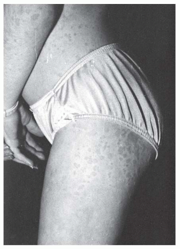


JIA Subtype


Exclusionsa


Inclusion Criteriab


Oligoarthritis


1-5


Persistent


<4 joints during disease course


Extended


>4 joints after the first 6 mo


Polyarthritis RF-negative


1-5


Arthritis affecting >5 joints during the first 6 mo


Polyarthritis RF-positive


1-3, 5


Arthritis affecting >5 joints during the first 6 mo, plus RF positivity on two occasions more than 3 mo apart


Systemic


1-4


Arthritis with or preceded by daily fever of at least 2 weeks’ duration, accompanied by one or more of the following: Evanescent, nonfixed erythematous rash Generalized adenopathy Hepatomegaly or splenomegaly Serositis


Psoriatic


2-5


Arthritis and psoriasis, or arthritis and at least two of the following:


a.  Dactylitis


b.  Nail abnormalities (pitting or onycholysis)


c.  Family history of psoriasis in a first-degree relative


Enthesitis-related


1, 4, 5


Arthritis and enthesitis, or arthritis or enthesitis with at least two of the following:


  1. SI joint tenderness and/or inflammatory spinal pain


  2. Presence of HLA-B27


  3. Family history of HLA-B27-associated disease in a first-degree relative


  4. Onset of arthritis in a male after the age of 6 yr


Undifferentiated


Children with arthritis of unknown cause that persists >6 wk Does not fulfill criteria for any of the other categories Fulfills criteria for >1 of the other categories


a Exclusions: 1, psoriasis in the patient or a first-degree relative; 2, arthritis in an HLA-B27 positive male beginning after the sixth birthday; 3, ankylosing spondylitis, enthesitis-related arthritis, sacroiliitis with IBD, Reiter syndrome, or acute anterior uveitis in a first-degree relative; 4, IgM RF on at least two occasions more than 3 mo apart; 5, presence of systemic JIA.

b Inclusion criteria for all subtypes: 1, age at onset <16 yr; 2, arthritis in one or more joints; 3, duration of disease is at least 6 wk. From Petty RE, et al. International League of Associations for Rheumatology classification of juvenile idiopathic arthritis: second revision, Edmonton, 2001. J Rheumatol 2004;31(2 ):390-392.


Oligoarthritis


Epidemiology.

Most children with oligoarthritis present before 4 years of age and girls outnumber boys by a ratio of 4 to 1. Whites are affected more often than other races. It is the most frequent subtype of JIA, accounting for up to 40% of cases (13, 14). Prevalence is estimated at 60 per 100,000 children (15).


Etiology.

The etiology of oligoarticular JIA is unknown, but associations with HLA-A2, DRB1*01, DRB1*08, DRB1*11, DRB1*13, DPB1*02, DQA1*04, and DQB1*04 have been reported (14, 16). Oligoarticular JIA is rarely familial. Approximately 70% of oligoarticular JIA patients are positive for antinuclear antibodies (ANA).


Clinical Features.

Approximately 50% of children with oligoarticular JIA present with a single affected joint, most commonly the knee, followed by ankles and small joints of the hands. The hips and shoulders are rarely affected. Early wrist involvement is uncommon and may portend progression to a polyarticular or extended oligoarticular course. At presentation, the majority of children have morning stiffness, gelling, and pain. However, up to 25% of children have painless arthritis at presentation (17).

Most children with oligoarticular JIA have a mild and remitting course. However, in untreated children with longstanding unilateral knee arthritis, there can be overgrowth of the affected limb, resulting in a marked leg-length discrepancy (18, 19). Temporomandibular joint (TMJ) arthritis is present in a majority of children at disease onset (20) and if untreated, may cause localized growth disturbances, micrognathia, malocclusion, and chewing difficulties (21—23). Chronic uveitis is the most common extra-articular complication seen in oligoarthritis, is associated with ANA positivity, and occurs in approximately 20% of children. Periodic screening for uveitis is necessary as the inflammation is typically asymptomatic and unable to be detected without the use of a slit lamp. Untreated uveitis may result in cataracts, band keratopathy, secondary glaucoma, and blindness.

Long-term, children with oligoarticular JIA have the greatest likelihood of remission of all JIA subtypes. In one study, 68% of persistent and 31% of extended oligoarticular JIA patients achieved long-term clinical remission off medication (24).


Polyarticular Arthritis


Epidemiology.

RF-negative polyarthritis can occur at any age, with the median age of onset at 6.5 years (25), with girls outnumbering boys by a ratio of 3:1. RF-positive polyarticular JIA occurs most frequently in adolescent girls and is indistinguishable from adult rheumatoid arthritis (RA). Polyarticular JIA is the second most frequent subtype of JIA, accounting for up to 22% of cases (13, 14). Prevalence is estimated at 40 and 10 per 100,000 children for RF-negative and RF-positive subtypes, respectively (15).


Etiology.

The etiology of polyarticular JIA is unknown. Multiple studies have examined the association of HLA genes and disease. RF-negative polyarticular JIA has been associated with HLA-A2, DRB1*08, DQA1*04, and DPB1*03. Associations of RF-positive polyarticular JIA with HLA-DQA1*03, DQB1*03, and DRB1*04, a gene also associated with adult RA, have been reported (14).


Clinical Features.

Polyarticular-onset JIA is characterized by the insidious, but occasionally acute, onset of symmetric arthritis in five or more joints. It can involve both large and small joints and frequently affects the cervical spine and TMJs. Mild systemic features such as low-grade fever, lymphadenopathy, and hepatosplenomegaly may be present at diagnosis. The fevers are not typically the high quotidian temperature spikes that are diagnostic of systemic arthritis, and rash is rarely seen (26).


This RF-negative subgroup may be ANA positive (40% to 50%), and this is associated with an increased incidence of uveitis (5%) (27). Children with RF-positive polyarticular JIA are more likely to have a symmetric small-joint arthritis, rheumatoid nodules, and early erosive synovitis with a chronic course. However, these children rarely develop chronic uveitis.

Children with RF-positive polyarticular JIA are at risk for a prolonged and destructive course. These children are typically older girls with involvement of multiple joints (20 or more) including the small joints of the hands and feet, early erosions, and rheumatoid nodules. The presence of hip arthritis has been shown to be a poor prognostic sign and may lead to destruction of the femoral heads (28). If polyarthritis persists longer than 7 years, remission is unlikely. In a recent study, only 5% of RF-positive and 30% of RF-negative polyarticular JIA patients achieved long-term remission off medication (24).


Systemic Arthritis


Epidemiology.

Systemic JIA is one of the least common JIA subtypes, accounting for approximately 10% of all JIA cases (13). Onset can occur at anytime during childhood but peaks between 1 and 5 years of age (25). Boys and girls are affected equally. Prevalence of systemic JIA is estimated at 10 per 10,000 children (15).


Etiology.

Etiology of systemic JIA is unknown. HLA associations that have been reported include DRB1*04, DRB1*11, and DQA1*05 (14). Non-HLA genetic associations have been found with macrophage migration inhibitory factor (30) and a variant of the interleukin-6 (IL-6) gene (8).


Clinical Features.

The fever of systemic JIA is typically daily or twice-daily, usually to 39°C or higher (31). In between fever spikes, the temperature is often below normal. Children frequently appear quite ill while febrile but recover in between fevers. The fever often responds poorly to nonsteroidal antiinflammatory drugs (NSAIDs) but will typically respond well to corticosteroids. In most children, the fever is accompanied by a characteristic rash that consists of discrete, transient, nonpruritic erythematous macules (Fig. 11-2) (32). The rash is typically more pronounced on the trunk but may occur on the extremities and the face. The most commonly involved joints are the knee, wrist, and ankle (33). Many children with systemic JIA will have extra-articular manifestations, including hepatosplenomegaly, pericarditis, pleuritis, lymphadenopathy, and abdominal pain. The extra-articular features may be present for weeks, months, and, occasionally, years prior to the onset of arthritis. Usually, the extra-articular manifestations of systemic JIA are self-limiting and will resolve spontaneously or with corticosteroid therapy. Occasionally, the pericarditis can result in tamponade.

The prognosis of systemic JIA is determined predominantly by the course of arthritis. Approximately 50% of children with systemic arthritis will have a mild oligoarticular course, and in most of these children, the arthritis will ultimately remit. The remaining half of the children with systemic onset will develop a polyarticular arthritis that can remit, but progresses in approximately 50% of the cases (25% of all systemic-onset JIA) to a severe, unrelenting, and destructive course despite all currently available therapeutic interventions (34). Chronic anterior uveitis is extremely rare in systemic arthritis. Systemic amyloidosis, usually presenting with the onset of proteinuria and hypertension, can occur as a result of any chronic inflammatory disease. Approximately 8% of European children with systemic JIA have been shown to develop this life-threatening complication (35). The incidence of amyloidosis in North America is significantly lower
than that seen in Europe. The reason for this discrepancy remains unclear.


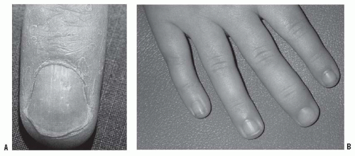
FIGURE 11-2. Juvenile psoriatic arthritis. A: Nail pitting associated with psoriasis. B: Swelling of a single DIP joint in a child with juvenile psoriatic arthritis.

Macrophage activation syndrome (MAS), also termed hemophagocytic lymphohistiocytosis, is a severe, potentially lifethreatening complication seen nearly exclusively in systemic arthritis. It is characterized by macrophage activation with hemophagocytosis and is associated with hepatic dysfunction, disseminated intravascular coagulation with a precipitous fall in the erythrocyte sedimentation rate (ESR) secondary to hypofibrinogenemia, and encephalopathy (36). It has been suggested that anti-inflammatory medications and viral infections can induce this syndrome. High-dose corticosteroids, cyclosporine A, and IL-1 inhibition have been shown to improve the outcome of MAS (37—39).


Psoriatic Arthritis


Epidemiology.

Psoriasis occurs in approximately 0.5% of the population (40), 20% to 30% of whom have associated arthritis (41, 42). There is a bimodal distribution of age of onset with a peak in the preschool years and again around 10 years of age. Girls are slightly more affected than boys. Psoriasis often begins after the onset of arthritis, usually within 2 years. The prevalence of psoriatic JIA is estimated at 15 per 100,000 children (15). Psoriatic arthritis accounts for 5% to 7% of JIA (13).


Etiology.

The etiology of psoriatic arthritis is unknown but genetic associations with HLA-Cw6, DRB1*01, and DQA1*0101 have been demonstrated (14, 43). There is often a strong family history of psoriasis or psoriatic arthritis in affected children.


Clinical Features.

The arthritis in psoriatic JIA is often an asymmetric mono- or polyarthritis affecting both large and small joints. At onset, patients may have pitting of the nails (67%) (Fig. 11-2) and a family history of psoriasis (69%) or dactylitis (39%), while less than one-half of the children have the rash of psoriasis (13% to 43%) (25, 44, 45). JIA criteria do not require the development of psoriasis to confirm a diagnosis of psoriatic arthritis (Table 11-2) (46). In children younger than 5 years, the presentation is often characterized by the involvement of a small number of fingers or toes that are relatively asymptomatic, but leading to marked overgrowth of the digit(s).

Children with psoriatic arthritis may have chronic lifelong arthritis that follows a relapsing and remitting course. Arthritis mutilans and severe distal interphalangeal (DIP) joint disease are unusual. However, many of the children will have prolonged polyarthritis that may result in irreversible joint damage (47). Amyloidosis has been reported in the European literature as having resulted in the deaths of at least three children (47, 48). Chronic anterior uveitis has been observed in up to 17% of the children (44, 45) and is associated with a positive ANA titer; the uveitis associated with psoriatic JIA is clinically indistinguishable from the uveitis in oligoarticular and polyarticular JIA.


Enthesitis-Related Arthritis


Epidemiology.

Unlike the other subtypes of JIA, ERA is more common in boys. Disease onset is typically after the age of 6 years. Prevalence is estimated at 50 per 100,000 children (15).


Etiology.

The presence of HLA-B27 is part of the diagnostic criteria for ERA. In these children, molecular mimicry is thought to contribute to the pathogenesis. Other HLA genetic associations that have been found are HLA-DRB1*01, DQA1*0101, and DQB1*05 (14).


Clinical Features.

ERA is often associated with enthesitis and arthralgias or arthritis long before any axial skeletal involvement is identified (50). Enthesitis is identified when marked tenderness is noted at the 6, 10, and 2 o’clock positions on the patella, at the tibial tuberosity, iliac crest, or the attachments of the Achilles tendon or plantar fascia (Fig. 11-3) (51). However, in ERA not all entheses are created equal; some entheses are more prone to trauma and mechanical damage such as in Sinding-Larsen-Johansson syndrome while other entheses are frequently tender in normal children such as the plantar fascia insertion into the metatarsal heads. One study suggested that “pathologic” enthesitis be defined as the presence of three tender entheses at the following sites: SI joints, inferior patellar pole, Achilles tendon insertion, and plantar fascia insertion into the calcaneus (52).


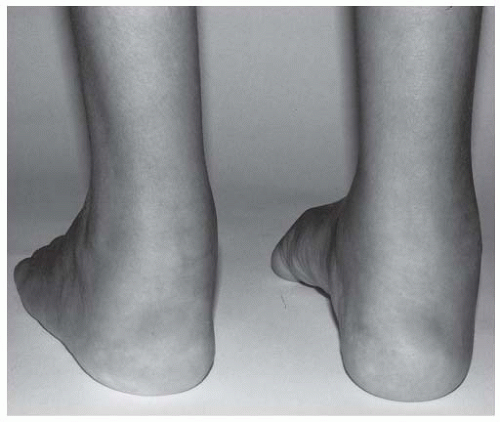
FIGURE 11-3. Achilles tendonitis and enthesitis in a child with enthesitis-related arthritis. (Courtesy of Dr. Ruben Burgos-Vargas.]

The primary extra-articular manifestation of ERA is acute anterior uveitis, which can occur in up to 27% of children with AS (53). The uveitis is manifested by an acute, painful, red, photophobic eye. ERA-associated uveitis may resolve with no ocular residua, but some of the children will have a persistent uveitis that is relatively resistant to therapy and can result in blindness (54, 55).


Juvenile Ankylosing Spondylitis


Epidemiology.

JAS most often presents in late childhood or adolescence. Boys outnumber girls by a ratio of 6 to 1 (56). There is a high frequency of JAS in Pacific Canada Indians (57) and a low incidence in African Americans (58).


Etiology.

The similarities between JAS and reactive arthritis, in which gastrointestinal and genitourinary infections trigger disease, suggest a role for infection. There is a strong genetic component to disease as AS occurs up to 16 times more frequently in HLA-B27-positive family members of patients with AS than in HLA-B27-positive individuals in the population at large (59). Further, children with JAS and SI involvement are frequently HLA-B27 positive (82% to 95%) (56).


TABLE 11-3 New York Criteria for AS


Clinical criteria


Limited lumbar motion in all three planes History or presence of lumbar spinal pain <2.5 cm of chest expansion at the 4th intercostal space


Definite AS


Grade 3 or 4 bilateral radiographic SI changes plus at least 1 clinical criterion


Grade 3 or 4 unilateral or grade 2 bilateral radiographic SI changes plus clinical criterion 1 or criteria 2 and 3


Probable AS


Grade 3 or 4 bilateral radiographic SI changes without any clinical criteria


Clinical Course.

Children with early JAS often fulfill the diagnostic criteria for ERA. Episodic arthritis of the lower extremity large joints, enthesitis, and tarsitis within 1 year of symptom onset predicts of progression to JAS (60). The presentation of JAS is most remarkable for the absence of axial involvement. Only 12% to 24% of children with JAS have pain, stiffness, or limitation of motion of the SI or lumbosacral spine at disease onset. A peripheral arthropathy or enthesopathy,
affecting predominantly the lower limb joints and entheses, is seen in 79% to 89.4%. These children tend to have fewer than 5 joints involved and rarely more than 10. At presentation, the pattern of involvement of the joints is usually asymmetric (61). Small joints of the toes are commonly involved in JAS but are seldom affected in other forms of JIA, with the exception of psoriatic arthritis. However, polyarticular and axial disease are usually evident after the 3rd year of illness (61). Children with long-standing JAS have been shown to develop tarsal bone coalition that has been termed ankylosing tarsitis (Fig. 11-4) (62).


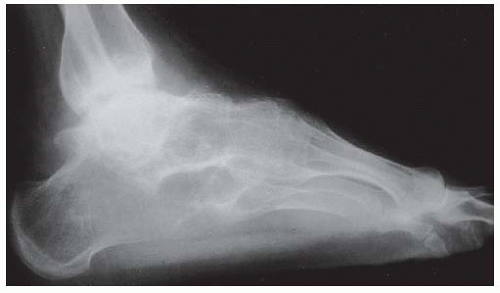
FIGURE 11-4. Ankylosing tarsitis, a complex disorder resulting in ankylosis of the foot in a child with JAS. (Courtesy of Dr. Ruben Burgos-Vargas.)

Outcome data for JAS are incomplete and at times contradictory. The prognosis of JAS has been reported as being worse according to some studies, and better according to others, than adult-onset AS (63, 64). Hip disease has been associated with a poor functional outcome (63, 65) and may require total hip arthroplasty.


Inflammatory Bowel Disease-Associated Arthritis

The frequency of arthritis in children with IBD has been reported to be 7% to 21%, and it usually occurs after the diagnosis of the bowel disease (66—68). Two different patterns of arthritis are seen (51). The most common type is oligo- or polyarticular arthritis of the lower limbs. This group is less likely to meet the criteria for ERA. This arthritis is often episodic, with exacerbation lasting 4 to 6 weeks or, rarely, for months. The activity of the peripheral arthritis is often related to the underlying bowel disease activity. The less common type of IBD-associated arthritis is an HLA-B27—associated oligoarticular arthritis of the lower limbs, with sacroiliitis and enthesitis, and no relationship to bowel inflammation (51). This form is more likely to persist and progress despite adequate control of the bowel disease. The clinical course is similar to that in other children with ERA.


TABLE 11-4 Differential Diagnosis of JIA


Monoarticular Arthritis


Polyarticular Arthritis


Febrile Syndromes


Oligoarthritis


Polyarthritis


Systemic arthritis


Psoriatic arthritis


Psoriatic arthritis


Malignancy:


Enthesitis-related arthritis


Enthesitis-related arthritis


Lymphoid


Sarcoidosis


Sarcoidosis


Neuroblastoma


Transient synovitis of the hip


Systemic lupus erythematosus


Systemic lupus erythematosus


Trauma


Juvenile dermatomyositis


Juvenile dermatomyositis


Hemophilia


Systemic vasculitis


Systemic vasculitis


Pigmented villonodular synovitis


Scleroderma


Infection (viral or bacterial)


Septic arthritis


Gonococcal septic arthritis


Inflammatory bowel disease


Reactive arthritis


Reactive arthritis


Reactive arthritis


DIFFERENTIAL DIAGNOSIS OF PAIN AND SWELLING IN THE JOINTS IN CHILDREN

A comprehensive differential diagnosis of arthritis in childhood is beyond the scope of this chapter as there are over 100 disorders in which arthritis may be a significant manifestation (69). The most common classes of disorders that must be considered in the differential diagnosis of JIA include infection, postinfectious phenomenon, inflammatory arthropathies, systemic autoimmune disease, mechanical or orthopaedic conditions, trauma, and pain disorders. Often, the differential diagnosis will be determined by whether the presentation is acute, subacute, or chronic, whether the child has monoarticular or polyarticular arthritis, and whether there are systemic signs such as fever (Table 11-4).


Infection-Related Arthritis


Septic Arthritis.

Septic arthritis generally affects a single joint and is associated with fever, elevated neutrophil count, ESR, C-reactive protein (CRP), and extreme pain. Synovial fluid analysis typically reveals white cell counts of >50,000 (70), neutrophil predominance, low glucose (<30 mg/dL), and a positive Gram stain. Oligoarticular JIA, in contrast, is seldom associated with systemic inflammation and joint effusions are often out of proportion to the reported pain. The most commonly infected joints in children are the knees, hips, ankles, and elbows. Gonococcal arthritis may present in a
sexually active adolescent as an oligoarticular, polyarticular, or migratory arthritis with significant tenosynovitis.


Lyme Arthritis.

Lyme arthritis may occur weeks to months after infection with the tick-borne spirochete Borrelia burgdorferi. Up to 60% of patients with untreated disease develop arthritis, which may be manifested by intermittent or continuous swelling (71). Many patients with untreated Lyme disease complain of migratory arthralgias or arthritis (72). In a recent retrospective study of 90 children with Lyme arthritis, Gerber et al. (73) noted that the majority (63%) had monoarticular disease, but no child had more than four joints involved. The knee was affected most often (90%), followed by hip (14%), ankle (10%), wrist (9%), and elbow (7%), whereas small joints were rarely involved. Most children with Lyme arthritis do not recall a tick bite or erythema migrans (73, 74). Lyme arthritis is typically an inflammatory synovitis with a very large and relatively painless joint effusion (Fig. 11-5). The ESR can be normal or elevated (73). The diagnosis should be confirmed with serologic testing, which includes an enzyme-linked immunosorbent assay (ELISA) and Western blot. There is a high rate of falsepositives with ELISA testing, so if the ELISA is positive, then a confirmatory Western blot should be performed. If the ELISA is negative, no further testing is needed. Synovial fluid analysis typically reveals white cell counts of 10,000 to 25,000. A small percentage of children may develop a persistent arthritis despite multiple courses of oral and/or intravenous antibiotics; persistence of swelling is associated with HLA-DR4 and HLA-DR2 alleles (75). In these patients, intra-articular corticosteroid injections are often helpful. Detection of Borrelia burgdorferi in the synovial fluid using polymerase chain reaction (PCR) can be confirmatory in seropositive patients. However, a positive PCR in the setting of negative serologies is likely to be a false-positive (76). Further a positive PCR is not proof of active infection as remnant DNA may persist for some time after Borrelia burgdorferi killing has occurred (76).


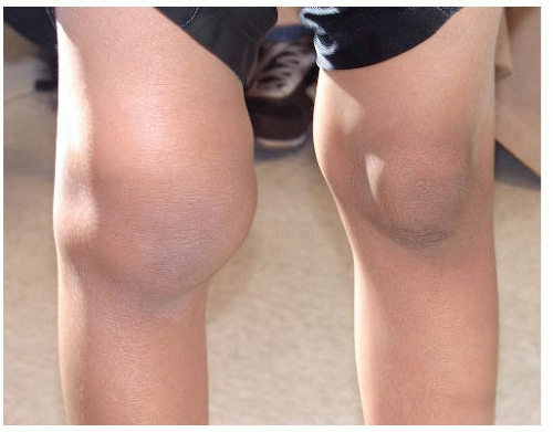
FIGURE 11-5. Right knee effusion in a child with Lyme arthritis.

Only gold members can continue reading. Log In or Register to continue

Stay updated, free articles. Join our Telegram channel

Jul 21, 2016 | Posted by in ORTHOPEDIC | Comments Off on Juvenile Idiopathic Arthritis

Full access? Get Clinical Tree

Get Clinical Tree app for offline access