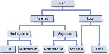
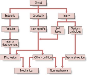
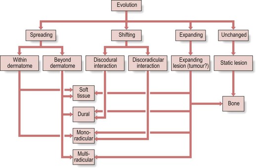
7 A standardized clinical examination enables the examiner to recognize clinical patterns. He/she will easily distinguish common patterns from uncommon ones and recognize so-called ‘warning signs’ (see Ch. 9). Neck, shoulder girdle and shoulder problems are differentiated and a distinction is made between ‘mechanical’ conditions, such as disc lesions or capsuloligamentous lesions, and ‘non-mechanical’ conditions, such as rheumatological, neurological or infectious disorders. The actual site of the pain is a first rough pointer. Pain may be localized or vague, and is felt either in the neighbourhood of the lesion or at a distance (see Referred pain, Ch. 1). Very localized pain, accurately indicated by the patient, is often a ligamentous or facet joint problem. Bony lesions also give rise to localized pain. Pain that is vaguely defined and spreads over a larger area is usually referred. It is then distal to the lesion (see Rules of referred pain, Ch. 1). Referred pain that is felt in a particular dermatome (segmentally referred) is often radicular in origin but may also result from any soft tissue lesion in the region of the neck. The source is usually an inflammation and/or compression of a mid- or lower cervical nerve root, giving rise to pain in the shoulder area (C4) or in the upper limb (C5–T2). It often indicates a discoradicular interaction, but other causes of root pain should also be considered, such as degenerative conditions or a space-occupying lesion in the radicular canal. Pain that is felt in several adjacent dermatomes at the same time is quite common and indicates a multisegmental type of reference (see Fig. 7.1). Multisegmental pain reference may be either the result of multiradicular involvement – which is extremely uncommon in the cervical spine and should immediately arouse suspicion (see Warning signs, Ch. 9) – or the result of a discodural interaction. In the latter case other dural symptoms may also be found and the functional examination shows a clinical picture of internal derangement (see Ch. 8). It should be stressed that dural symptoms are mostly discogenic in origin but may also occur in any space-occupying lesion in the spinal canal interfering with the sensitivity or the mobility of the dura mater. Dural pain is usually felt at the lower neck, in the trapezius area and the upper scapular and interscapular region, either centrally or unilaterally. The pain may spread further, upwards to the head, face and upper neck or the mid-scapular, pectoral and axillary regions. Pain may come on suddenly, gradually or as the result of an injury. Pain that starts suddenly is activity-related. It is a manifestation of sudden internal derangement of an intervertebral joint, mostly the result of the displacement of a discal fragment. It is then usually accompanied by sudden twinges when moving. It comes and goes in an irregular way and tends to recur. Functional examination shows the articular involvement. Pain that comes on gradually is not very informative because many different conditions begin in that way. If the pain is related to specific activities, a mechanical condition (see Ch. 8) is probable; if such a relation between symptoms and movements or postures is not found, a non-mechanical condition should be considered (see Ch. 9). If an injury is responsible for the development of the patient’s symptoms, further technical investigation will be necessary to exclude serious disorders such as fractures and luxations.
Interpretation of the clinical examination of the cervical spine

Interpretation of the history
Pain
Localization
Onset (Fig. 7.2)
Interpretation of the clinical examination of the cervical spine