Internal Impingement of the Glenohumeral Joint
Christopher M. Jobe MD
The orthopedic surgeon might add “the dynamic” to Moore’s definition of surgery. In orthopedics, as in other fields of surgery, mechanical problems are frequently related to an anatomic structure or a structural abnormality, but in the shoulder, mechanical problems are frequently related to a dynamic event. The shoulder patient often has an exaggeration of an event in the shoulder that is normal. These exaggerations are in magnitude or in frequency of a normal activity, necessitating different terms in discussing the normal and abnormal. For example, the normal shoulder has a certain amount of laxity, while an abnormal and symptomatic laxity is termed instability. In the subacromial space we see contact between the rotator cuff and the undersurface of the coracoacromial arch, termed “buffering” by Flatow and Soslowsky.2 In the pathologic or symptomatic setting, this is called subacromial (or external) impingement. In like fashion, internal impingement of the glenohumeral joint is an exaggeration of a normally occurring event that becomes abnormal or symptomatic when it is performed with increased force or increased frequency.3
In medical texts we usually begin with a description of the pathogenesis of diseases and proceed to their clinical picture. Because there are rival pathomechanical explanations, we should begin with the known portion of the clinical picture and then proceed to the possible pathomechanics and a more detailed clinical description.
The groups of patients with similar pathology described in the literature thus far include: overhead-throwing athletes, nonathletes with interior impingement on a repetitive or traumatic basis or anterior-internal impingement, and athletes with loss of internal rotation.
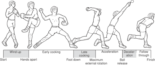
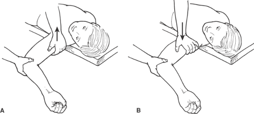
The most studied patient is the overhead-throwing athlete.4,5,6,7,8 The patient best known to the sports medicine physician and trainer is the thrower. The most frequent presenting complaint is a posterior pain in the shoulder at the initiation of the acceleration phase of throwing (Fig. 6-1). The most common physical finding is a positive relocation test with 90 degrees abduction and maximum external rotation and horizontal abduction producing the pain. In the second phase of the relocation test posterior pressure on the humerus relieves the pain (Figs. 6-2A,B and 6-3A,B).
The majority of overhead-throwing athletes can be treated by removing them from throwing for a period of time during which there would be strengthening of the muscles, total conditioning of the body, and finally correction of kinematic rhythm. Most importantly, because of the high energies involved, correction of the body mechanics or “style” (Fig. 6-4) is needed. A minority of throwers will develop recurrent symptoms on return to throwing and will be found to have a surgical (i.e., structural) lesion (Table 6-1).
Further study by professional trainers has shown that there is a prodromal phase before the thrower becomes truly symptomatic. During that phase the athlete complains of some stiffness and slowness to warmup. By resting and correcting mechanics in this earlier phase, the time away from play can be cut in half.
Any pathomechanical explanation would therefore include: features of the athletes or other patients’ anatomy and function, a dynamic event that can be aggravated by factors local or remote to the shoulder, correction by training, and later a surgical lesion or lesions beyond the reach of physical therapy.
Pathomechanics of the Overhead Athlete
The rival explanations for this condition involve one of two common features of overhead sports: the first is the extreme angulation of the joint achieved at the initiation of acceleration and the second is the tendency for overhead athletes to develop a tight posterior capsule.
Superimposed on one or both of these mechanisms is the transfer of enormous energy in overhead activities. Most of these activities consist of a controlled fall. The thrower or racquet player falls toward a target, turning his body into a large body rotating over his ipsilateral foot. He stops the fall with the contralateral foot and guides the retained kinetic energy into his torso. In like fashion the energy is guided into smaller and therefore more rapidly moving segments of the body. The large transfer we are discussing here occurs at the initiation of acceleration (Fig. 6-1). After releasing the ball or striking it with a racquet the athlete must dissipate the energy retained within his or her body in order to prevent injury. This is most safely done by a follow-through motion that converts the athlete back to a large forward moving object, this time rotating over the contralateral foot. As he or she is now a large object moving more slowly, there is more time in which to dissipate the retained energy with the large muscles of the pelvis and lower limbs.
Pathomechanics of Internal Impingement
There are several limitations on the normal range of motion of the glenohumeral joint. The bony dimensions of the glenoid limit the spinning motion of the humeral head. It is one of the unfortunate but mathematical ironies of geometry that small decreases in the bony limits on humeral motion result in large decreases in the bony stability of the joint calculated as area of coverage is a geometric function.
The capsule also limits motion of the humeral head in rotation. For most positions of the shoulder the capsule is lax and only becomes taut in end-range positions. For instance, in the abduction-external rotation (ABER) position the anterior-inferior capsule tightens toward the end of the range, resulting in a slight posterior translation of the humeral head. For most positions of the glenohumeral joint, alignment and stability are provided by muscle, mainly the
rotator cuff. The rotator cuff in a dynamic fashion functions to protect the capsule from stretch in the end-range positions.
rotator cuff. The rotator cuff in a dynamic fashion functions to protect the capsule from stretch in the end-range positions.
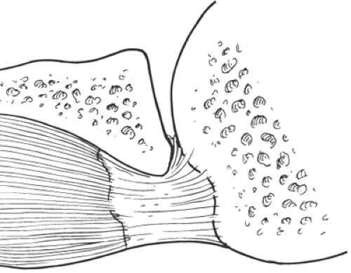
 Fig. 6-3. Impingement of the rotator cuff on the posterosuperior glenoid labrum during humeral abduction and maximum external rotation. Cross-section from a cadaver. |
The superior part of the glenohumeral joint is where the glenohumeral structures can make contact in elevation. The two positions where this contact may occur are ABER and forward flexion-internal rotation. In the ABER position the greater tuberosity approaches the posterior-superior glenoid. The structures between these two bones, the labrum and the internal fibers of the rotator cuff, are compressed between these two bones.9,10 An additional structure at risk is the anterior-inferior capsule that is stretched in this position (Table 6-1). Full forward flexion produces a similar compression of the superior labrum.11
In the forward flexion-internal rotation position it is the lesser tuberosity that approaches the anterior-superior glenoid. The fibers of the rotator cuff, in this case the subscapularis tendon, are pushed against the anterior-superior labrum (Table 6-2).12,13,14,15 The superior glenohumeral ligament and the biceps pulley can be damaged as well. The contact is against subscapularis below 90 degrees of elevation, and against the biceps tendon and pulley above 90 degrees.
Stay updated, free articles. Join our Telegram channel

Full access? Get Clinical Tree


