Injury to the Spinal Cord


Injury to the Spinal Cord


Peter G. Thomas

Neil R. Malhotra

M. Sean Grady


Spinal cord injury (SCI) remains one of the most costly aspects of trauma currently, not just in terms of the financial burden but more importantly, in terms of the physical and emotional burden of the patient. Approximately 40 new cases of SCI are reported per million people each year. This equates to 11,000 cases per year and does not include those who died from their injuries at the scene.1,2 These numbers represent data collected in the 1970s. No new nationwide incidence study has been conducted since then and as such, it is unknown if the incidence of SCI has changed. Significant improvement has however, been made in the survival rate of these patients. In 1970, 38% of patients with SCI died before hospital arrival.3 This rate decreased to 15.8% between 1997 and 2000.4 This reduction in injury has been attributed to increased seat belt and helmet use, improved auto design, improved swimming pool design, and improvements in the training of the prehospital personnel.2,5,6 Advanced Trauma Life Support (ATLS) mandates that trauma patients be presumed to have a SCI until proved otherwise. Early and safe control of the airway, while protecting from further movement of a SCI, has been a major advance in the last 30 years along with complete spinal immobilization with rigid cervical collars and securing the patient to a full-length backboard.7

The age of the patient with SCI has risen over the last 3 decades. In the early 1970s, the average age of injury was approximately 29 years. Currently, the average age of injury is 38 years. In addition, the percentage of people older than 60 years of age with SCI has increased. Before 1980, 4.7% of the patients with SCI were older than 60 years of age but since 2000, this has increased to 11.5%.1,2 Males comprise approximately 80% of SCI.1 In 2000, motor vehicle crashes were responsible for 47% of the SCI followed by falls (21%), acts of violence (11%), and recreational sports (10%).1,8,9 This order has been consistent throughout the last 30 years with the exception of the mid-1990s when gun violence caused more SCI than falls.10


ANATOMY

The spinal cord starts at the level of the distal brainstem and continues to the cauda equina, which usually terminates at L1-L2. It is divided into three regions: cervical, thoracic, and lumbosacral. The nerve roots from the cervical cord have a different function in the neck and arms at each level. The thoracic spinal cord supplies the nerve roots for the muscles of the chest and abdominal wall. The lumbosacral spinal cord and nerve roots are responsible for the functions of the pelvis, legs, bowel, bladder, and sexual function (see Fig. 1).

There are several different types of nerves within the spinal cord and each has a different function and hence a different injury pattern and treatment plan. The spinal cord contains both upper motor neurons (UMNs) and lower motor neurons (LMNs). UMN lesions (SCI) are located above the anterior horn cells and result in a spastic type of paralysis. LMN lesions (peripheral nerve injury) occur at or below the level of the anterior horn cells and these lesions tend to cause flaccid paralysis. UMN lesions carry a worse prognosis. There are also sensory nerves that are carried in the cord. These relay pain, temperature, vibration, and proprioception from the skin to the brain. For the purposes of this book, there are two main columns that relay this information. The spinothalamic column relays pain and temperature and it is located in the middle of the spinal
cord. The dorsal columns relay position and sensation or light touch and are located in the posterior portion of the cord (see Fig. 2).


Figure 1 Structures affected by spinal nerves (From Timby B, Smith N. Instructor’s resource CD-ROM to accompany essentials of nursing: care of adults and children. Philadelphia: Lippincott Williams & Wilkins, 2005.)

The spinal cord also contains the autonomic nervous system (ANS). The ANS comprises the sympathetic and parasympathetic nervous systems. The sympathetic nerve fibers are located from the first thoracic nerve root to the third or fourth lumbar root. Two of the main functions of the sympathetic nervous system are to cause vasoconstriction of blood vessels and increase heart rate. The parasympathetic nerves are located in the cranial outflow tract and the sacral nerve roots. The cranial outflow tract includes the oculomotor, facial, glossopharyngeal, and vagus nerves. The parasympathetic system is responsible for vasodilating blood vessels and slowing the heart rate. Both systems help control the respiratory, digestive, and cardiovascular systems.1,11,12 The anatomy of the spine will be discussed in Chapter 28.


EVALUATION/DIAGNOSIS OF INJURY

Physical examination is crucial to correctly determine the level of SCI. Whereas bony vertebral column injury is diagnosed with radiographs (see Chapter 28), SCI is diagnosed with physical examination and supplemented with magnetic resonance imaging. As stated in the preceding text, all patients with a suspected SCI should be immobilized with a rigid cervical collar and spinal backboard. Rigid longboards should be removed as soon as possible and hard surfaces should be padded during resuscitation and in the operating room. Keep in mind that although a patient may have ambulated at the scene of the accident, it does not mean that serious cord or potential cord injury is not present. Patients may have either a spinal column injury or an SCI and each is neither inclusive nor exclusive of the other.

As with all trauma patients, ATLS protocol should be followed. The patient’s airway must be secured, breathing must be adequate, and circulation intact. Full spinal immobilization is continued to prevent any further injury to the cord. Next, a thorough neurologic evaluation should be done. This evaluation should include a sensory and motor examination, a mental status examination, a reflex examination, and cranial nerve testing. The mental status can quickly and efficiently be evaluated with the Glasgow Coma Scale (GCS). The motor score is the patient’s best movement of the upper extremity. If the patient is unable to move any extremity, but is following a command such as “stick out your tongue,” the GCS would be incomplete or guarded with further more exact description of the deficit needed. The vertebral column is palpated feeling for deformities and step-offs. If the patient is alert, tenderness at a particular segment can be indicative of injury. Each extremity should be evaluated individually and each neurologic level assessed. The motor strength should be recorded according to the American Spinal Injury Association/International Medical Society of Paraplegia (ASIA/IMSOP) scoring system. The scale is from 0 to 5. Normal strength is given 5 points whereas some movement against resistance is given 4. Only within the 4 level of strength should a + or – be used to indicate near-normal strength or only slightly more resistance than gravity, respectively. Movement against gravity but not resistance receives 3 points and movement with gravity being eliminated is given 2. Finally, being able to feel muscle twitching is given 1 point and no movement is given 0 points.13 Some key muscle groups can be quickly tested to determine the level of injury; C5 elbow flexors, C6 wrist extensors, C7 elbow extensors, C8 finger extensors, T1 small finger abductors, L2 hip flexors, L3 knee extensors, L4 ankle dorsiflexion, L5 long toe extensors, and S1 ankle plantar flexors (see Table 1).14 Sensory testing is somewhat subjective. The scale is 0 to 2 with intact sensation being 2, decreased or hyperesthetic being 1 and no sensation being 0. The location of the dermatomes is shown in Fig. 3. A rectal examination is also a crucial part in evaluating the integrity of the spinal cord. The examination is done for two reasons. The first is to determine motor function of the sphincter muscle. The second is to determine perianal sensation. Presence of either of these demonstrates that there is sacral sparing, which indicates
that there is at least some integrity of the spinal cord because the sacral fibers travel along the periphery of the cord.


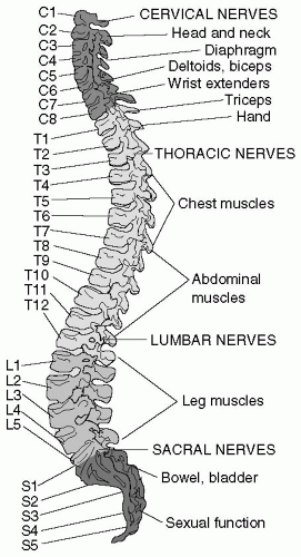
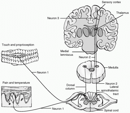

Figure 2 Pathways of ascending tracts. Sensory neurons enter the cord at the dorsal horn. Axons of sensory neurons for touch and proprioception ascend in the dorsal columns to the medulla, where they synapse with second-order projection neurons that cross (decussate) to the opposite side before ascending to the thalamus in the tract called the medial lemniscus. First-order neurons for pain and temperature enter the dorsal gray matter of the cord, where they synapse with second-order projection neurons that cross to the opposite side and ascend in the lateral spinothalamic tract to the thalamus. Third-order neurons connect both pathways from thalamus to the sensory cortex. (From Morton PG, Fontaine D, Hudak CM, et al. Instructor’s resource CD-ROM to accompany critical care nursing: a holistic approach, 8e. Philadelphia: Lippincott Williams & Wilkins, 2005.)


TABLE 1 CHART OF MUSCLE GROUPS AND NERVE AND NERVE ROOT SUPPLY


Muscle


Nerve Root


Nerve


Cervical flexors


C1-C4


Cervical extensors


C1-C4


Trapezius


Cranial nerve XI


Sternocleidomastoid


Cranial nerve XI


Arm abduction


0 to 15 degrees, supraspinatus


C4-C6


Suprascapular


15 to 90 degrees, deltoid


C5-C6


Axillary


>90 degrees, trapezius and serratus anterior


C5-C7


Long thoracic


Biceps


C5-C6


Musculocutaneous


Forearm supination


C5-C6


Musculocutaneous


Forearm pronation


C6-C7


Median


Wrist flexors


C7-C8, T1


Median


Wrist extensors


C6-C8


Radial


Hand intrinsics


C7-T1


Median and ulnar


Hip flexion


L1-L3


Femoral


Hip extension


L4-S1


Sciatic


Thigh abduction


L4-S2


Superior gluteal


Thigh adduction


L2-L4


Obturator


Leg flexion


L4-S2


Sciatic


Leg extension


L2-L4


Femoral


Foot plantar flexion


L5-S1


Superficial peroneal and tibial


Foot dorsiflexion


L4-L5


Deep peroneal


Great toe extension


L4-L5, S1


Deep peroneal


Foot inversion


L4-L5


Deep peroneal


Foot eversion


L5-L5


Superficial peroneal


Rectal sphincters


S2-S4


Pudendal


(From Welch WC, Donaldson WF, Marion DW. Injuries to the spinal cord and spinal column. The trauma manual 2e. Philadelphia: Lippincott Williams & Wilkins; 2002.)

Only gold members can continue reading. Log In or Register to continue

Stay updated, free articles. Join our Telegram channel

Oct 17, 2016 | Posted by in MANUAL THERAPIST | Comments Off on Injury to the Spinal Cord

Full access? Get Clinical Tree

Get Clinical Tree app for offline access