Special Considerations in Trauma in Children


Special Considerations in Trauma in Children


Thane A. Blinman

Michael L. Nance


There is no greater health concern in the pediatric population than trauma. Trauma is the leading cause of death among children between 1 and 18 years of age, greater than all other causes combined.1 Injury-related death is responsible for approximately 30% of all years of potential life lost.1 For every one injury-related death in the pediatric population, there are 12 children hospitalized for injury and more than 60 treated for an injury in an emergency department (ED) (see Fig. 1).1 Despite well-intentioned safety laws and apparently better safety products, the problem persists.

Care of the injured child differs in important ways from routine care of the injured adult. In addition to the anatomic differences, injury patterns and physiologic responses diverge for adult and pediatric trauma patients. The biomechanics of pediatric patients differ from adults in discreet ways, as follows:


  • Decreased mineralization of bone means thatbone offers less protection to structures in the central nervous system (CNS), thorax, and abdomen.


  • Decreased muscle strength per unit volume means not only diminished protection of the cervical spine and abdomen, but decreased Starling effect in the heart.


  • Increased surface area relative to body mass means dramatically increased vulnerability to radiative and evaporative loss of heat and fluid.

Not only are the patterns of injury different than in adults (e.g., pulmonary contusion in the absence of rib fracture) but the physiologic responses to hemorrhage and resuscitative interventions differ substantially from those exhibited by adults (e.g., increased heart rate [HR] instead of preload-recruitable stroke work in order to maximize delivery). Failure to consider these differences can lead to treatment that is incorrect either in terms of therapeutic choice (e.g., early splenectomy for spleen laceration) or of degree (e.g., over- or under-resuscitation of the child in shock or traumatic brain injury [TBI]). In addition, the largely nonoperative nature of pediatric trauma has lulled some into believing that no injured children need surgical consultation, a myth easily dispelled by the case logs of any major children’s trauma center.2,3

The objective of this chapter is to describe clinically important differences in the care of the injured child with emphasis on resuscitation and surgical care of specific injuries. Orthopaedic injuries, burns, and exposures are excluded.


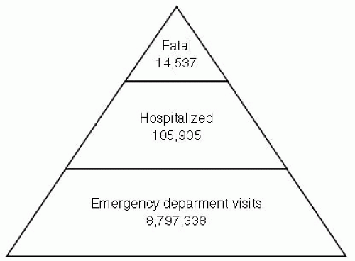
Figure 1 Injury pyramid demonstrating fatal injuries, injuries requiring hospitalization and injuries resulting in an emergency department visit for children aged 0 to 18 years, 2004. (Data from National Center for Injury Prevention and Control. Available at: http://www.cdc.gov/ncipc/wisqars/; Accessed May 4, 2007.)


TRAUMA SYSTEMS

Trauma centers were developed to provide the best care to the injured patient. By concentrating resources in identified institutions, and by directing the most severely injured patients to these centers, outcomes were optimized.4 The American College of Surgeons (ACS) has established guidelines to accredit hospitals as trauma centers. Similar guidelines were established to verify pediatric trauma centers as well. Most states have adopted these (or locally modified) ACS guidelines as a foundation for a trauma system. Despite the existence of pediatric resource centers for trauma, more than three fourths of pediatric trauma care occurs outside of such centers.5 Of concern, the overall survival of the younger (age younger than 10 years) and most injured (Injury Severity Score [ISS] greater than 15) patients treated in a freestanding pediatric hospital was improved compared to adult hospitals and non-freestanding pediatric hospitals.6 Pediatric trauma centers have also shown improved outcomes compared to nonpediatric trauma centers for specific injury types including head and spleen injuries.7,8 However, the fact remains that given the limitations of geography or availability of resources, most injured children will be treated outside of pediatric trauma centers. Therefore, it is imperative that all practitioners treating pediatric patients (up to 25% of typical trauma center volume) are comfortable with and equipped to provide care for the injured child. The basic equipment recommended by the American Academy of Pediatrics and American College of Emergency Physicians to care for injured children was found in only 5.5% of hospitals in the United States.9 Equipment essential for treatment of the pediatric trauma patient is listed in Table 1.


PREHOSPITAL CARE

There is evidence that paramedics deliver good prehospital care to children as well as to adults.10 Although properly sized equipment is often absent on a standard rescue ambulance, paramedics appear to improvise and adapt existing equipment well to protect the injured child. Response times and extrication methods are independent of patient age, of course. Heavy use of helicopter evacuation and transport (in patients who tend to have less severe injuries) is perhaps evidence that emergency medical service (EMS) providers prefer to err on the side of caution.11

Nevertheless, prehospital providers struggle with some important aspects of pediatric care, especially the pediatric airway, intravenous (IV) access, and C-spine immobilization. Recent evidence indicates that airway management short of endotracheal intubation (in particular, oral airway and bag-valve-mask ventilation) is superior to unsuccessful attempts at field intubation, especially in a child with traumatic brain injury (TBI).12,13 Meanwhile, complications are seen in as many as half of prehospital intubations and attempts, with unrecognized esophageal intubations alarmingly common. Current recommendations are to avoid attempts at endotracheal intubation in the field and to transport using oral airway and bag-valve-mask ventilation with chin-lift/jaw-thrust unless these maneuvers are demonstrably inadequate. An exception may be the pediatric patient requiring prolonged transport in whom a stable airway is preferable. EMS staff who must intubate a child should clearly understand the anatomic differences of the pediatric airway (see subsequent text).

IV access in the pediatric patient is frequently challenging. A high percentage of pediatric trauma patients arrive in the ED with no IV access. Intraosseus (IO) catheters seem underused, despite their demonstrated effectiveness in children. The thin sternum of a child makes this location contraindicated; practitioners should preferentially use the tibia if an IO is used. The risk of infection remains, and definitive IV access including central venous access should be established as soon as feasible in the hospital setting.

C-spine precautions are often misapplied or not employed. Pediatric patients whose C-spines are immobilized often suffer from one of three errors: incorrectly sized C-collar (almost always so large that it slips over the chin, simultaneously obstructing the airway and providing no C-spine protection); absent C-collar (usually with the possibly effective but inadequately tested work-around of a rolled towel and tape); inadequate padding beneath the thoracic spine to compensate for the prominent occiput of the child (which produces flexion at the neck instead of neutral positioning).

Other prehospital issues, such as “hypotensive resuscitation” or the use of intentional hypothermia have been inadequately studied in the pediatric population to make thoughtful recommendations.


ACUTE CARE


Primary Survey

Early hospital care of the pediatric trauma patient parallels standard Advanced Trauma Life Support (ATLS) protocols, and the usual rubric of primary survey (Airway, Breathing, Circulation, Disability, and Exposure [ABCDE]), secondary survey, and tertiary survey provides a good structure for discussion, with emphasis on clinically important anatomic and physiologic differences.


Airway

Safe control of the airway is paramount in pediatric trauma resuscitation. Success here prevents secondary injury from hypoxia, but success depends upon a clear understanding of the indications for intubation, an organized clinical approach, and facility with anatomic differences of the airway of a child.


TABLE 1 GUIDELINES FOR EQUIPMENT AND SUPPLIES FOR USE IN PEDIATRIC PATIENTS IN THE EMERGENCY DEPARTMENT (ED)a


Monitoring equipment


▪ Cardiorespiratory monitor with strip recorder


▪ Defibrillator with pediatric and adult paddles (4.5 and 8 cm) or corresponding adhesive pads


▪ Pediatric and adult monitor electrodes


▪ Pulse oximeter with sensors and probe sizes for children


▪ Thermometer or rectal probe


▪ Sphygmomanometer


▪ Doppler blood pressure device


▪ Blood pressure cuffs (neonatal, infant, child, and adult arm and thigh cuffs)


▪ Method to monitor endotracheal tube and placementb


▪ Stethoscope


Airway management


▪ Portable oxygen regulator and canisters


▪ Clear oxygen masks (standard and nonrebreathing-neonatal, infant, child, and adult)


▪ Oropharyngeal airways (sizes 0-5)


▪ Nasopharyngeal airways (12F through 30F)


▪ Bag-valve-mask resuscitator, self-inflating (450- and 1,000-mL sizes)


▪ Nasal cannulae (child and adult)


▪ Endotracheal tubes: uncuffed (2.5, 3.0, 3.5, 4.0, 4.5, 5.0, 5.5, and 6.0 mm) and cuffed (6.5, 7.0. 7.5, 8.0, and 9.0 mm)


▪ Stylets (infant, pediatric, and adult)


▪ Laryngoscope handle (pediatric and adult)


▪ Laryngoscope blades: straight or Miller (0, 1, 2, and 3) arid Macintosh (2 and 3)


▪ Magill forceps (pediatric and adult)


▪ Nasogastric/feeding tubes (5F through 18F)


▪ Suction catheters-flexible (6F, 8F, 10F, 14F, and 16F)


▪ Yankauer suction tip


▪ Bulb syringe


▪ Chest tubes (8F through 40F)c


▪ Laryngeal mask airway (sizes 1, 1.5, 2, 2.5, 3, 4, and 5)


Vascular access


▪ Butterfly needles (19-25 gauge)


▪ Catheter-over-needle devices (14-24 gauge)


▪ Rate limiting infusion device and tubingc,d


▪ Intraosseous needles (may be satisfied by standard bore needle aspiration needles)


▪ Arm boardsd


▪ Intravenous fluid and blood warmersc


▪ Umbilical vein cathetersce (size 5F feeding tube may be used)


▪ Seldinger technique vascular access kitc


Miscellaneous


▪ Infant and standard scales


▪ Infant formula and oral rehydrating solutionsc


▪ Heating source (may be met by infrared lamps or overhead warmer)c


▪ Towel rolls, blanket rolls, or equivalent


▪ Pediatric restraining devices


▪ Resuscitation board


▪ Sterile linenf


▪ Length-based resuscitation tape or precalculated drug or equipment list based on weight


Specialized pediatric trays


▪ Tube thoracotomy with water seal drainage capabilityc


▪ Lumbar puncture


▪ Pediatric urinary catheters


▪ Obstetric pack


▪ Newborn kitc


▪ Umbilical vessel cannulation suppliesc


▪ Venous cutdownc


▪ Needle cricothyrotomy tray


▪ Surgical airway kit (may include a tracheostomy tray or a surgical cricothyrotomy tray)c


Fracture management


▪ Cervical immobilization equipmentc,b


▪ Extremity splintsc


▪ Femur splintsc


Medical photography capability


aAdapted from Middleton KR, Burt CW. Committee on Pediatric Equipment and Supplies for Emergency Departments, National Emergency Medical Services for Children Resource Alliance. Adv Data. 2006;28(367):1-16; United States, 2002-03.


b Many types of cervical immobilization devices are available, including wedges and collars. The type of device chosen depends on local preferences and policies and procedures. Chosen device should be stocked in sizes to fit infants, children, adolescents, and adults. Use of sandbags to meet this requirement is discouraged, because they may cause injury if the patient has to be turned.

c Equipment that is essential but may be shared with the nursery, pediatric ward, or other inpatient service and is readily available to the ED.

d Equipment or supplies that are desirable but not essential.

e Ensure availability of pediatric sizes within the hospital.

f Available within hospital for burn care.

g Suitable for hypothermic and hyperthermia measurements with temperature capability from 25°C to 44 °C.

h May be satisfied by a disposable CO2 detector of appropriate size for infants and children. For children 5 years or older who are >20 kg in body weight, an esophageal detection bulb or syringe may be used additionally.

i To regulate rate and volume.


(Adapted from Care of Children in the Emergency Department: Guidelines for preparedness. Pediatrics. 2001;107:777-781.)


All trauma centers should have an established protocol for pediatric intubation, a pediatric intubation cart stocked with appropriate sizes of tubes and laryngoscopes, and a guide (such as a Broselow tape) that facilitates choice of tubes and dosing of key medications.

Every institution should maintain a rapid sequence intubation protocol (see Fig. 2). Any unstable child should be intubated by the most experienced person present; unstable trauma patients cannot tolerate multiple unskilled attempts.

The pediatric airway differs from the adult in (at least) six discreet ways, as follows (see Fig. 3):


  • Shorter trachea (even relative to body size)


  • Anteriorly displaced epiglottis


  • Prominent occiput


  • High larynx


  • Proportionately large tongue and smaller mouth


  • Narrowed at the cricoid cartilage

These differences mean that a different strategy for intubation of the child is required. In particular, the short trachea necessitates caution to avoid a mainstem intubation. One strategy is to carefully observe the double lines on the endotracheal tube (ETT), when these have just passed the cords, the tube is likely in good position. Careful auscultation using the axilla and observation of the chest rise can confirm good tube position. The anterior epiglottis can be compensated for by less aggressive head tilt (to be avoided in any case if the C-spine is not clear), opting instead for a gentle “sniffing position.” Meanwhile, an assistant can direct the epiglottis into a more favorable position with careful cricoid pressure, even maneuvering the larynx left or right slightly as needed. The prominent occiput can be obviated by gentle padding behind the shoulders (not the neck). The small mouth and large tongue are handled by having reliable suction at the ready, using a
correctly sized laryngoscope (especially the narrower Miller blade), and having the assistant pull gentle traction at the side of the mouth. Finally, the typical narrowing of the cricoid cartilage has traditionally translated into use of uncuffed tubes for children in order to avoid subglottic stenosis from balloon pressure injury, and while some have recently questioned this practice in the intensive care unit (ICU) setting, it remains true that a correctly sized (a classic heuristic is to choose a tube with the same external diameter as the pinky finger) uncuffed tube is easier to place. Finally, all of these anatomic differences mean that esophageal intubation is common but easily detected with use of end-tidal CO2 devices now widely available.


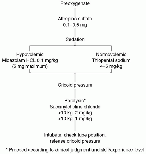
Figure 2 Algorithm for rapid sequence induction of the pediatric trauma patient. (From American College of Surgeons, 1997.)


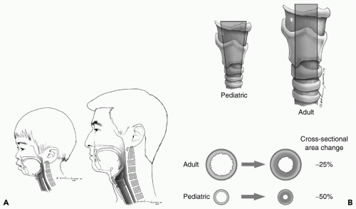
Figure 3 A: Anatomic differences between the pediatric and adult airway. B: Anterior view of airway demonstrating narrowing at the level of the cricoid cartilage in the pediatric airway and proportionately greater impact on airway diameter due to edema (Courtesy Brian Denham, MD).

Indications for intubation include the following:


  • Inability to spontaneously protect the airway (e.g., Glasgow Coma Scale [GCS] <8, overdose, drugs, facial trauma)


  • Inability to ventilate (e.g., flail chest)


  • Inability to oxygenate (e.g., pulmonary contusion, smoke inhalation, and cardiac instability)


Breathing

Several factors can inhibit breathing in infants and children in ways not seen in adults. Aside from the airway considerations listed in the preceding text, clinicians should remember that infants are obligate nose-breathers; tubes, debris, and blood in the nose impede air movement in these patients. Calculations for minute ventilation are somewhat different as well. Children require higher respiratory rates and lower tidal volumes, especially during bag-valve-mask ventilation. Bradypnea/respiratory arrest is the most common cause of cardiac arrest in babies. The clinician should give rapid (approximately 30 to 40 bpm) breaths using a pressure valve, taking care to not exceed peak inspiratory pressures greater than 25 to 30 mm Hg. Higher pressures are unlikely to improve tidal volume but more likely to inflict barotrauma/volutrauma. In addition, because gastric distension is one of the chief causes of respiratory embarrassment in the younger pediatric trauma patient, the overflow ventilation at higher pressures is more likely to diminish tidal volumes. Early recognition and tube decompression of a distended stomach in a crying, injured child can prevent unnecessary ventilation failure.


Circulation

The total circulating blood volume of the child can be surprisingly small. For young children, blood volume is approximately 80 mL per kg; therefore a 5-kg baby has just 400 mL of total blood volume, and has sustained a 40% hemorrhage (class IV shock) with a loss of only 160 mL (just over 5 oz). Similarly a 25-kg child with a total blood volume of 80 × 25 = 2,000 mL can quietly lose 25% of his blood volume in little time from a scalp wound that bleeds just 0.5 L in an hour, or 8.3 mL per minute. The small starting volumes can fool the complacent clinician who ignores these seemingly small but persistent sources of blood loss in the resuscitation suite. Pediatric patients have a remarkable ability to compensate for blood loss, however. The initial response to hypovolemia (i.e., hemorrhage) is tachycardia. In the active trauma resuscitation area, it would not be surprising to see tachycardia in a frightened, injured
child. Distinguishing the source of the tachycardia can be challenging. Often, the pediatric patient will maintain a normal blood pressure until 40% or more of the estimated blood volume has been lost. Hypotension in the pediatric patient is always concerning.

Control of the circulation begins with good venous access. According to ATLS protocol, two attempts should be made to place large-bore IV cannulas in the upper extremities. If this is unsuccessful, an IO line can be placed in the tibia. Large volumes can be readily infused through IO lines, and they can be used for the usual blood products and IV medications. However, they should be regarded as temporary and infection prone; rarely should a child leave the trauma bay with IO catheters in place.

In children, the elements governing oxygen delivery as defined by the oxygen delivery (DO2) equation are the same as adults. However, the relative contribution of each of these to oxygen delivery differs importantly in children. Oxygen delivery depends on blood oxygen content multiplied by cardiac output (CO). This section considers only CO, which is governed not by one equation, but by two simultaneous equations:14


CO = HR × SV = HR × EF × EDV

and


CO = (MAP – CVP)/SVR

So the elements of CO are:


HR (heart rate)

SV (stroke volume)

EDV (end diastolic volume, i.e., “preload”)

EF (ejection fraction, i.e., “contractility”)

MAP (mean arterial pressure)

SVR (systemic vascular resistance, i.e., “afterload”)

The mainstay of resuscitation in adult trauma patients has been to maintain circulating volume, and to take advantage of preload (EDV) recruitable increases in CO. In other words, aggressive volume resuscitation in an otherwise healthy adult trauma patient could be shown to boost CO and oxygen delivery as far as EDV could be pushed upward. Although enthusiasm for “supraphysiologic” DO2 (appropriately) has waned, it is largely true that large volumes can be administered to young adults with little negative effect.

The same is not true for babies and children. Starling’s law of the heart does not function over nearly the same range of preload, and children rely more on scaling up HR in order to increase CO. Rather than responding to increased stretch with increased stroke work, the pediatric heart will more quickly yield to edema (which increases wall stiffness) and begin to fail. Consequently, clinicians must use more caution in children to avoid not only under-resuscitation but over-resuscitation. Frequently, children in shock require earlier use of pharmacologic agents to alter SVR/afterload or EF/contractility rather than the aggressive fluid volume resuscitation that succeeds in adults and teens.

This has implications for choice of resuscitation fluid as well. Recommendations for the sick pediatric trauma patient include limiting crystalloid volume resuscitation (and the importance of using warm fluids is even more pronounced in children; see section “Exposure” in the subsequent text) to just two boluses of 20 mL per kg, then switching to O(-) packed red blood cells (PRBCs) for ongoing hemodynamic instability. In this way, circulating volume is more likely to approach “euvolemia” using a fluid that also increases oxygen carrying capacity, thereby maximizing DO2.

As always, children benefit from “just right” interventions, for example, specific treatments continued just until clinical endpoints are achieved. Endpoints for resuscitation include the following:


  • Normalization of HR (to age-appropriate levels)


  • Increasing pulse pressure


  • Improved skin color and extremity warmth


  • Clearing sensorium (as measured by GCS)


  • Increasing MAP


  • Production of urine (2 mL/kg/hour in infants, 1 mL/kg/ hour in adolescents)


Disability

A modified GCS has been validated in children (see Table 2).15 As in adults, a GCS of 8 or less is an indication for intubation. Importantly, a low GCS is also an independent risk factor for other major injury. Unlike
adults, children are more likely to have seizures associated with injury and therefore to have transient altered mental status from postictal states. The routine use of prophylactic antiseizure medication, however, has not been shown to be efficacious.16 In addition, children seem to suffer more mental status degradation from shock and therefore better recovery from resuscitation.


TABLE 2 GLASGOW COMA SCALE REVISED FOR USE IN PEDIATRIC POPULATION


Best Response


Pediatric GCS


Score


Eye


No eye opening


1


Eye opening to pain


2


Eye opening to speech


3


Eyes open spontaneously


4


Verbal


No vocal response


1


Inconsolable, agitated


2


Inconsistently consolable, moaning


3


Cries, but is consolable, inappropriate interactions


4


Smiles, oriented to sounds, follows objects, interacts


5


Motor


No motor response


1


Extension to pain


2


Flexion to pain


3


Withdrawal from pain


4


Localizing to pain


5


Obeys commands


6


GCS, Glasgow Coma Scale.


Children are more prone to severe TBI. The head makes up a larger fraction of body mass than in adults and is less well supported by the neck, and the brain is less protected by the bony skull. More importantly, children suffer disproportionately more from transient deficiencies in cerebral oxygen delivery, either from hypoxia or hypotension. Even brief episodes of either measurably increase the risk of death and disability in severe TBI.


Exposure

“Exposure” in the pediatric trauma patient should be understood to mean the following:


  • Unobstructed visualization and palpation of the entire body


  • Determination of exposure to toxins (CO, acid, petroleum distillates, etc.)


  • Protection from exposure in the trauma bay in particular, protection from hypothermia

While complete exposure and thorough examination of the injured child is crucial, so is protecting the child from hypothermia in the trauma bay, computed tomography (CT) scanner, and operating room (OR). After the traditional “strip and flip,” early effort to covering the child with warm blankets as much as possible greatly protects against iatrogenic heat loss. Covering or bundling can also decrease anxiety in the awake child. Meanwhile, a warm ambient temperature in the trauma bay, overhead warming lights and/or underbody warmers, and warm fluids should be employed to prevent hypothermia. Efforts should also be made to remove all trauma patients, but especially children, from hard spine boards expeditiously (when clinically appropriate). It is demonstrable that remaining on a hard board causes pain in <30 minutes, and can cause skin injury within an hour.17


Secondary Survey

During the head-to-toe secondary survey, a number of other measures should be considered:

Chest X-ray (CXR): The ordinary anterior-posterior chest x-ray (AP-CXR) is a rich source of information and is fast. It is a good practice to have a plate positioned on the table before the child arrives, and to shoot the film during the primary survey. Often, problems with airway or breathing (mainstem intubation, pneumothorax, gastric distension, etc.) are identified or confirmed by the film.

Electrocardiogram Monitoring, Pulse Oximetry: They are routinely used, as in adults.

Placement of Central Venous Catheter: This should be considered for any unstable child. However, central lines in children can be challenging to those who lack experience. Landmarks are subtly different, the hardware smaller, and the mechanical response of the needle and wire in the vein misleading. If there are two large-bore peripheral IV catheters, central access can be delayed until later in the resuscitation or in the OR or ICU.

Placement of Bladder Catheter: During inspection of the perineum, the rectal examination is commonly omitted in children because the anal aperture is very small compared to most examiners’ fingers, and the information gained from the examination is limited. Similarly, the classic “pelvic rock” is to be avoided, or performed at most once, and gently. If visual, gentle physical examination of the perineum and urethral meatus show no evidence of injury, a size-appropriate bladder catheter can be placed. Resistance or blood indicates the need for urethrogram, especially in the setting of pelvic fracture demonstrated by x-ray.

Placement of Nasogastric (NG): The NG tube is useful to decrease the risk of aspiration of gastric contents, to administer oral contrast, and to diminish abdominal respiratory compromise from gastric distention. Gastric distension can be profound in a crying, aerophagic child, or in a child undergoing bag-valve-mask respiration. In this case, the ventilatory compromise can produce hypoventilation severe enough to lead to cardiac arrest. NG tubes are contraindicated in cases of basilar skull fracture or midface instability; an orogastric (OG) tube can be substituted in this case. Common sense indicates that gastric tubes are not to be inflicted on every patient arriving in the trauma bay.

Laboratory Studies: Recent literature indicates that few laboratory studies (e.g., liver function tests [LFTs], metabolic panel) are useful for directing care. Little effort should be made at attempting blood draws in injured children, except for complete blood count (CBC) and a type and cross.18 An arterial blood gas may add some information in the critically injured child because the admission base deficit reflects injury severity and predicts mortality. Mortality increases in children with a base deficit <-8 mEq per L, and is strong evidence of potentially lethal injuries or uncompensated shock.19 In patients with significant head injury, thrombin and partial thromboplastin times should be drawn to help determine the need for correction of coagulation abnormalities.

Re-evaluation: Resuscitation is a dynamic process. The practitioner must frequently reassess the physiologic response to resuscitation to ensure goals have been achieved. At the same time, the trauma team leader should anticipate a plan and destination for the child (e.g., CT scanner, pediatric intensive care unit [PICU], OR, or transfer to a higher level of care (e.g., pediatric trauma center). It is the trauma team leader’s principal responsibility to drive the patient toward execution of a treatment plan efficiently.


DIAGNOSTIC PROCEDURES

Unstable patients with an obvious source of ongoing hemorrhage (e.g., hard signs of arterial injury, hemothorax, etc.) need to go directly to the OR. The primary decision to be made once ABCs are secured is operative approach and incision. But these circumstances are uncommon in the pediatric patient, and the surgeon frequently benefits from this additional information. Several diagnostic tools are available to help with this diagnostic process.


Plain X-rays

Plain films are useful screening tools, but are probably less helpful overall than in adults because of the variable sizes and extent of ossification of bony structures. The anterior-posterior chest film, ideally taken immediately on patient arrival, is probably the most helpful. The AP-CXR may reveal pneumothorax/hemothorax, pneumoperitoneum, foreign objects, widened mediastinum, and misplaced tubes (especially ETTs).

Other films may not be as useful. Pelvic x-rays are both less sensitive and less specific for fractures in children. C-spine films generally show bony deformity, but should be considered screening tools only: Positive findings always require further definition (usually with CT and/or magnetic resonance imaging [MRI]), but even negative results should be viewed with suspicion in a child with a suggestive mechanism of injury or tenderness on physical examination. Injuries to the spinal cord without bony injury are much more common in children.


Ultrasonograph/Focused Abdominal Sonography for Trauma

Most centers report that Focused Abdominal Sonography for Trauma (FAST) is reliable in discerning the following:


  • The presence or absence of fluid in the abdominal cavity


  • The presence or absence of fluid in the thorax


  • The presence of fluid in the pericardium (where it is the test of choice for tamponade)

FAST does not, however, give one important piece of information-the actual source of fluid in the abdomen. In experienced hands, FAST is reported to be useful to exclude the abdomen as a source of bleeding in the unstable child. However, it is critical to validate physician-performed diagnostic accuracy before employing FAST as a basis for operative decisions.20


Computed Tomography Scanning

CT scanning has become the workhorse of trauma diagnostic information. Although there are reports of measurable, albeit very small, lifetime increases in neoplastic risk from CT radiation, these worries should be discounted in the face of immediate injury.21,22 Rather, scanning protocols that employ weight-specific radiation dosing should be utilized. The rapid scan acquisition and processing made possible by multislice scanners and modern software mean few children need to be restrained or sedated in the scanner.

IV contrast is contraindicated in initial head scans. For abdominal CT scans, IV contrast is required to optimally visualize the solid organs, and in particular, to determine active extravasation: a “blush” from a liver, spleen, or kidney signifies arterial bleeding that may not be suitable for nonoperative protocols. For most injuries, CT angiography has supplanted more invasive methods of detecting injury to thoracic and carotid vessels, and can even exclude some extremity vascular injuries.23 Meanwhile, oral contrast is often omitted during initial abdominal CT scans because administration of contrast takes time, typically elicits vomiting in children, and usually does not have time to travel to the distal bowel. Oral contrast may, however, help identify proximal injuries such as duodenal hematomas. CT scanning reliably detects bony injuries, but children can sustain severe injuries to brain, spinal cord, lung, and genitourinary (GU) tract without fractures because of the decreased mineralization and stiffness of the bony structures protecting these organs. In particular, CT is poor at detecting spinal cord injuries without radiologic abnormality (SCIWORA); MRI is the modality of choice here, but only in the stabilized patient. Currently, MRI has no role in the acute workup.


Diagnostic Peritoneal Lavage

Diagnostic peritoneal lavage (DPL) is rarely used in children now as the information it provides is similar (fluid or no fluid) to that gleaned from other sources (e.g., FAST). It can give some information about source of fluid that FAST cannot (e.g., presence of vegetable fibers implies viscus injury). However, DPL is more difficult to perform (particularly in an awake child), takes more time than other tests, evaluates only the peritoneum, not the retroperitoneum, and has greater associated risk (e.g., bleeding, perforation, or infection).

Nevertheless, it is still conceivable that DPL could be useful in detecting hemorrhage in a trauma patient when FAST or CT was unavailable. DPL is performed as in adults, with the following few differences:


  • Decompress the bladder and stomach.


  • Use a supraumbilical incision to avoid the high-riding pediatric bladder, and use the open or seldinger technique to direct a catheter toward the pelvis.


  • Gross blood is a positive result. If no blood is seen, instill 10 mL per kg (up to 1 L) of warmed Ringers lactate solution and allow it to drain. As in adults, with microscopic analysis, a positive test is given by: >100,000 RBC per mm3, >500 WBC per mm3, bile, urine, or food material.


Emergency Department Thoracotomy

Emergency department thoracotomy (EDT) has a poor track record in all trauma patients, but in children it has been particularly unhelpful. As early as 1987, it was apparent that in pediatric blunt trauma, EDT had no influence on survival.24 Later, it was demonstrated that if pediatric patients had no signs of life (SOL) in the field, they never survived, even with EDT. Worse, there have been no neurologically intact survivors among pediatric blunt trauma patients who presented to the trauma bay without SOL.25 Even those with limited SOL on presentation had a survival of only 23.5% with the risk of fatality after cardiopulmonary resuscitation (CPR) increased for children with a systolic blood pressure below 60 mm Hg on arrival. Although children with penetrating injury had better survival, for all survivors of traumatic arrest, 64% had at least one impairment in the functional activities of daily living.26 These findings and other data have consistently demonstrated that the potential for survival in pediatric trauma arrest is quite poor, contrary to the popular perception of extraordinary resilience in the pediatric patient. EDT may have value only in cases where arrest occurs in the trauma bay, regardless of mechanism.

Oct 17, 2016 | Posted by in MANUAL THERAPIST | Comments Off on Special Considerations in Trauma in Children

Full access? Get Clinical Tree

Get Clinical Tree app for offline access