Injury to the Brain


Injury to the Brain


Jose L. Pascual

Vicente H. Gracias

Peter D. LeRoux


Central nervous system (CNS) trauma is the most common cause of death from injury and accounts for a third of all trauma deaths.1 Recent data show that in the United States, approximately 2% of the population (5.3 million citizens) lives with disability as a result of traumatic brain injury (TBI).2 Approximately 50,000 deaths1 and 1 million hospital visits each year are associated with head injury in North America.3 Most patients are victims of a motor vehicle collision (MVC) and are aged between the ages of 16 and 30 years.4 Gunshot wounds to the head also remain a common cause of brain injury.5 During the last decade, greater awareness of the epidemiology of head injury and better understanding of its pathophysiology has led to the development of management guidelines. Implementation of these guidelines in patient care protocols appears to have improved outcomes.


ANATOMY

The brain is surrounded by several distinct layers (see Fig. 1). The bony skull is covered with muscle, subcutaneous fat, and skin. Beneath the skull are the leptomeninges including the outer dura mater (a fibrous membrane that adheres to the internal surface of the skull), the arachnoid, and the inner pia mater (which is intimately attached to the outer surface of the brain). Cerebrospinal fluid (CSF) circulates between the arachnoid membrane and pia mater in the subarachnoid space.

The cranial vault is divided into three regions: the anterior region covered by the frontal bone, the middle section or parietal region covered by the parietal and temporal bones and separated from the frontal bone by the coronal suture, and the posterior or occipital region covered by the occipital bone that contains the occipital lobe and in the suboccipital region, the cerebellum and lower brainstem. The bone is particularly thin in the temporal region and thick in the occiput.

The brain is formed by the cerebrum, which is divided into two hemispheres each with four lobes (frontal, parietal, temporal, and occipital), the cerebellum, and the brainstem. The brainstem is further divided into the midbrain, the pons, and the medulla. The reticular activating system (responsible for state of alertness) is within the midbrain and upper pons. The cardiorespiratory center resides in the medulla. Small lesions in the brainstem can cause profound neurologic deficit.


PHYSIOLOGY

To understand TBI management, two essential aspects of brain physiology must be understood—the Monro-Kellie Doctrine6,7 and cerebral autoregulation. The Monro-Kellie Doctrine states that the total volume of intracranial contents is constant in an intact rigid cranial vault. Consequently, when there is an expanding mass lesion such as an ongoing hemorrhage, or edema, intracranial pressure (ICP) will only remain normal if there are concurrent reductions in the volume of other contents of the cranial vault, mainly CSF and blood.

When there is brain injury, cerebral edema usually ensues. This adds further to intracranial volume. This volume shift will continue until a reduction in blood/CSF volume can no longer occur, at which point ICP will begin to rise sharply. As ICP increases (>20 mm Hg), brain tissue begins to herniate from areas of high pressure to areas of low pressure.

ICP is sensitive to cardiac and respiratory functions that help to define its waveform when measured. Two other concepts are allied to ICP—elastance and its inverse, compliance. These concepts, in particular compliance, describe
how the intracranial contents are able to compensate for changes in volume. When compliance is reduced, small changes in volume lead to large changes in ICP. It is important to understand this rightward shift of the pressure-volume index (PVI), (see Fig. 2) because even when ICP is normal, very small changes in volume can have disastrous effects by causing herniation through a rapid increase in ICP.


Figure 1 Sagittal magnetic resonance image (T1) of the skull and brain.

The concept of cerebral autoregulation refers to the ability of the brain to maintain a constant cerebral blood flow (CBF) despite changes in cerebral perfusion pressure (CPP) (see Fig. 3). CPP is defined as mean arterial pressure (MAP) minus intracranial pressure (CPP = MAP − ICP) and normal CBF is approximately 50 mL/100 g brain/minute. CBF <20 mL/100 g brain/minute can lead to cerebral ischemia and CBF <5 mL/100 g brain/minute will result in cell death. There are both metabolic and pressure autoregulation. Metabolic autoregulation is influenced by cellular requirements. Pressure autoregulation refers to changes in vasomotor tone, which keep CBF constant over a range of mean arterial blood pressure. As CPP decreases or increases in the normal individual, vasoconstriction or dilation of the cerebral vasculature help maintain CBF constant across a broad range of perfusion pressures, which are believed to be between 50 and 150 mm Hg. Beyond these “breaking points,” changes in CBF parallel changes in CPP and can result in inadequate or excessive (luxuriant) tissue oxygen and glucose delivery. Autoregulation is frequently impaired during the first 2 to 3 days following TBI. This can have deleterious effects on CBF but also allows certain therapies to be effective. When ICP equals CPP, CBF can no longer be maintained and vascular autoparalysis occurs.


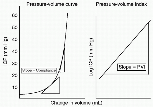
Figure 2 Brain compliance. After any injury, the brain will compensate for increases in pressure through elastance and compliance. As compliance is lost, small changes in volume can lead to large changes in pressure. In a patient who is on the right side of the curve, even with normal intracranial pressure (ICP), small changes in volume can precipitate large rises in pressure.


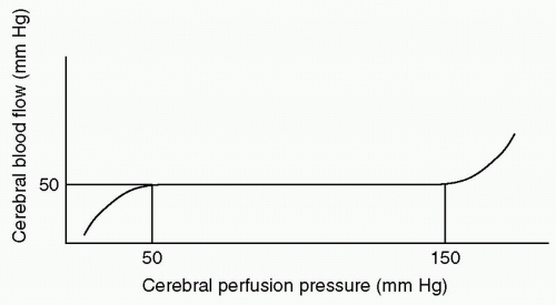
Figure 3 Cerebral autoregulation.


PATHOLOGY OF HEAD INJURY

Head injury can be classified as focal or diffuse. Focal injuries are typically associated with direct blows to the head and may cause fractures, hematomas, or contusions. The morbidity of focal injuries depends on their location as well as the magnitude of the applied force. Diffuse injury to the brain, often called diffuse axonal injury (DAI), is caused by inertial forces imparting acceleration/deceleration shearing within the brain tissue. Head injury can also be classified as open or closed. This depends on whether there is a fracture of the skull or sinuses that communicates with the external environment. A critical determinant of outcome and management of an open injury is whether the dura is intact or not.

Head injury can lead to brain injury through primary or secondary mechanisms. Primary brain injury results from the direct mechanical damage that occurs at the time of trauma. However, brain injury does not cease with the initial trauma but progresses over the subsequent hours, days, and even months. This continuing cerebral damage, known as secondary brain injury, is important and its identification and management are the basis of much of critical care for TBI. Several substances can contribute to secondary brain injury including excitatory amino acids, cytokines, and oxygen free radicals released during systemic inflammation.8,9 In addition, systemic derangements can contribute to and aggravate secondary brain injury. In particular, hypotension and hypoxemia increase the risk of poor outcome following TBI.10,11 A single hypotensive episode (systolic blood pressure [SBP] <90 mm Hg) results in a fourfold increase in the mortality risk of patients with TBI.12 Other systemic abnormalities that contribute to secondary cerebral injury include hyperglycemia, hyperthermia, hypercapnea, and hypocapnea. In addition, seizures can contribute to secondary neuronal injury.


PATHOLOGIC SUBTYPES OF HEAD INJURY


Epidural Hematomas

Epidural (or extradural) hematomas (EDHs) are found in <1% of all patients with head injuries and 10% of those who are comatose.13 On head computed tomography (CT) scan acute EDHs are typically hyperdense, lenticular, or biconvex in shape, and are formed by a pressure-driven separation of the dura mater from the overlying skull (see Fig. 4). The most common type of EDH occurs in the temporal region and is caused by a bleeding middle meningeal artery lacerated by a skull fracture. An EDH may present with a period of normal consciousness after an initial loss of consciousness. This can be followed by profound coma, that is, the “talk and die” phenomenon.


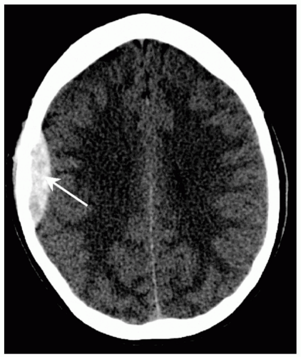
Figure 4 Right temporal epidural hematoma (arrow) on head computed tomography (CT). Note its smooth lentiform shape.


Subdural Hematomas

Acute subdural hematomas (SDHs) are the most common traumatic mass lesions and occur in 30% of patients with severe head injuries.13 They usually result from a shearing disruption of the subdural bridging veins. Acute SDHs are most often crescent shaped, hyperdense on head CT, and are caused by passive filling of the subdural space with blood (see Fig. 5). In addition, SDHs are frequently associated with underlying brain injury (e.g., contusion, intracerebral hematomas [ICHs], or DAI) unlike EDH where there is generally no brain injury. It is the underlying brain injury that often determines the outcome of SDH and also helps explain why outcome is much worse after SDH than EDH. Chronic SDH may occur in individuals with brain atrophy (e.g., the elderly and alcoholics). Again, the subdural bridging veins are torn even after minor trauma. These bleeds can be subclinical initially but as the hematoma liquefies over the course of subsequent weeks, they can become symptomatic. These lesions are also crescenteric in shape but are usually iso- or hypodense on head CT scan. However, there can be mixed densities on head CT scan suggesting episodes of rebleeding within the same hematoma (i.e., acute or subacute on chronic SDH).


Intracerebral Hematomas and Contusions

ICHs and contusions are bleeding areas within the brain itself that appear as mass lesions. Brain tissue can be seen interspersed with hemorrhage on head CT scan (salt and pepper) in contusions. By contrast, the blood forms a
coalesced mass in an ICH on CT scan. Contusions are often found in the frontal and temporal lobes because this tissue “glides” over the underlying rough bony surface of the skull base during impact and acceleration/deceleration of the head (see Fig. 6). The brain may also impact the inner table of the skull across from the area of impact, thereby causing a contusion or hemorrhage. This is known as a coup-contre-coup lesion. ICH may also occur with penetrating injuries (e.g., stab wounds or gun shot wounds, see subsequent text). Finally, ICH may develop or progress ≥24 hours after injury (delayed traumatic ICH) and therefore, patients at risk require careful follow-up and repeat imaging.14,15


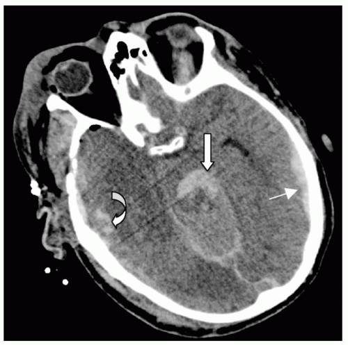
Figure 5 Subdural hematoma (SDH). Acute left temporal parietal subdural hematoma (arrow) and right temporal intraparenchymal contusion (curved arrow). In addition there is blood (subarachnoid) layering on the tentorium and involving the brainstem (large arrow).


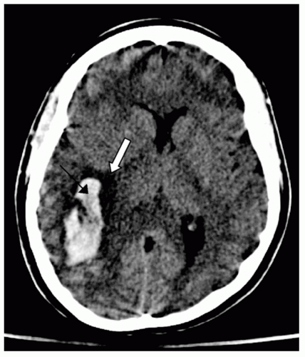
Figure 6 Intracerebral hemorrhage. Right temporal occipital lobe intracerebral hemorrhage (arrow) with associated vasogenic edema (block arrow). Note the compressed right occipital horn of the lateral ventricle.


Traumatic Subarachnoid Hemorrhages/Intraventricular Hemorrhages

Subarachnoid hemorrhage (SAH) is often found following moderate or severe head injury. It is usually found in a diffuse pattern over the convexities and in the subarachnoid space. It is hyperdense on head CT scan (see Fig. 7), and is often associated with other intracranial pathology, in particular SDH or ICH. When SAH is found on imaging, it is important to determine whether it is traumatic or it arose from rupture of an aneurysm that then led to loss of consciousness, fall, and subsequent injury. This is important because SAH resulting from aneurysmal rupture may lead to vasospasm (delayed narrowing of cerebral vessels), which can cause delayed cerebral infarction and poor neurologic outcome.16 For reasons that remain unknown vasospasm is not seen following traumatic SAH.

Intraventricular hemorrhage (IVH) is blood within the ventricles. This may occur with SAH, ICH, or SDH. Isolated IVH may also be seen in DAI. Patients with IVH are at increased risk for acute posttraumatic hydrocephalus17 and more chronic hydrocephalus (i.e., enlargement of the ventricles).


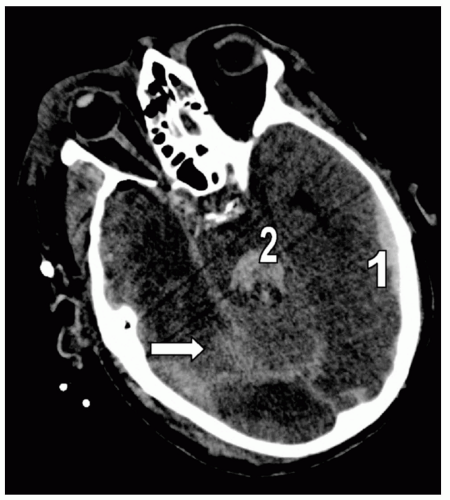
Figure 7 Subarachnoid hemorrhage (SAH). Different detail from the patient in Fig. 5 demonstrating extensive right posterior SAH (arrow). Note the accompanying left temporal subdural hematoma (1) and fourth ventricle blood (2).


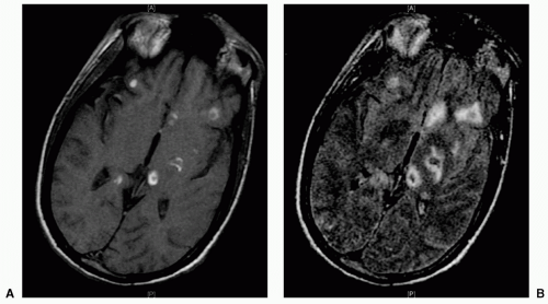
Figure 8 Diffuse axonal injury (DAI). A: T1 magnetic resonance imaging (MRI) demonstrating susceptibility artifact at the gray-white junction of the left thalamus, left internal capsule, right splenium, and corpus callosum. B: Equivalent image on flair MRI.


Diffuse Axonal Injury

DAI is considered the most common pathology in TBI. It is seen in mild, moderate, or severe TBI. It results from shearing forces that affect the axons.18 These axons undergo sequential focal changes, which result in axonal cytoskeletal dysfunction followed by axonal swelling and disconnection in the hours that follow injury.19 The corpus callosum, brainstem, white matter, and reticular activating system may be affected. The admission head CT scan may be normal but may also show small hemorrhages in the white matter, corpus callosum, deep basal ganglia, or brainstem. IVH may also occur. Acute SDHs are often associated with DAI. Depending on its severity, the prognosis of DAI can be poor and it is the leading cause of disability among TBI survivors. Brain magnetic resonance imaging (MRI) helps identify characteristics typical of the condition (see Fig. 8).


CLINICAL EVALUATION


Primary Survey

TBI requires prompt management but always after ensuring a secure airway with proper oxygenation and circulation (Airway, Breathing, and Circulation [ABCs]).20 During the primary survey, one must determine whether all extremities are moving and if the pupils are equal and reactive. Pupils are an important element in the examination of patients with head injuries. A dilated pupil that does not react to light suggests ipsilateral uncal herniation and compression of the third cranial nerve.

The Glasgow Coma Scale (GCS)21 is a standard score with excellent interobserver variability, which should be applied to all patients (see Table 1). The postresuscitation GCS is essential for subsequent management and prognostication and the motor score is the most predictive variable following TBI. When there is asymmetry in either eye opening or motor scores, the best score is used. The GCS should be obtained early in a patient’s evaluation and preferably
before administration of sedatives and/or paralytics. A GCS of ≤8 indicates coma and airway intubation, and ventilation should be considered early. If necessary, an emergency surgical airway (cricothyroidotomy, tracheostomy) may be required.


TABLE 1 GLASGOW COMA SCALE


Signs


Score


Eye Opening


Spontaneous


4


To verbal command


3


To pain


2


No response


1


Best motor response


Obeys verbal commands


6


Localizes to pain


5


Withdraws to pain


4


Flexion response to pain


3


Extension response to pain


2


No response


1


Best verbal response


Orientated


5


Confused


4


Inappropriate words


3


Nonspecific sounds


2


No response


1


TABLE 2 CLASSIFICATION OF TRAUMATIC BRAIN INJURY (TBI)


Mild TBI


Moderate TBI


Severe TBI


GCS


13-15


9-12


≤8


Proportion of all TBI


80%


10%


10%


Sensorium


Transient confusion and retrograde amnesia, follows commands


Confused, can follow some commands


Unable to follow commands


Prognosis/mortality


Excellent/<1%


Good/<5%


Poor/30%-40%


Long-term recovery


Headaches, memory problems mostly in first month


Cognitive and behavioral deficits lasting months or permanently. Usually good recovery with treatment


Persistent coma, vegetative state; severe disability frequent


Secondary Survey

Once the primary survey is completed and immediate life-threatening conditions are managed or excluded, the secondary survey acts as a complete physical examination. A proper neurologic examination to evaluate motor and sensory deficits, reflexes, cranial nerves, and rectal tone should be included.


CLASSIFICATION OF TRAUMATIC BRAIN INJURY

TBI is categorized as mild, moderate, or severe according to the level of neurologic dysfunction at the time of initial evaluation (GCS 13 to 15; 9 to 12, and ≤8, respectively) (see Table 2). An advantage of using the GCS is that it can be repeated frequently to determine if there is any change in the patient’s condition.


MILD AND MODERATE HEAD INJURY

The vast majority of head injuries are mild. Despite their names, both mild and moderate head injuries are not trivial conditions. The postconcussive symptoms following mild TBI are thought by some to be associated with shear stress at the gray-white cortical interface following sudden deceleration. The pathophysiologic correlation of these injuries may represent a part of the spectrum of DAI, and follow a similar mechanism of injury and pathology.

Only gold members can continue reading. Log In or Register to continue

Stay updated, free articles. Join our Telegram channel

Oct 17, 2016 | Posted by in MANUAL THERAPIST | Comments Off on Injury to the Brain

Full access? Get Clinical Tree

Get Clinical Tree app for offline access