Initial Resuscitation and Diagnosis


Initial Resuscitation and Diagnosis


Andrew Rosenthal

Grace S. Rozycki


During the initial hospital phase, the injured patient is rapidly assessed and the treatments are prioritized based on the mechanism of injury and the patient’s vital signs. The goal of the resuscitation is to improve organ and tissue perfusion by rapidly identifying and simultaneously treating life-threatening conditions. In most cases, the initial resuscitation of the patient is conducted in the trauma resuscitation area (the design of which is covered in Chapter 12 of this book), but there are select patients who should bypass the trauma resuscitation area and be taken directly to the operating room for lifesaving interventions (see Table 1). In either case, advance planning is needed so that all of the essential equipment and materials are immediately available to execute the American College of Surgeons’ Advanced Trauma Life Support (ATLS) primary and secondary surveys.1 Standard universal precautions (e.g., face mask, eye protection, water-impervious gown, leggings, and gloves) should be used even when evaluating patients who have seemingly minor injuries.1 Additionally, because life-threatening procedures may need to be performed during the initial resuscitation, prepackaged trays with sterile contents such as airway, tube thoracostomy, open chest, and minor and major suture trays should be well stocked and immediately available. The trays should be easily accessible, consistently in the same place, and color-coded so that they can be identified and accessed instantly.

Medications frequently used in the trauma resuscitation area are listed in Table 2. Although the principles of resuscitation are based on the primary and secondary surveys, the resuscitating physician should keep in mind that treatment priorities are established on the basis of the patient’s mechanism of injury, obvious injuries (on presentation or diagnosis soon thereafter), clinical examination, and vital signs. Furthermore, the resuscitating physician and trauma team members identify and treat life-threatening conditions simultaneously, but continually assess and reassess the patient’s status.
PRIMARY SURVEY

A rapid assessment following the A B C D Es of trauma care provides a systematic method for simultaneously identifying and treating life-threatening conditions.


Airway (with cervical spine immobilization and protection)

Ensuring adequate oxygenation, ventilation, and protection from aspiration are the cornerstones of airway management and the first priority when treating trauma patients. All patients should receive supplemental oxygen by a secured
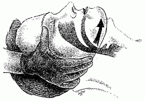
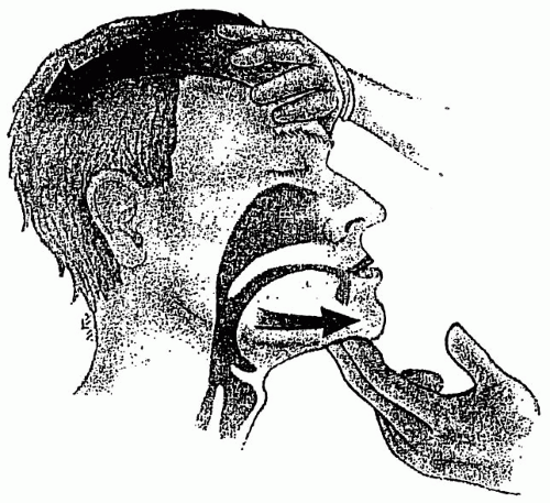
oxygen reservoir face mask with a high flow rate. A pulse oximeter is applied to the patient’s digit or ear lobe as a noninvasive method to measure oxygen saturation. If the patient is able to speak easily, then the airway is patent. A patient with facial fractures or tracheal/laryngeal injuries may have a patent airway but may have difficulty with secretions and therefore, may require a definitive airway in the form of a cuffed orotracheal or nasotracheal tube or by way of a surgical airway. In the unconscious patient, the tongue may fall backwards and obstruct the airway. While maintaining cervical spine immobilization, the chin-lift and jaw-thrust maneuvers (see Figs. 1 and 2) may be employed to relieve this obstruction. When performing the chin-lift maneuver in a trauma patient, the head is not tilted and the cervical spine is maintained in a neutral position. If the patient is not responsive, an oropharyngeal airway should be inserted to assist in bag-valve mask ventilation. The nasopharyngeal airway is reserved for the more awake patient with an intact gag reflex, but is not used as a definitive airway. Although other devices, such as the multilumen esophageal airway device or the laryngeal mask airway (LMA), may be used for temporary ventilation and oxygenation, they are not considered definitive airways because they do not provide secure long-term airway stabilization.


TABLE 1 INDICATIONS FOR DIRECT TRANSPORT TO THE OPERATING ROOM


Systolic blood pressure ≤80 mm Hg


Penetrating torso trauma


Major limb amputation or mangled extremity


Extensive soft tissue wounds


Severe maxillofacial injury


Rhodes M, Brader A, Lucke J, et al. Direct transport to the operating room for resuscitation of trauma patients. J Trauma. 1989;29:907-915.2


TABLE 2 MEDICATIONS FOR THE TRAUMA RESUSCITATION AREA


Antitetanus


Tetanus may occur after minor woundsa


0.5 mL intramuscular tetanus toxoid vaccine for those immunized >10 years ago


250 U intramuscular tetanus immunoglobulin for the never-immunized and for grossly dirty wounds


Antibiotics


Generally a first-generation cephalosporin (e.g., 1 g cefazolin) intravenously, or clindamycin 600-900 mg IV for penicillin-allergic patients, for the following:


Open fractures and joints


Grossly contaminated wounds, major soft tissue injury


Immunocompromised patients


Patients with prosthetic cardiac valves


Analgesics (dose and frequency vary depending on patient status)


Morphine 0.1 mg/kg intravenously


Fentanyl 1 µg/kg intravenously


Demerol 0.25-0.50 mg/kg intravenously


Anxiolytics (dose and frequency vary with patient status)


Midazolam 2-4 mg intravenously


Haloperidol 5-20 mg intravenously


a American College of Surgeons Committee on Trauma. Advanced trauma life support program for doctors, 7th ed. Chicago: American College of Surgeons Committee on Trauma; 2004; Rhee P, Nunley MK, Demetriades D, et al. Tetanus and trauma: A review and recommendations. J Trauma. 2005;58:1082-1088.3,4


Figure 1 Trauma jaw thrust. (From Sanders MJ, ed. Paramedic textbook, 2nd ed, chapter 11, figure 1, St. Louis: Mosby; 2000: 397.)5

The absolute indications for immediate emergency intubation are the following:


  • Airway obstruction unrelieved with basic interventions (e.g., chin-lift and jaw-thrust maneuvers)


  • Apnea or near apnea


  • Respiratory distress


  • Severe neurologic deficits, such as a Glasgow Coma Score <9, or high (C1,C2) spinal cord injury

Urgent indications for intubation are the following:


  • Penetrating neck injury


  • Persistent or refractory hypotension


  • Chest wall injury or major pulmonary dysfunction such as flail chest with pulmonary contusion


  • Impending upper airway obstruction such as severe oral maxillofacial injury or laryngeal edema


Figure 2 Chin lift. (From Sanders MJ, ed. Paramedic textbook, 2nd ed, chapter 11, figure 2, St. Louis: Mosby; 2000:397.)6


TABLE 3 DIFFICULT AIRWAY CART


Suction catheter


Endotracheal introducer and tube changer


Pediatric and adult laryngeal mask airway (sizes 1, 1.5, 2.0, 2.5, 3, 4, and 5)


Lighted laryngoscopes (Miller sizes 0, 1, 2, 3, and 4 and Macintosh sizes 1, 2, 3, and 4)


Pediatric and adult endotracheal tubes (sizes 4.0 mm, 5.0 mm, 6.0 mm, 6.5 mm, 7.0 mm, 7.5 mm, and 8.0 mm)


Assorted Shiley tracheostomy tubes (sizes no. 4 and no. 6)


Oral airway


Nasopharyngeal airway


McGill forceps


14-gauge angiocatheter


10-mL syringe for cuff inflation


End-tidal CO2 detector


Surgical airway tray (Table 5)


American College of Surgeons Committee on Trauma. Advanced trauma life support program for doctors, 7th ed, chapter 2, Airway and ventilatory management. Chicago: American College of Surgeons Committee on Trauma; 2004:53-68.7


In addition to standard intubation equipment, each trauma resuscitation area should have additional instruments that are used for “the difficult airway.” The contents of a “difficult airway cart” are listed in Table 3. Rapid-sequence intubation is the accepted standard process for endotracheal intubation in a multitrauma patient who is assumed to have head injury, cervical spine injury, and a full stomach. The guidelines for rapid-sequence intubation are listed in Table 4. A surgical airway, that is, a cricothyroidotomy, is performed when there is an edematous glottis, severe oropharyngeal hemorrhage, or simply an inability to obtain an orotracheal airway expeditiously. A cricothyroidotomy is performed by making a small transverse incision through the cricothyroid membrane. Either the handle of the scalpel or a hemostat may be used to dilate the opening so that a small tracheostomy or endotracheal tube can be inserted (see Fig. 3). After the performance of a definitive airway, a CO2 detector is used to ensure that the airway tube is within the tracheal lumen. Bilateral chest rise and breath sounds should be present. Additionally, the position of the tube is confirmed by a chest x-ray. Table 5 lists the equipment helpful in obtaining a surgical airway.


TABLE 4 RAPID SEQUENCE INTUBATION PROCEDURE


Preoxygenation with 100% oxygen


Cricoid cartilage pressure (Sellick maneuver) to compress the esophagus


Sedation (midazolam 3-5 mg or etomidate 0.3 mg/kg intravenously)


Succinylcholine (1-2 mg/kg, usually 100 mg dose intravenously)


Orotracheal intubation with inline stabilization of the cervical spine


Cuff inflation


Tube placement confirmation (ausculate, visualize chest rise, and confirm CO2 return from exhaled air)


Release cricoid pressure


Ventilate and oxygenate the patient


American College of Surgeons Committee on Trauma. Advanced trauma life support program for doctors, 7th ed, chapter 2, Airway and ventilatory management. Chicago: American College of Surgeons Committee on Trauma; 2004:47-50.8

Only gold members can continue reading. Log In or Register to continue

Stay updated, free articles. Join our Telegram channel

Oct 17, 2016 | Posted by in MANUAL THERAPIST | Comments Off on Initial Resuscitation and Diagnosis

Full access? Get Clinical Tree

Get Clinical Tree app for offline access