Fig. 7.1
(a) Preoperative radiograph of a 49-year-old patient with hip hemophiliac arthropathy. An osteopenic acetabulum and a funnel-shaped femoral canal can be observed. (b) Radiograph of the same patient at the first postoperative week with a cementless press-fit cup and a cementless stem. The bearing surface is an alumina-on-alumina couple. (c) Radiograph of the same patient done during the second postoperative year. Note the radiological signs of osteointegration of both acetabular and femoral cementless components

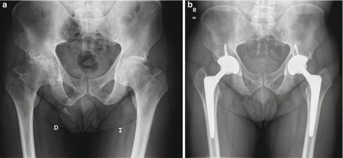
Fig. 7.2
(a) Anteroposterior pelvic radiograph of a 56-year-old male patient showing hemophilic arthropathy of both hips. Note deformity in both femoral heads, osteopenic acetabulum, and dysplastic cylindrical femur. (b) Anteroposterior pelvic radiograph during the third and second postoperative year after bilateral alumina-on-alumina cementless total hip arthroplasty of the same patient. These cementless cups needed screw for a proper primary fixation
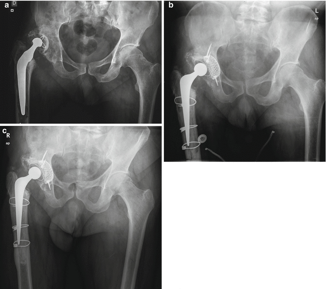
Revision hip surgery remains a challenge for the orthopedic surgeon, particularly when a major bone defect is present [22]. Thus, young age of hemophilic patients is another concern regarding the best reconstruction for their hips. The incidence of this surgery is increasing since better medical management has been developed and physical activity remains a higher level than some decades ago. Management of bone loss is the most critical issue in these young adult patients, and impaction grafting, for both acetabular and femoral sides, is one of the best options for these patients [23, 24] (Fig. 7.3)


Fig. 7.3
(a) Radiograph showing aseptic loosening of a cemented low-friction arthroplasty of a 62-year-old hemophilic patient at the tenth postoperative year. There was an intraoperative segmental and cavitary bone defect in both acetabular and femoral sides. (b) Sixth postoperative week radiograph of the same patient after reconstruction hip surgery with impaction bone grafting and cemented implants. A medial mesh was needed in the acetabulum prior to grafting and an extended femoral osteotomy in the femur to remove the implant and the cement. (c) Second postoperative year radiograph of the same patient. Bone remodeling and stable fixation can be observed on both acetabular and femoral sides
7.3 Muscular Disorders of the Hip in the Hemophilic Patient
Other characteristics of the pathology that affect the musculoskeletal system in hemophiliac patients are muscular disorders. Goodfellow et al. described a series of 24 patients diagnosed with classical hemophilia with the typical symptoms of iliacus hematoma [25]. Pain in the groin in the absence of a significant trauma was the most important finding, and it could be acute or insidious. Flexion contracture in external rotation with preservation of movements of the hip joint but extension was also other frequent sign. Another finding observed was femoral nerve palsy due to compression and a mass in the iliac fossa.
The appearance of muscle hematomas can be spontaneous, and hematological treatment is required before further complications develop [26]. Since this process may take several weeks, a hemophilic pseudotumor can develop due to the appearance of new bleedings. This finding can be the source of new disorders in the musculoskeletal system, and because it may destroy bone and soft tissue, resection can become very difficult, particularly for a pelvic pseudotumor [27]; however, with proper management including a multidisciplinary approach, a satisfactory outcome can be obtained. A typical site for hematomas in patients with hemophilia is within the iliopsoas muscle [28]. The anatomy and size of this muscle motivate the large volume of the mass, and, to date, muscular and femoral nerve involvement has been frequently reported, as well as re-bleeding. Groin pain and contractures are frequently seen, and management of a hematoma can be conservative: rest, physical therapy, and long-term hemostatic therapy. However, the clinician must be alert to the possibility of muscular affectations in order to prevent complications. Ultrasound-guided percutaneous drainage is a current option before surgery for some hematomas, although recurrence is still relatively frequent [29]. At the long term, the appearance of heterotopic ossification can develop into a myositis ossificans that will affect the joint function and may necessitate surgery.
7.4 Conclusions
Although hip affectation is relatively rare in hemophilia, when it occurs it generally requires a THA to safeguard their mobility and quality of life. Current implants and alternate bearing surface may improve clinical and radiological outcome of these patients. While hip pain and functional impairment are usually due to arthropathy, hemophilic patients can also present muscular disorders with similar symptoms and can produce serious complications. These patients require close monitoring by a multidisciplinary team to successfully manage the musculoskeletal affectation of the hip.


