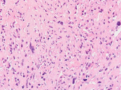
Fig. 15.1
AP radiograph of the left humerus (a) of a 29-year-old female with a high-grade surface osteosarcoma shows a mixed density, partially mineralized mass involving the mid and proximal humeral diaphysis with mineral pattern indicative of osteoid matrix. Axial soft tissue (b) and coronal bone (c) CT images nicely illustrate that the lesion is attached to the cortex of the humerus and is markedly heterogeneous with areas of osteoid and soft tissue attenuation. Although somewhat more heavily mineralized near the attachment to the bone, the mineral is scattered throughout the mass. Coronal T1-weighted images (d and e) show preservation of the marrow fat signal in the adjacent humeral diaphysis and, therefore, confirm lack of tumor involvement in the medullary canal. Axial fat-suppressed T2-weighted MR images (f and g) show the anatomic extent of the mass in the soft tissues with large heterogeneous bulky tumor masses posteriorly and medially
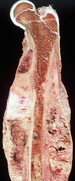
Fig. 15.2
Large high-grade surface osteosarcoma encircling most of the underlying femur. There is no intramedullary invasion