8 Hand Fractures
Overview
 Anatomic Features
The skeleton of the hand consists of eight small carpal bones (wrist), five metacarpals (palm), fourteen phalanges (fingers), and two sesamoid bones (Plate 8.1).
The carpus is made up of eight carpal bones, which are arranged into two rows: a proximal and distal row. The proximal row from lateral to medial contains: the scaphoid, lunate, triquetral, and pisiform bones; all of these except the pisiform bone are part of the radiocarpal joint. The distal row contains, in the same order: the trapezium, trapezoid, capitate, and hamate bones, which are all involved in the formation of the carpometacarpal joints. The metacarpus consists of five cylindric bones, each of which is made up of three parts: a body, base, and head. There are 14 phalanges on each hand, three for each finger, and two for the thumb. Each finger has a proximal, middle, and distal phalanx except the thumb, which only has a proximal and distal phalanx.
 OTA Classification and Coding System for Hand Fractures
Based on the Orthopaedic Trauma Association (OTA) classification for fractures, a hand fracture is coded as the number “7,” and the numeric codes for fractures of each individual bone are as follows: 71: lunate; 72: scaphoid; 73: capitate; 74: hamate; 75: carpal bone on the ulnar side (triquetral and pisiform bones); 76: carpal bone on the radial side (trapezium, and trapezoid bones); 77: metacarpal bone; 78: phalanx bone; and 79: multiple hand fractures (Plate 8.2).
 Clinical Epidemiologic Features of Hand Fractures
A total of 8913 patients with 10973 hand fractures were treated at our trauma center over a 5-year period from 2003 to 2007. All cases were reviewed and statistically studied; the fractures accounted for 14.79% of all patients with fractures and 16.81% of all types of fractures. Among these 8913 patients, there were 839 children with 964 hand fractures, accounting for 8.81% of pediatric patients with fractures, and 9.79% of all types of fractures in children. The rest of the 8074 adult patients had 10009 fractures, representing 15.91% of adult patients with fractures, and 18.06% of all types of fractures in adults.
Epidemiologic features of hand fractures are as follows:
• more males than females
• more right-side than left-side injuries
• the high risk age group is 21–30 years—the same age group for males, whereas the high-risk age group for females is 31–40 years
• phalanx fractures are the most common fractures of the hand.
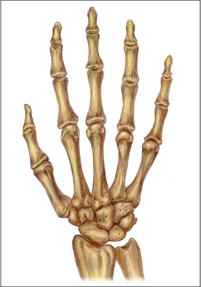
Plate 8.1
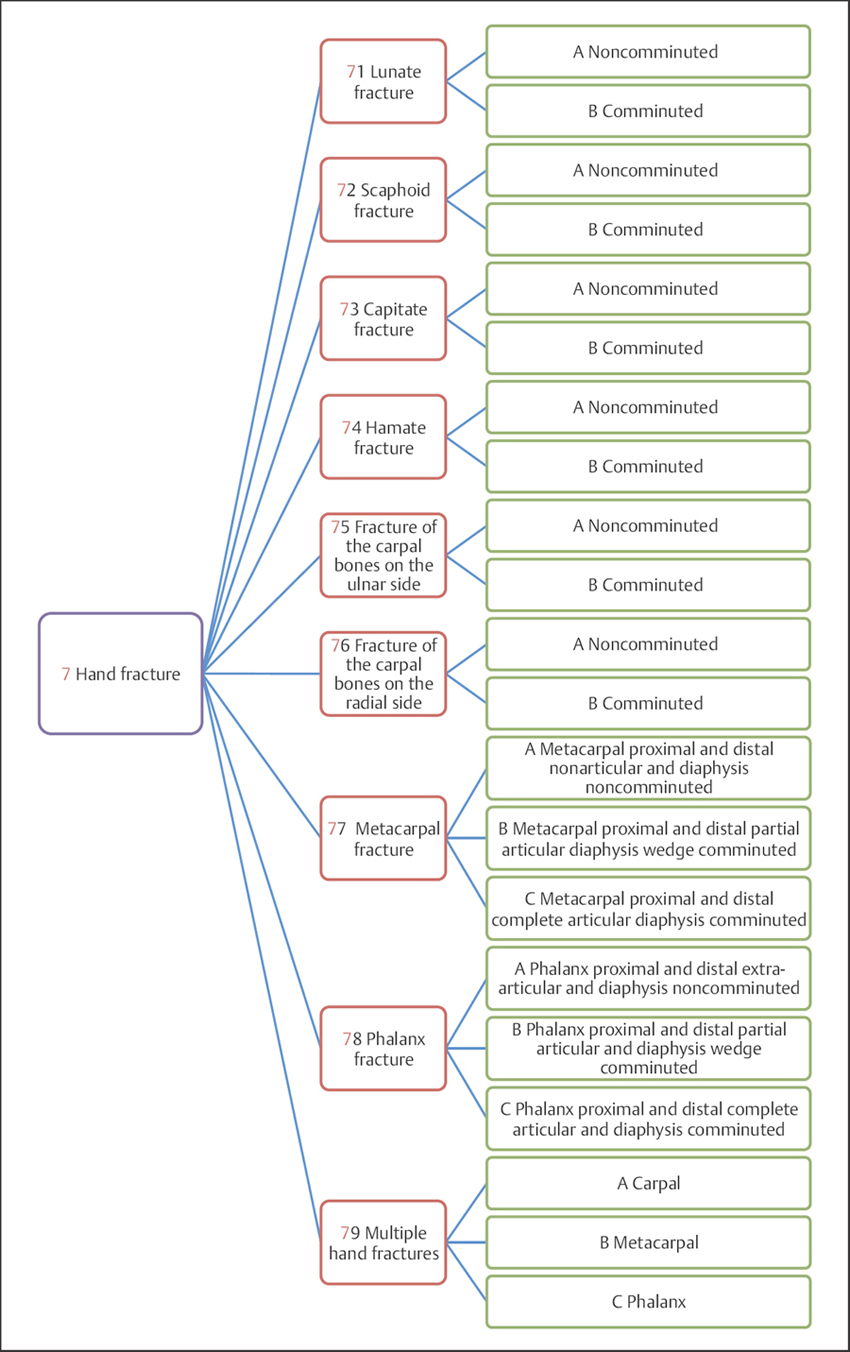
Plate 8.2
 Hand Fractures by Sex
Table 8.1 Sex distribution of 8913 patients with hand fractures
| Sex | Number of patients | Percentage |
| Male | 7004 | 78.58 |
| Female | 1909 | 21.42 |
| Total | 8913 | 100.00 |
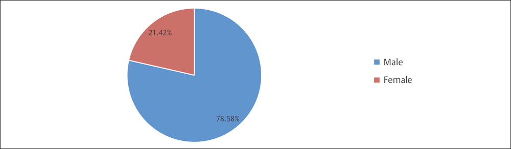
Fig. 8.1 Sex distribution of 8913 patients with hand fractures.
 Hand Fractures by Fracture Side
Table 8.2 Fracture side distribution of 8913 patients with hand fractures
| Fracture side | Number of patients | Percentage |
| Left | 4203 | 47.16 |
| Right | 4697 | 52.70 |
| Bilateral | 13 | 0.15 |
| Total | 8913 | 100.00 |
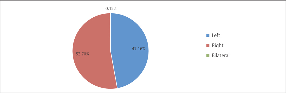
Fig. 8.2 Fracture side distribution of 8913 patients with hand fractures.
 Hand Fractures by Age Group
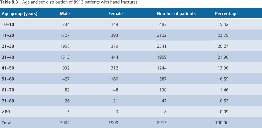
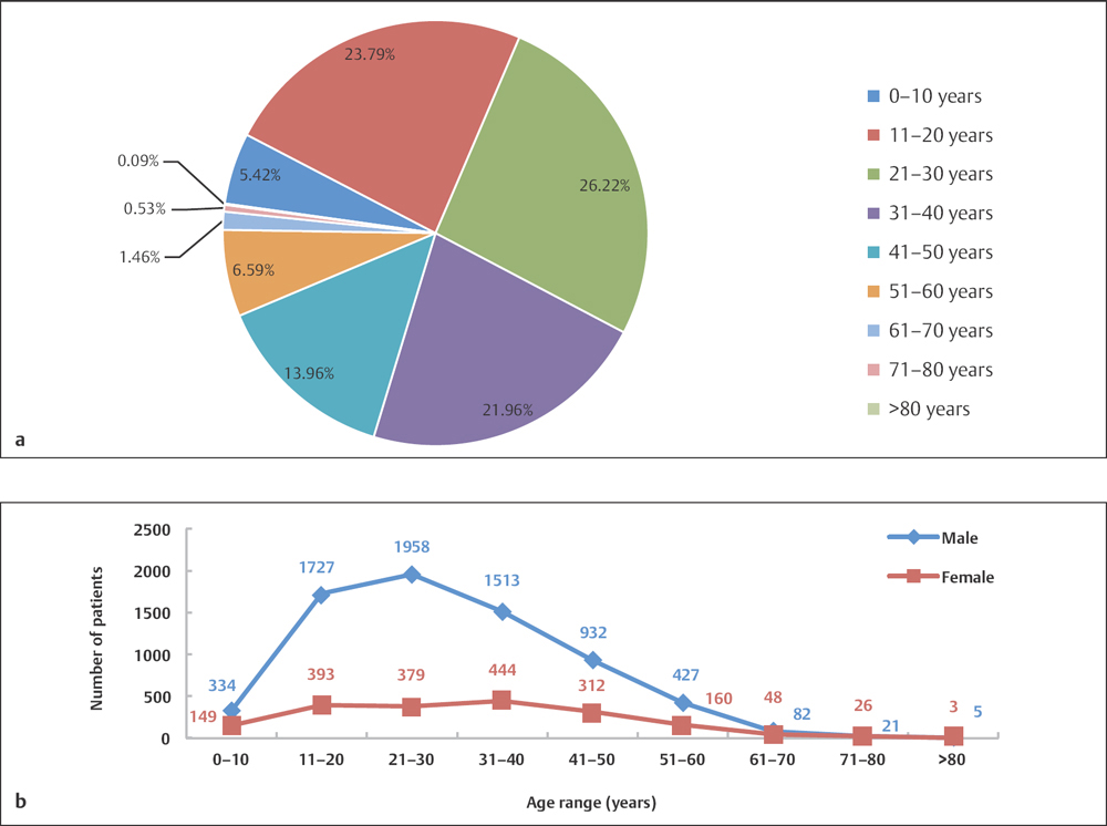
Fig. 8.3 a, b
a Age distribution of 8913 patients with hand fractures.
b Age and sex distribution of 8913 patients with hand fractures.
 Hand Fractures by Fracture Location
 Hand Fractures by Locations in Adults Based on OTA Classification
Table 8.4 Fracture location distribution of 10009 hand fractures in adults
| Fracture location | Number of fractures | Percentage |
| 71–76 (carpals) | 450 | 4.50 |
| 77 (metacarpals) | 1978 | 19.76 |
| 78 (phalanges) | 7581 | 75.74 |
| Total | 10009 | 100.00 |
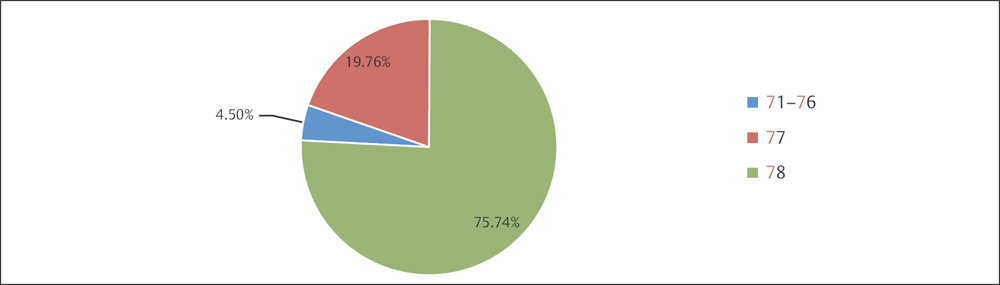
Fig. 8.4 Fracture location distribution of 10009 hand fractures in adults.
 Hand Fractures by Locations in Children
Table 8.5 Fracture location distribution of 964 hand fractures in children
| Fracture location | Number of fractures | Percentage |
| Carpals | 14 | 1.45 |
| Metacarpals | 184 | 19.09 |
| Phalanges | 766 | 79.46 |
| Total | 964 | 100.00 |
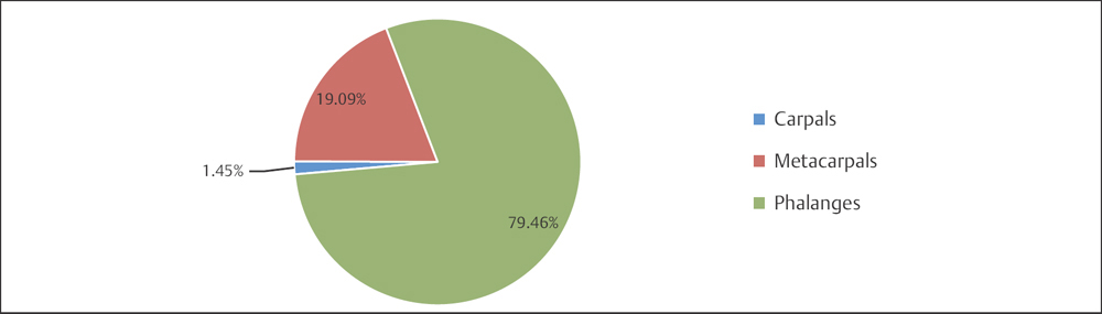
Fig. 8.5 Fracture location distribution of 964 hand fractures in children.
Carpal Fractures (Segments 71–76)
 Anatomic Features
There are eight carpal bones, arranged in two rows. Those of the proximal row, from lateral to medial, are the scaphoid, lunate, triangular, and pisiform; those of the distal row, in the same order, are the trapezium, trapezoid, capitate, and hamate. From the proximal row, the superior articular surfaces of the scaphoid, lunate, and triangular bones are connected by ligaments, present a convex surface, and articulate with the inferior surface of the radius and articular disk, forming the radiocarpal joint; the distal row of carpal bones articulates with the proximal bases of the five metacarpal bones, forming the carpometacarpal joints.
Carpals are short bones; each bone (except the pisiform) has six surfaces. The anterior and posterior surfaces, which make ligamentous attachment, are rough. The surfaces where the carpal bones make contact with contiguous bones are all articular, thus covered with articular cartilage, and are involved in the formation of the joint. The construction of these short bones provides complex but limited movement.
 OTA Classification of Carpal Fractures
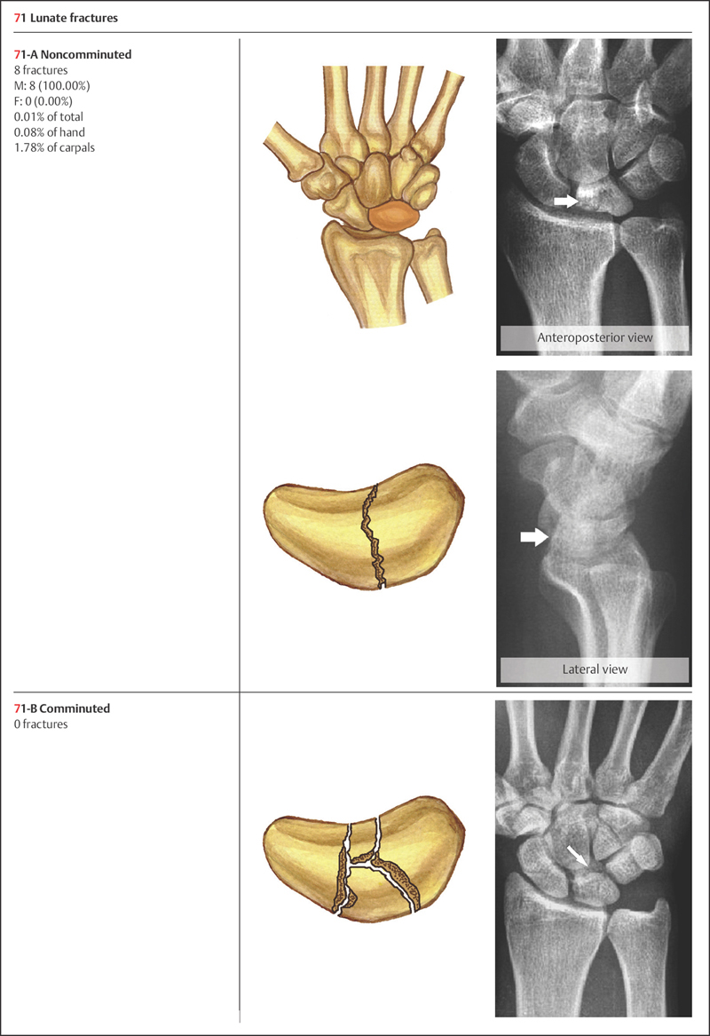
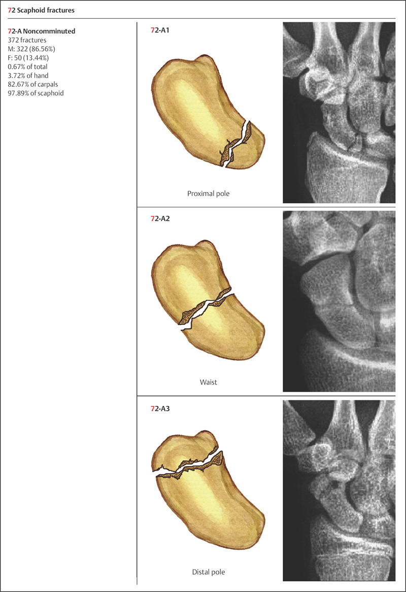
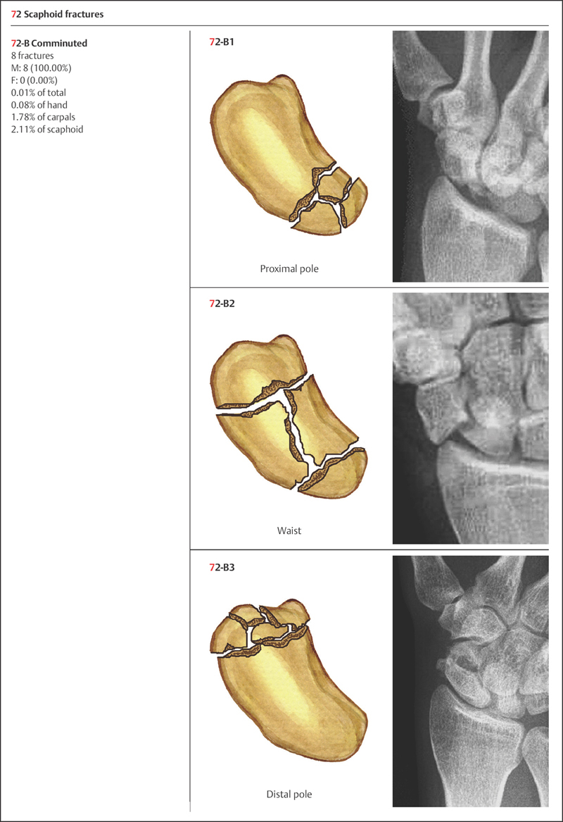
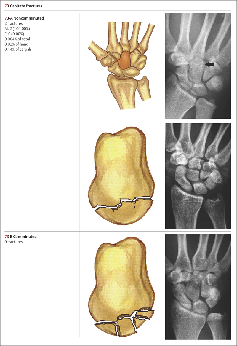
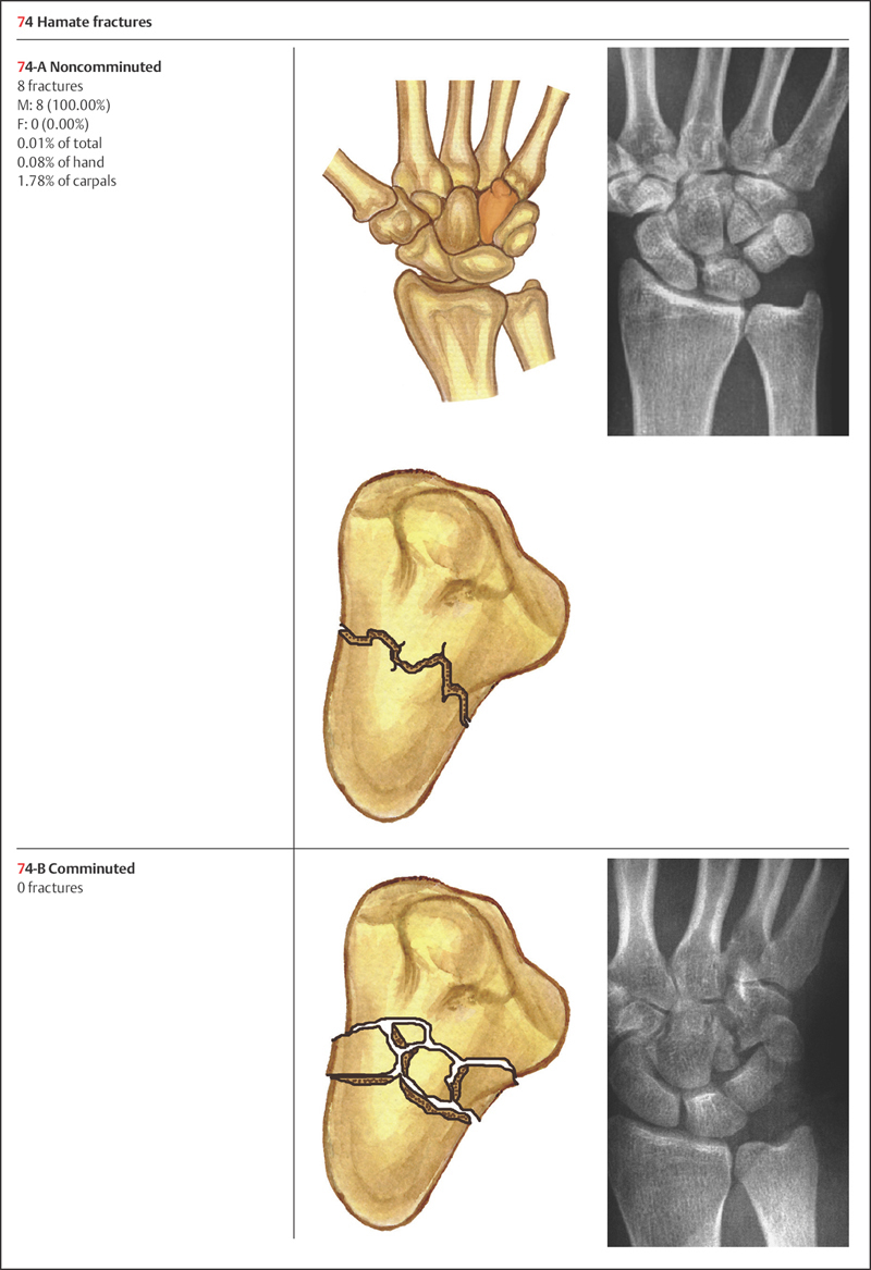
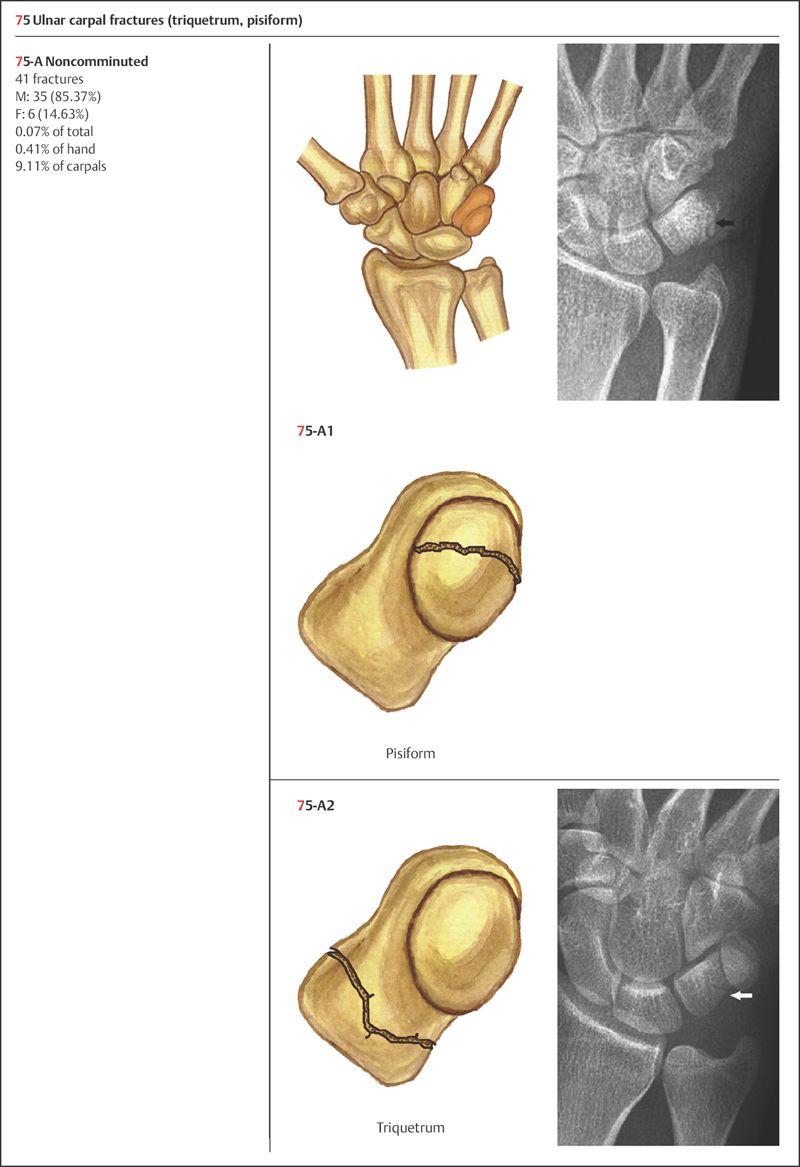
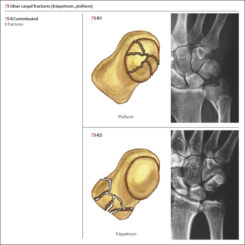
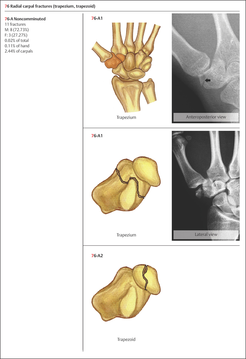
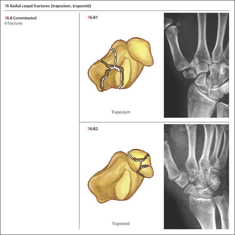
Carpal fractures are classified based on OTA classification as follows: 71: lunate; 72: scaphoid; 73: capitate; 74: hamate; 75: ulnar carpal bones; and 76: radial carpal bones (Plate 8.3).
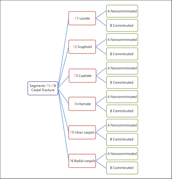
Plate 8.3
 Clinical Epidemiologic Features of Carpal Fractures (Segments 71–76)
A total of 450 carpal fractures in 442 adult patients were treated at our trauma center over a 5-year period from 2003 to 2007. All cases were reviewed and statistically studied; the fractures accounted for 4.50% of hand fractures in adults. Their epidemiologic features are as follows:
• more males than females
• the highest-risk age group is 21–30 years—the same age group for males, while there is no apparent high-risk age group for females
• scaphoid fractures (72) are the most common carpal bone fractures (71–76).
 Carpal Fractures (Segments 71–76) by Sex
Table 8.6 Sex distribution of 442 patients with carpal fractures (segments 71–76)
| Sex | Number of patients | Percentage |
| Male | 383 | 86.65 |
| Female | 59 | 13.35 |
| Total | 442 | 100.00 |
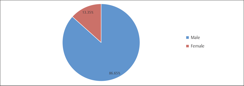
Fig. 8.6 Sex distribution of 442 patients with carpal fractures.
 Carpal Fractures (Segments 71–76) by Age Group
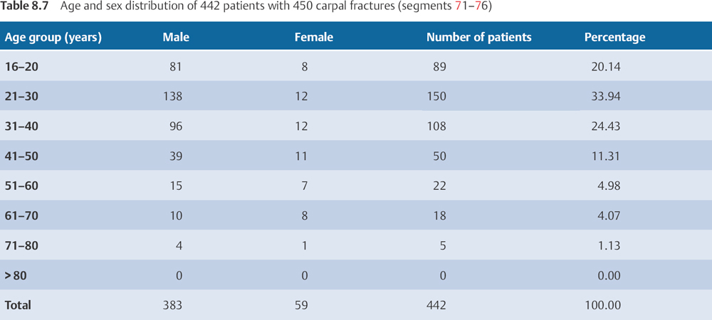
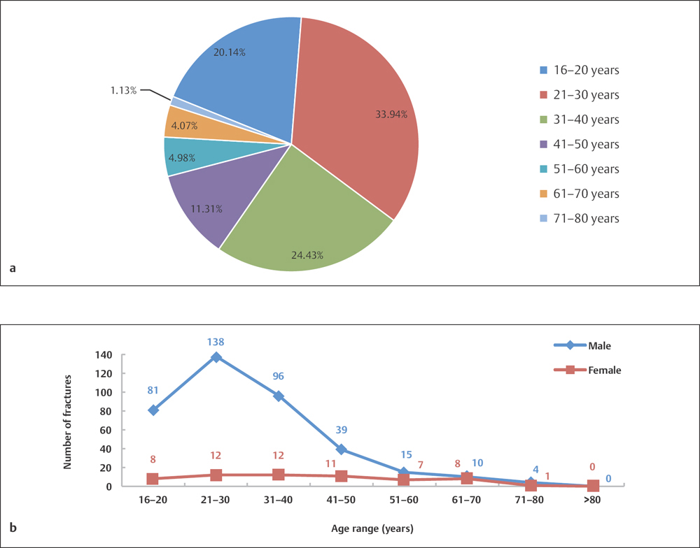
Fig. 8.7 a, b
a Age distribution of 442 patients with carpal fractures.
b Age and sex distribution of 442 patients with carpal fractures.
 Carpal Fractures (Segments 71–76) by Fracture Type
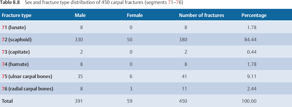
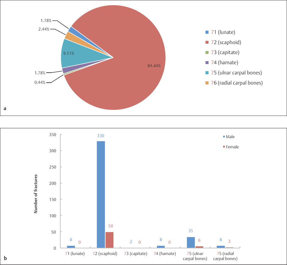
Fig. 8.8 a, b
a Fracture type distribution of 450 carpal fractures (segments 71–76).
b Sex and fracture type distribution of 450 carpal fractures (segments 71–76).
 Injury of Mechanism
Most carpal fractures are a result of axial loading on the outstretched palm and an extended wrist, for example from a fall on an outstretched hand or motor-vehicle collision. A direct blow to the dorsum of the hand, a crush injury, or cutting through the dorsum of the hand can also cause this type of injury.
 Diagnosis
Most patients present with history of a fall on an outstretched hand, or a traumatic event like a motor-vehicle accident. If palpation of each carpal bone and the intercarpal ligaments elicits pain and apparent local tenderness, one should strongly suspect the presence of fractures. Where carpal fracture is suspected, X-rays of anteroposterior (AP), lateral, and oblique views are needed. Bone scans and computed tomography (CT) scans are sometimes helpful if the plain X-ray is inconclusive for fracture.
 Treatment
Most carpal fractures can be treated with nonsurgical intervention, except scaphoid fractures. The indications for nonsurgical treatment are as follows:
• nondisplaced carpal fracture
• stable wrist joint injury, with less than 2 mm fracture displacement
• stable wrist joint injury, with less than 1 mm intra-articular fracture step-off
• isolated ligamentous rupture, in elderly low-demand patients
• hamate fracture with the hook intact
• pisiform fracture.
Related posts:


Stay updated, free articles. Join our Telegram channel

Full access? Get Clinical Tree


