Fig. 34.1
Glomus tumor of a finger. Macrophotography showing a well-delimited dark brown nodule
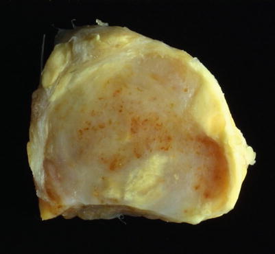
Fig. 34.2
Glomus tumor of a finger. Macrophotography of the cut surface of another tumor showing partial enclosure by a fibrous pseudo-capsule and a smooth tan-white surface
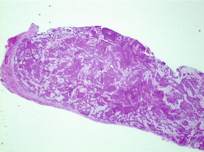
Fig. 34.3
Panoramic microscopic view of a glomus tumor. Tumor cells proliferate around small vascular channels
Fig. 34.4
Medium- and high-power view of a glomus tumor. Cells proliferate around compressed vascular lumina (a). Cells present eosinophilic cytoplasm, round nuclei, and well-delimited cell membranes (b)
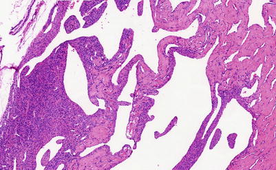
Fig. 34.5
Low-power microscopic view of glomangioma. In this picture vascular channels predominate over cellular areas
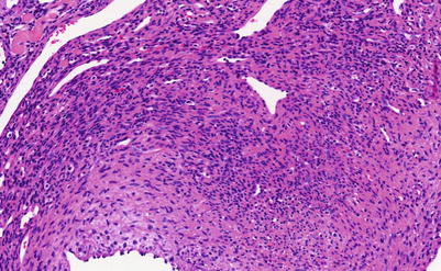
Fig. 34.6
Medium-power microscopic view of a glomus tumor depicting somewhat more elongated cells. Possible glomangiomyoma
Fig. 34.7
(a and b) Medium-power microphotographs of a glomus tumor of uncertain malignant potential involving soft tissue and bones of the ankle. There is marked cellular and vascular proliferation but does not present all the cytologic criteria of malignancy
Fig. 34.8
(a and b) Immunohistochemistry stain of two different glomus tumors. Strong staining with SMA
Fig. 34.9


Glomus tumor at the end of a distal phalanx. (a) X-ray shows a well-delimited lytic lesion in its most typical location. (b) Panoramic microscopic view shows a well-circumscribed cellular lesion, with clefts corresponding to lumina of blood vessels and pink hyaline areas
Related posts:


Stay updated, free articles. Join our Telegram channel
Full access? Get Clinical Tree
