Chapter 17 Fractures of the Distal Humerus
Plating Techniques
Introduction
Severe comminution, bone loss and osteopenia predispose distal humerus fractures to unsatisfactory results due to inadequate fixation. Poor outcomes include contracture, secondary to prolonged immobilization thought necessary to protect the fixation, and non-union. In an effort to reproducibly obtain stable fixation in the presence of osteoporosis or comminution, we have developed an improved fixation technique for fractures of the distal humerus based on principles that enhance fixation in the distal fragments and provide compression at the supracondylar level.1–4 The key to the stability achieved with this fixation construct is that it combines the features and stability of an arch while locking the two columns of the distal humerus together.
Background
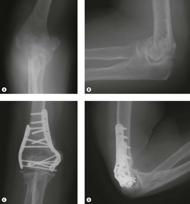
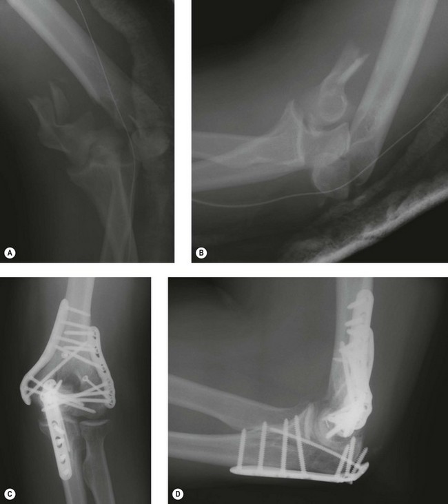
The critical concept being presented here is the idea that stability of the distal humerus is achieved by the creation of an architectural structure. The bone fragments rely for stability on their integration with the structure, rather than on fixation by screw threads. The concept is borrowed from modern architecture and the application of civil engineering principles to surgery. The interdigitation of screws within the distal segment rigidly attaches the articular fragments to the shaft by linking the two columns together. This permits stability to be achieved in such cases as low transcondylar (Fig. 17.1) or severely comminuted (Fig. 17.2) fractures.
Surgical techniques
Exposure
I prefer to perform this operation with the patient in the supine position. If assistance is limited, the lateral decubitus position with the arm over a bolster or arm holder may be easier. A sterile tourniquet is inflated only for dissection of the ulnar nerve, which is transposed anteriorly. An olecranon osteotomy provides the greatest exposure and is recommended in the setting of intra-articular comminution. The TRAP (triceps-reflecting anconeus pedicle) approach provides adequate exposure for a surgeon experienced with the technique.5 This involves combining the Bryan–Morrey and modified Kocher approaches to reflect the triceps in continuity with the anconeus.
Principle-based fixation technique
Stability is optimized by achieving eight technical objectives derived from the principles of (1) maximizing fixation in the distal fragments and (2) ensuring that all fixation in the distal segment contributes to stability at the supracondylar level. Six of these objectives concern the screws in the distal fragments and two concern the plates (Fig. 17.3).

Summary Box 17.1 Technical objective checklist
Concerning screws in the distal fragments (articular segment):
Concerning the plates used for fixation:
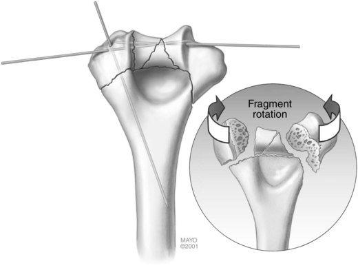
Step 1: articular surface reduction (Fig. 17.4)
The first step is articular surface reduction. The proximal ulna and radial head can be used as a template for the reconstruction of the distal humerus. Large articular fragments are provisionally fixed with smooth K-wires (Fig. 17.4). In cases with extensive comminution, fine threaded wires (1 mm) are used, then cut off and left in and used as ‘dowels’. It is necessary that these wires be placed close to the subchondral level, so as not to interfere with the passage of screws from the plates into the distal fragments. No screws are placed in the distal fragments until the plates are applied.


Stay updated, free articles. Join our Telegram channel

Full access? Get Clinical Tree


