Fig. 62.1
(a) Roentgenograph of the proximal phalanx of the hand showing a dense soft tissue mass with a periosteal reaction. (b) Sagittal MRI evidences of the soft tissue swelling and the periosteal reactive reaction of the phalanx
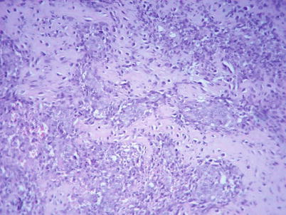
Fig. 62.2
Microphotograph showing woven osteoid bone trabeculae rimmed by active osteoblast. The bone marrow stroma is hypercellular, and some cells have a bizarre aspect
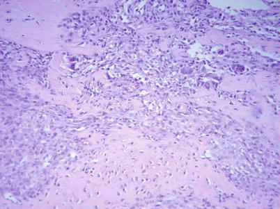
Fig. 62.3


Microphotograph showing a histology similar to the former image with the presence of multinucleated giant cells, osteoclastic type
Related posts:


Stay updated, free articles. Join our Telegram channel
Full access? Get Clinical Tree
