Fig. 34.1
Pincer fluoroscopy image (a), before and after rim trimming (b). Intraoperative fluoroscopic check before (c) and after femoroplasty (d), confirming complete CAM resection
CAM-type impingement treatment is possible without traction and a 45° hip flexion. It is sometimes indicated to perform a longitudinal capsulotomy to better reach the head-neck junction. The scope is inserted from the anterolateral portal, while the instruments (shaver, burr) are inserted from the mid-anterior portal, but switching is common and very useful for a better three-dimensional assessment (Fig. 34.1c, d). Lateral-based lesions are challenging due to the intimate location of the retinacular vessels; thus, proper attention should be given to the vascular anatomy. Osteochondroplasty should include all pathologically appearing cartilage, but shall not go higher or more proximally to the epiphyseal scar, which can be confirmed fluoroscopically. It is also fundamental to perform a dynamic evaluation to ensure an adequate decompression, and this should be the last gesture before arthroscopy can be considered completed.
34.5 Technical Notes
Consider preoperative planning to evaluate the amount of acetabular trimming and femoroplasty (3D CT dynamic reconstruction) |
Careful attention in portal placement when entry into the joint (labrum penetration, femoral head scuffing) |
If rim trimming is not appreciated on intraoperative fluoroscopic imaging, direct arthroscopic visualization, dynamic testing, and preoperative x-rays should guide further resection |
Divergent suture anchor placement orientation is recommended to prevent screw penetration of the acetabulum |
“T” capsulotomy is useful in case of a wide femoral bump |
Address lateral retinacular vessels before starting femoral decompression |
Fluoroscopy and dynamic evaluation are mandatory to confirm the amount of bone resection |
34.6 Rehabilitation
After hip arthroscopy, athletes wish to return to a fully active lifestyle and to practice their preferred sport as soon as possible. Currently, the best evidence for postoperative rehabilitation is based upon few scientific production; thus, communication with the specialist is vital to the treating physical therapist in order to give an individualized and evaluation-based program [23, 24].
Before starting rehabilitation, it is fundamental to know the exact procedure and operative findings, to plan a truly customized rehabilitation program both for simple and complex procedures (Table 34.1, Fig. 34.2).

Table 34.1
Classification based on the complexity of the surgical procedure
Simple | Diagnostic |
Removal loose body | |
Labral debridement | |
Ligamentum teres debridement | |
Intermediate | CAM decompression |
Iliotibial band release | |
Iliopsoas release | |
Complex | Acetabular rim trimming + labral repair + CAM decompression |
Microfracture | |
Very complex | Acetabular rim trimming + labral repair + CAM decompression + capsular plication |

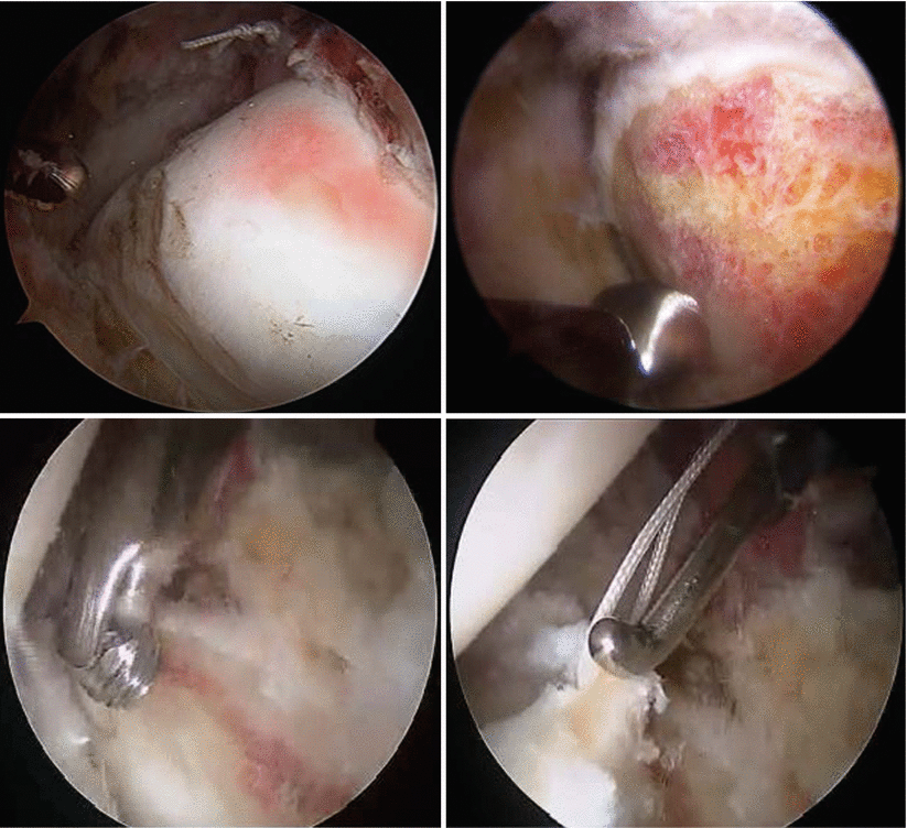
Fig. 34.2
Arthroscopic CAM image before (a) and after femoroplasty (b). Acetabuloplasty (c) and labrum refixation after rim trimming (d)
Rehabilitation can be divided into four phases. The timeline for each phase is based on clinical findings. If clinical presentation meets the established criteria, the athlete may move to the next phase. Progression in terms of the type and intensity of the workout should be function based, not time based (Table 34.2).
Table 34.2
Schedule of the rehabilitation program
Phase I | Start mobilization and isometric exercises; avoid swelling |
Phase II | Continue recovery of range of motion and isometric exercises |
Phase III | Recovery of full strength |
Phase IV | Recovery of balance and neuromuscular control |
Phase V | Functional recovery and return to sport |
It would be helpful to see the player preoperatively to prepare the affected joint and explain process and timescales involved. It should also be mandatory to give written rehabilitation indications at discharge.
Time recovery for a full activity is usually 4 months, but it may last longer depending on operative findings or prolonged rehabilitation.
It is fundamental not to force recovery [25]. Possible risks of a premature return to sports activity are:
Persistent pain
Prolonged rehabilitation time
Low performance
Reinjury (new labral tear, articular cartilage lesion)
New injuries
34.7 Outcome and Return to Play
Several published articles have been written on athletic patient population after hip arthroscopy.
Results of this studies show that athletes with FAI can return to high-level competitive sport following this procedure.
Philippon has published a cohort study of 28 professional hockey players who underwent hip arthroscopy for FAI. The return to sport was 3.8 months (range, 1–5 months) with MHHS of 95 at follow-up. Patients with symptoms lasting less than 1 year returned to sport at 3 months, but patients who delayed surgery over 1 year returned to sport at 4.1 months [26].
Brunner et al. in 2009 reported that values return to full sporting activity in 68.8 % of cases [27].
Byrd in 2009 reported its results with a mean follow-up of 27 months. In 90 % of professional and 85 % of college athletes, there was a return to full sports activity [28].
Another study by Nho et al. estimated the return to sport in patients undergoing hip arthroscopy up to 83 % [29].
A recent systematic review showed a high rate of return to pre-injury activity level in athletes treated for FAI. Results achieved a 92 % rate return to activity, observed in athletic populations across a variety of sports, with 88 % of athletes returning to pre-injury activity levels of participation [30].
Arthroscopic management among athletes is very favorable, but often performed when an important damage has occurred. Substantial secondary damage is frequently present that cannot be completely reversed. In fact FAI is very often not recognized, leading to a delay for a precise diagnosis. Early recognition and treatment have been demonstrated to have a tremendous impact on outcome; Philippon has showed that patients with symptoms lasting less than 1 year returned to sport at 3 months, but patients who delayed surgery over 1 year returned to sport at 4.1 months. Because early treatment is the only change for full recovery, athletes that decide to delay treatment should be aware of the risk [31].
Stay updated, free articles. Join our Telegram channel

Full access? Get Clinical Tree


