CHAPTER 32 Extrinsic Contracture: Lateral and Medial Column Procedures
INTRODUCTION
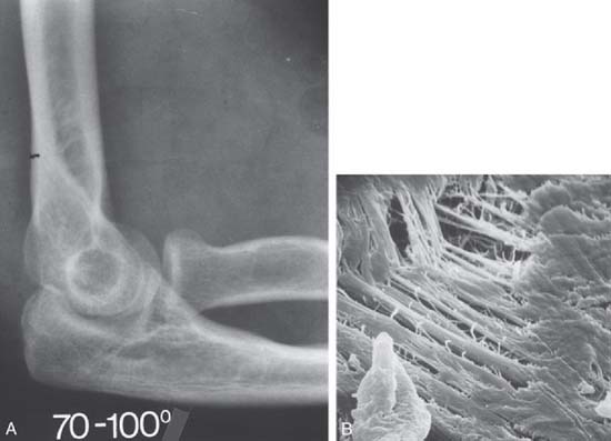
Of the numerous potential causes for elbow stiffness, the causes and pathophysiologic mechanisms dictate treatment and affect prognosis. Extrinsic contracture typically involves only the soft tissues around the elbow, sparing the joint space (Fig. 32-1).45 Post-traumatic stiffness is one of the most frequent causes of this kind of contracture47; however, it can also occur in association with other causes, such as congenital or developmental disease, osteoarthritis or inflammatory arthritis, burns, and head injury. Intrinsic contracture is associated with joint articular involvement and is not discussed here (see Chapters 33 and 69).
Several options have been proposed for treatment of elbow contracture. Conservative treatment sometimes gives good results if the contracture is of short duration4,6,13,15,16,21,23,38,46; however, its efficacy is unpredictable. With failure of nonoperative treatment, surgical release may be indicated. Most employ an open procedure, and several have been described.1,3,7,9–12,17,19,22,26,27,31–34,39,43,51,53,55,56,58–64 There is, however, an increasing interest in using an arthroscopic procedure.5,29,35,49,52,54,57
ETIOLOGY AND INCIDENCE
An extrinsic contracture usually involves the periarticular soft tissue without involving the articulating surface. Contracture may involve the capsuloligamentous structures or muscle tissue. Ectopic ossification is also considered an extrinsic condition. Bone may form a bridge across the joint or form in the capsule or in the muscle crossing the joint. Trauma is the major cause of extrinsic stiffness, especially elbow dislocation, with or without fracture.30,41 The brachialis muscle that crosses the anterior capsule36 tears with dislocation, developing scar tissue or ectopic bone when healing,49 often associated with contracture of the capsule.25,47,65 Pain, swelling, limited motion, and contracture after this type of elbow trauma then leads to the irreversible changes that constitute extra-articular ankylosis. Collateral injuries can contribute to elbow ankylosis from permanent contracture.8,24,28 In trauma, length of immobilization has also been recognized as a major contributor to postinjury contracture. The precise incidence of elbow stiffness after trauma is difficult to identify and is as much a function of the severity of injury as of the initial treatment. In adults, nontraumatic elbow contractures are usually caused by a primary inflammatory process. With osteoarthritis, a mild inflammatory synovitis occurs with periarticular fibrosis and osteophytic new bone formation.50 The articular surface of the joint is intact, but osteophytes are present at the tip of the olecranon and at the tip of the coronoid process.3 Hemophilia,14 juvenile rheumatoid arthritis, acute or chronic septic arthritis, and periarticular new bone formation after head injury18,37,42 can produce ankylosis of the elbow but often involve the joint space. Congenital stiffness is rare and is often associated with bone malformation or soft tissue dysplasia.2
PRESENTATION AND CLASSIFICATION
Generally, the patient initially notices loss of full extension but no limitation of activity. The first complaint is pain in terminal extension. Concurrent with this is the recognition that midarc motion typically is not painful, a finding that confirms the extrinsic character of the stiffness. Occasionally, full flexion also produces pain. Flexion contracture develops progressively.
In addition to classifying elbow contracture according to extrinsic and intrinsic lesions, age of the patient, severity of the stiffness, and distribution of the contracture are also important for evaluating what might be expected from the surgery. Thus, the stiffness may be graded as very severe, severe, moderate, or minimal, depending on data on the amount of residual arc of flexion.1,17,48 The stiffness is considered very severe when the total arc is 30 degrees or less, severe when it is between 31 and 61 degrees, moderate between 61 and 90 degrees, and minimal when it is greater than 90 degrees. Based on the functional arc of motion described by Morrey and colleagues,48 the distribution of the contracture referable to the 30- to 130-degree arc is also considered.
CONTRAINDICATIONS
Limited involvement and limited soft tissue contracture argue against these procedures. An inadequate period of an appropriate splint program is also a contraindication. Intrinsic lesions are not absolute contraindications, but a lower level of improvement has to be expected in these cases.39
PREOPERATIVE PLANNING
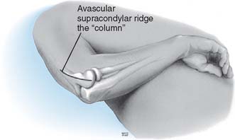
Before surgery, the decision must be made to approach the capsule from the lateral or medial aspect. If the ulnar nerve is to be addressed or there is extensive medial or coronoid arthrosis, the medial approach is of value. If the radiohumeral joint is involved or if a simple release is all that is required, the lateral “column” procedure is carried out (Fig. 32-2).
TECHNIQUE: COLUMN PROCEDURE
DISADVANTAGES
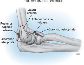
The column procedure consists of open arthrolysis of the elbow through a limited lateral approach, with the aim of releasing the anterior capsule safely but also of releasing the triceps attachment and posterior capsule, if necessary. The exposure employs a limited skin incision of only about 6 cm, but this is sufficient to remove coronoid or olecranon osteophytes (Fig. 32-3).
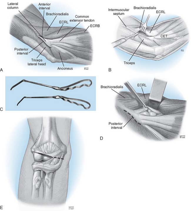
To release the anterior capsule with minimal disruption of normal tissue, the origin of the distal fibers of the brachioradialis and the extensor carpi radialis longus are identified (Fig. 32-4A). Release of the fleshy origin of the extensor carpi radialis longus and the distal fibers of the brachioradialis from the humerus provides direct access to the superior aspect lateral of the capsule (see Fig. 32-4B). The capsule is entered anteriorly at the radiohumeral joint, an approach that allows identification of the thickness and orientation of the capsule. The brachialis muscle is swept from the anterior capsule with a periosteal elevator. A specially designed retractor protects the brachialis muscle, the median nerve, and the brachial artery (see Fig. 32-4C). The anterior capsule is grasped and excised, at least to the level of the coronoid. The medial most aspect of the capsule is often difficult to visualize clearly but can be palpated. It is incised to complete the release (Fig. 32-4D). At this juncture, if extension is full or within 10 degrees of normal and the radiographs reveal no olecranon spur, no additional release is needed and the wound is closed. If scarring is extensive and adhesions involve the posterior capsule, the triceps is elevated from the posterior aspect of the humerus. The posterior capsule is released, and the olecranon fossa is cleaned of soft tissue (see Fig. 32-4E). The tip of the olecranon is excised when osteophytes are present. The range of flexion and extension at the elbow is assessed. If flexion is essentially normal, nothing more need be done posteriorly.

FIGURE 32-4 A limited Kocher skin incision measuring 6 cm is made over the lateral epicondyle. A, The anterior and posterior aspects of the lateral column are identified. B, The extensor carpi radialis longus (ECRL) and distal fibers of the brachioradialis are elevated. C, A special retractor facilitates exposure and protection of the anterior structures. D, The anterior capsule is isolated from the brachialis and identified by arthrotomy at the anterior radiohumeral joint. E, The lateral capsule is excised as widely as possible, and the remaining medial capsule is incised. The posterior capsule is identified (D) and excised as necessary by elevating the triceps from the lateral osseous column. The posterior aspect of the ulnohumeral joint is exposed and the capsule incised (see Fig. 32-3). ECRB, extensor carpi radialis brevis.


