Searches
Methods
Results
1
Exp Synostosis/or radio-ulnar synostosis.mp.
7976
2
Congenital.mp. or exp Congenital Abnormalities/
583,195
3
Exp Radius/or radius.mp.
34,880
4
Exp Ulna/or ulna.mp.
9703
5
3 or 4
38,991
6
1 and 2 and 5
403
7
Limit 6 to English language
283
Fig. 34.1
Flow chart of search methodology
What Are the Causes and Natural Progression of Disease?
Embroyology
At 26 days after conception, the upper limb bud arises from the body wall. Elbow is first identifiable at 35 days. Three connected cartilaginous anlagen that will eventually progress to become humerus, radius and ulna are still present at this stage [6]. By 42 days the three bones are separated by condensation of tissue but joints are still yet to form. At this stage, the forearm remains to be in neutral position and pronation occurs by week 8. Longitudinal segmentation with separation starts distally and progress proximally. Various factors either as a result of abnormal genetics or environmental factors can interrupt subsequent morphogenesis of the elbow joint at this point leading to failure of segmentation. The final specific defect is influenced by differences in development timing and can result in complete bony synostosis (Fig. 34.2) or smaller area of synostosis if joint development continues before development arrest [3].
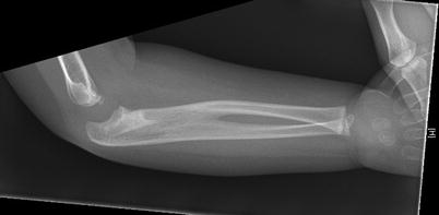
Fig. 34.2
Plain x-ray showing complete bony radio-ulnar synostosis
Genetic Associations
The actual etiology of the condition remains unknown. However, a lot of authors had suggested a familial presentation and genetic background. Disorders such as Apert and Klinefelter syndromes have been known to be associated with CRUS.
Catena et al has suggested in their proposed classification system that CRUS to be related to Type IV Poland Anomaly. The classic Poland Anomaly consists of unilateral agenesis/hypoplasia of pectoralis major muscle with associated ipsilateral hand or upper limb anomalies [7].
Gaspar et al also identified an association of CRUS with Giuffre-Tsukahara syndrome (GTS) which appeared to be a X-linked dominant inheritance. The other features of the syndrome included, short stature, microencephaly, scoliosis and mental retardation [8]. This report was also supported by Zhu et al in their paper with a three generation Chinese family with similar associated features suggested to be a variant of GTS due to a lack of microencephaly in the family [9].
There seems to be also multiple reports of CRUS being linked with bone marrow failure. Sola et al and Thompson et al have also identified association with amegakaryocytic thrombocytonpenia as a result of HOXA11 gene mutation. Thompson et al also reported in his case series of two families that there is an autosomal dominant disorder presenting with CRUS, congenital amegakaryotic thrombocyptopenia, aplastic anemia, clinodactaly, syndactaly, hip dysplasia and sensorineural hearing loss [10–11]. Castillo-caro et al. also reported on a case in infant associated with amegakaryotic thrombocytopenia without HOXA11 mutation [12].
Natural Progression of Disease
There seems to be a male preponderance in the incidence of congenital radio-ulnar synostosis. As previously mentioned, some patients might have a familial predisposition reported to be 13 % or other associated phenotypes suggesting congenital syndromes. Patients commonly presents at an average age of 2 and a half years old with majority noted by parents due to their functional abnormalities [5]. Children tend to compensate well using ipsilateral shoulder and wrist when deformity is mild resulting in minimal noticeable disability. Most patients present with hand fixed in various degrees of pronation (Fig. 34.3). With a bony synostosis (the most common type) there is no true pronation or supination, although adaptive ligamentous laxity at either the wrist or elbow can allow some apparent motion. The position of the wrist is variable ranging from being fixed in full supination to full pronation, with restriction in function dependent on the position. Quite frequently patients modify their activities and adapt to compensate for restricted rotation of forearm and rarely complained about restriction of function. In general an unilateral mid-pronated position is least debilitating, but with functional deficiencies were more commonly mentioned in patients with bilateral involvement and in more severe degrees of hyperpronation, in which some patients try the ‘backhanded’ technique in getting objects to mouth or receiving change [1–3].
Fig. 34.3
Radio-ulnar synostosis clinical photograph, arrow showing radial head prominence
Patients with CRUS can experience sudden onset of acute flexion contracture secondary to hyperflexion injury. Wang et al explained that this could be due to overgrown dislocated radial head becoming trapped under hypertrophied annular ligament-type tissue.
Classification
Wilkie and Davenport et al developed a classification for the condition in 1914. They suggested two different types of synostosis based on radiological appearance with either complete synostosis or partial union just distal to proximal radial epiphysis associated with radial head dislocation [13].
In 1985, Cleary and Omer et al noticed in their case series 4 different types of radiological appearance which conforms with the theory that timing of embryological arrest maybe reflected by both fixed position of forearm and degree of bony development at elbow. However, there were no functional differences noted amongst different radiological appearance hence it was not particularly helpful in determining management of patients (Table 34.2) [14].
Table 34.2
Cleary and omer classification
1. Fibrous synostosis |
2. Bony synostosis |
3. Osseous synostosis associated with posterior dislocation of a hypoplastic radial head |
4. Osseous synostosis with associated anterior dislocation of mushroom-shaped radial head |
What Is the Evidence Behind the Current Treatment Options?
As mentioned before, patients can compensate remarkably well despite deformity and reduction in forearm rotation which explained why the condition remain under diagnosed. Hence, in majority of patients CRUS can be treated non surgically especially if the arm is in a neutral position and there is a reasonable degree of compensatory wrist rotation.
Related posts:
 Evidence-Based Treatment of Flexible Flat Foot in Children
Evidence-Based Treatment for Congenital Dislocation of the Knee
Evidence-Based Treatment for Congenital Femoral Deficiency
Evidence-Based Management of Limb Length Discrepancy
Physeal Injury, Epiphysiodesis and Guided Growth
Evidence-Based Treatment for Feet Deformities in Children with Neuromuscular Conditions
Evidence-Based Treatment of Flexible Flat Foot in Children
Evidence-Based Treatment for Congenital Dislocation of the Knee
Evidence-Based Treatment for Congenital Femoral Deficiency
Evidence-Based Management of Limb Length Discrepancy
Physeal Injury, Epiphysiodesis and Guided Growth
Evidence-Based Treatment for Feet Deformities in Children with Neuromuscular Conditions
Stay updated, free articles. Join our Telegram channel

Full access? Get Clinical Tree


