Anatomy and biomechanics
Anatomical difference to adult spine
Consequence
Greater head to body size ratio
• Higher rates of upper cervical spine injuries
• Neutralisation of spinal alignment on spinal board required with torso boosting pad or occipital recess to prevent neck hyperflexion, airway closure and/or neurological deterioration
• SCIWORA much more common
• Higher proportion of TL junction injuries where rigid thorax meets elastic lumbar spine
• Greater risk of intra-abdominal/intra-thoracic injuries
• Injury patters more adult like ≥8 years
• Normal variants found on imaging
Relative paraspinal muscle immaturity
Shallow, horizontally oriented facet joints
Greater ligamentous elasticity
Incomplete vertebral ossification
Paediatric spine reaches adult like maturity at around 8–9 years-of-age
Nucleus pulposus has increased water and decreased collagen content
Greater elasticity and ability to dissipate force with less osseous fractures
Spinal cords ends at L3 at birth migrating caudally to end at L1 or L2 by adolescence
Spinal cord injury possible more distally secondary to trauma or surgical fixation
Vertebral physis/apophysis persists
Salter Harris I fractures possible with traction injuries (excellent prognosis with conservative management)
Apophyseal fracture and herniation possible
Wedge shaped small vertebral bodies
Compression fractures more common
Multilevel fractures more often
Developing body more sensitive to radiation
Avoid excess radiation (CT’s) where possible; increased overall risk of cancer
Ossification
Ossification centers and synchondroses (Fig. 28.1) can be troublesome when interpreting imaging and are therefore important to take into consideration. The atlas has three ossification centers, one anterior arch and two posterior arches. The anterior and posterior ossification centers are separated by the right and left neuro-central synchondroses which usually fuse by 7 years of age [2]. The posterior synchondrosis separates the posterior ossification centers and usually closes by 3 years of age. Beware; the posterior synchondrosis can remain unfused giving the appearance of a fracture [4, 5].
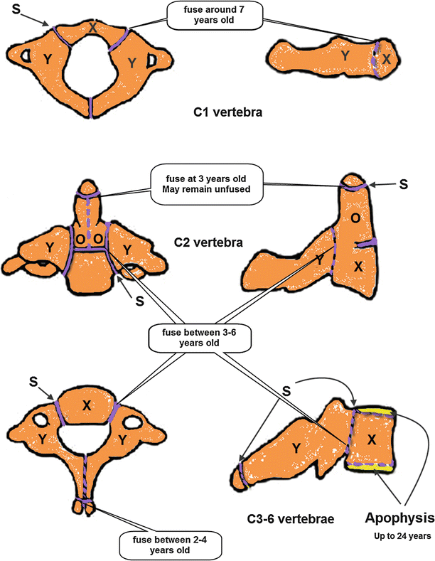

Fig. 28.1
Diagram of the ossification centres and synchondroses of the cervical vertebrae. Top images: Lateral and axial views of C1 and its three ossification centres. Middle images: Lateral and coronal views of the five ossification centres of C2. Bottom images: Axial and lateral views of a typical subaxial cervical vertebrae (thoracic and lumbar vertebrae have similar ossification patterns) X anterior ossification centre, Y posterior ossification centre, O odontoid ossification centre, Purple dashed line/gaps between ossification centres = synchondroses, yellow line = apophysis
The axis has a total of five primary ossification centers, one for each neural arch, one for the body, and two for the odontoid process. The odontoid is separated from the body of the axis by a cartilaginous physis that fuses between 3 and 6 years of age. It is situated below the level of C1–C2 facet joints and can therefore be mistaken for a fracture up to 11 years of age [2, 4]. The posterior arches fuse in the midline by 2–3 years and fuse to the body by 3–6 years of age.
Subaxial cervical, thoracic and lumbar vertebrae are all typical vertebrae and have a similar ossification pattern: one ossification center for the body and one for each neural arch. These fuse in the midline between 2 and 4 years and the neuro-central synchondroses close at 3–6 years. With aging, these ossification centers enlarge and the ratio of bone to cartilage increases. Apophyseal rings on the upper and lower surfaces of the vertebral bodies begin to ossify late in childhood and complete their fusion to the vertebral body by 25 years of age. Fractures of the apophyseal ring (Salter Harris type I) have been reported and are more common in the inferior endplates.
Cause of Injury
Spinal injury should be suspected in all children who are severely injured or involved in a high-energy accident. Children less than 8 years old are more likely to have spinal injuries resulting from automobile versus pedestrian accidents, motor vehicle collision (MVC), a fall from a height or non-accidental trauma (NAT) [2, 3, 6]. NAT is a common cause of injury in patients less than 2 years old and should not be overlooked in this age group [7]. Motor Vehicle Collisions (MVCs) are the major cause of injury across all age groups causing a high number of cervical and thoracolumbar fractures from inappropriate seat belt use. In older children and adolescents, one would expect to see more sporting injuries and motorcycle accidents. In general, serious head-injury and multi-injury trauma are commonly associated with spinal injuries. Patients with cervical spine injuries commonly have other associated traumatic injuries to the head, face, chest, abdomen or limbs [1, 8].
Important features to elicit from the history include conditions that predispose to spinal instability: e.g. Down’s syndrome (15 % have atlanto-axial instability), previous cervical spine injuries, arthritis or previous spinal surgery.
Birth trauma resulting in spinal injury or cervical instability has a high mortality rate and is more often than not, fatal. The hallmark for upper cervical spinal cord injury at birth is apnoea with flaccid quadriplegia following either breech or prolonged forceps manipulation. This should be treated with immobilisation for presumed C-spine injury: the length of immobilisation remains arbitrary however [2, 7].
Pre Hospital Immobilisation
The aim of immobilisation is to prevent further damage or injury whilst the child is transported to an appropriate care facility. As with adults, all children with suspected spinal injuries should be immobilised at the scene as soon as possible. However, unlike adults, immobilisation of a child on a spinal board requires specific paediatric modifications.
Due to the relatively large size of a child’s head compared to their body, evidence suggests that children less than 8 years of age should have thoracic elevation or an occipital recess to achieve better neutral alignment of their spine (25 mm on average) [9], (Figs. 28.2 and 28.3).
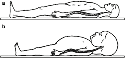
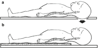

Fig. 28.2
The effect of relatively large size of a child’s head. (a) The relatively large size of a child’s head compared to their body results in cervical flexion when placed on a rigid spinal board (compared with adults (b)) (Reproduced by kind permission of Elsevier from Nypaver and Treloar [10])

Fig. 28.3
Occipital recess and thoracic elevation allow for neutral alignment of the cervical spine. Use of (a) an occipital recess or (b) thoracic elevation (25 mm) allows for neutral alignment of the cervical spine (Reproduced by kind permission of Elsevier from Nypaver and Treloar [10])
In general, the type of immobilisation should take into account the child’s age and physical maturity. Ideally there should be a combination of a spinal board, rigid collar and torso tape. One must remember however that these may negatively influence the child’s respiratory function, which should be monitored vigilantly [11, 12].
Safe removal of every patient from a spinal board must be an early priority after the primary survey and resuscitation phase. This prevents the complications of prolonged immobilisation as well as avoiding the agitation of a strapped down child.
Imaging
Figure 28.4 displays the criteria employed by the 2014 NICE guideline (CG176) for determining whether cervical spine imaging is required [13]. Although this is taken from a guideline that primarily manages children with a head injury, the algorithm is derived from the Canadian C-Spine Rule [14] which has successfully been used to reduce the amount of inappropriate radiation in children. There is some evidence that great caution is advised when applying these to children less than 11 years of age [15, 16].
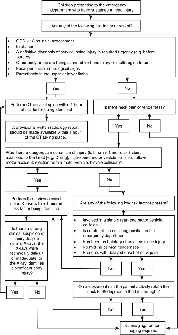

Fig. 28.4
NICE Clinical Guideline 176: Selection of children for imaging of the cervical spine (algorithm 4)
Whilst is it important to consider the different anatomical and physiological differences in a growing child, health professionals must also be aware of the significant risks involved in exposing children to high doses of radiation. CT confers 90–200 times more thyroid radiation than standard plain films with an increased risk in children less than 4 [17]. Children have developing tissues which are significantly more sensitive to radiation and a longer life expectancy than adults, resulting in a greater time period to express such radiation damage.
As discussed previously, the spinal injury patterns that occur in young children differ from those in adults. The diagnostic tests and imaging required to exclude spinal injury also differ to those required in adults. Interpretation of paediatric radiographs requires a sound knowledge of age related ossification and anatomical variations and, more often than not, will require discussion with a musculoskeletal radiologist with specialised paediatric experience. (See Tables 28.2 and 28.3 for common normal variants found on cervical spine radiographs and their satisfactory measurements [2, 5, 18].)
Table 28.2
Common normal variants found on cervical spine radiographs
Common normal variants | Comments |
|---|---|
Absence of cervical lordosis | Seen in children up to 16 years of age |
Prevertebral soft-tissue thickening | Can result from expiration, especially if the child is crying |
‘Pseudospread’ of atlas | Results from a discrepancy between the “neural” growth pattern of the atlas and the “somatic” pattern of the axis |
Increased atlanto-dental interval (ADI) | Potentially reflects incomplete ossification of the dens and laxity of the transverse ligament |
Overriding arch of C1 above the odontoid | Can be mistaken for atlantoaxial instability, normal in 20 % of children <8 years |
Incomplete ossification of posterior arch of C1 | |
Anterior wedging of vertebrae (up to 3 mm) | Can be present until endplates fuse. |
Pseudosubluxation (anterior displacement of C2 on C3) | Seen as a normal variant in up to 40 % of young children |
Odontoid and body of C2 synchondrosis mistaken for fracture |
Table 28.3
Acceptable measurements for cervical spine radiographs
Parameter | Normal value (mm) |
|---|---|
C1 facet-occipital condyle distance | ≤5 |
Atlanto-dens interval (ADI) | ≤4 (3 mm in adults) |
Space available for the cord (SAC) | ≥14 |
Pseudosubluxation of C2 on C3 (most common misinterpretation) | ≤4 |
Pseudosubluxation of C3 on C4 | ≤3 |
Retropharyngeal space | ≤8 (at C2) |
Retrotracheal space | ≤14 (at C6, under age 15 years) |
Cervical spine imaging is not recommended in all children who have experienced trauma. It is vital, however, to image the whole spine in the presence of a fracture as there is a significant (8–24 %) risk of a non-contiguous fracture [5, 9, 19]. Unexplained hypotension should always raise suspicion of spinal cord injury.
Table 28.4 demonstrates the most up to date evidence available for imaging of the cervical spine in children [12].
Table 28.4
Recommendations of radiological investigations
Level I evidence | • Use CT to determine the condyle-C1 interval (CCI) for children with suspected atlanto-occipital dislocation (AOD) |
Level II evidence | • Cervical spine imaging is not recommended in children who are >3 years of age, who have experienced trauma and who: – Are alert – Have no neurological deficit – Have no midline cervical tenderness – Have no painful distracting injury – Do not have unexplained hypotension – Are not intoxicated • Cervical spine imaging is not recommended in children who are <3 years of age, who have experienced trauma and who: – Have a Glasgow Coma Scale (GCS) >13 – Have no neurological deficit – Have no midline cervical tenderness – Have no painful distracting injury – Are not intoxicated – Do not have unexplained hypotension – Do not have motor vehicle collision (MVC), a fall from a height > 10 ft, or non-accidental trauma (NAT) as a known or suspected MOI • Cervical spine radiographs or CT are recommended for children who have experienced trauma and who do not meet either set of criteria above • Use three-position CT with C1–C2 motion analysis to confirm and classify the diagnosis in atlantoaxial rotatory fixation (AARF) |
Level III evidence | • Use AP, lateral and open mouth cervical spine radiographs or CT to assess the cervical spine • Use CT scan with attention to the suspected level of injury to exclude occult fractures or to evaluate regions not adequately visualized on plain radiographs • Flexion and extension cervical radiographs or fluoroscopy are recommended to exclude gross ligamentous instability when cervical instability remains a possibility • Use MRI to exclude spinal cord or nerve root compression, assess ligamentous integrity, or provide information regarding neurological prognosis |
When plain radiographs are indicated, an adequate cervical spine series must include:
- 1.
Lateral cervical spine x-ray to include the base of skull and the junction of C7 and T1.
- 2.
Antero-posterior cervical spine x-ray to include C2 to T1.
- 3.
Adequate peg view if attainable*.
*Due to a child’s age and difficulty with cooperation, current evidence suggests that open-mouth views only need to be obtained if the child is ≥9 years of age [4, 5, 12].
Unfortunately, there are no well-established, evidence-based recommendations for suspected thoracolumbar spine injuries following trauma. However, AP and lateral plain radiographs are useful initial imaging techniques along with MRI for those children presenting with neurological deficit. Children who have sustained significant poly-trauma or high-energy accidents will commonly have CT imaging. A high clinical suspicion of spinal injury exists in the presence of: neurological deficit, high energy mechanism of injury, spinal step-off or crepitus, concurrent head or facial injuries, abnormal GCS and children who are non-compliant with examination [20].
Types of Injury
Cervical Spine Injuries
Occipital Condyle Fractures
These are rare fractures that require a high degree of suspicion. They can be associated with high-speed blunt traumas and may result in cranial nerve palsies and cervical spine instability. Use CT scan of the cranio-vertebral junction to promptly confirm the diagnosis.
A number of different classification systems have been developed over the years to help guide treatment options: reported evidence to date suggests that these classifications contribute little to management [2].
Below is the classification identified by Anderson and Montesano in 1988:
Type I – Impaction fracture.
Type II – Basilar skull fracture with extension into the condyle.
Type III – Avulsion of the alar ligament off the infero-medial portion of the condyle.
Appropriate choice of treatment is therefore based on the CT assessment of the fracture with or without MRI, rather than relying on fracture type alone [21, 22].
Management should involve occipito-cervical arthrodesis or halo fixation for those cases with occipito-cervical malalignment. If malalignment is not present, treat with immobilisation in a cervical orthosis with follow up imaging in a clinic setting [18, 23, 24]. C1–C2 transarticular screws, or C2 pedicle screws with rigid loops and plate or rods, have been successful in pediatric patients as young as 11 months old [2].
Atlanto-Occiptal Dislocation (AOD)
This is a very unstable injury and usually fatal. It results from sudden deceleration leading to hyperflexion and cranio-vertebral separation (Fig. 28.5). This causes disruption of the alar ligaments, articular capsules and tectorial membrane (ligamentous injury). Level I evidence recommends CT imaging to determine the condyle-C1 interval (CCI) [12] but requires a high index of suspicion: there is frequent association with head and face injuries following high-energy traumas [2]. Surgical stabilisation with occiput to C2 fusion or halo immobilisation is required [25].
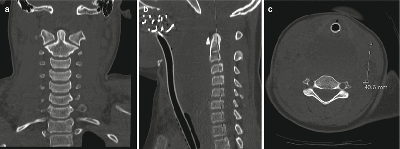

Fig. 28.5
Atlanto-occiptal dislocation. (a) Coronal, (b) sagittal and (c) axial (at C4 level) MRI images demonstrating significant atlanto-occiptal dislocation with associate soft tissue swelling
Atlas Fractures (Jefferson Fractures)
These injuries result from rupture or avulsion of the transverse ligament caused by axial compression. The ring of C1 is commonly broken at more than one site. Greater than 6.9 mm of combined lateral overhang of the lateral masses on AP radiograph is diagnostic. As mentioned previously, up to 6 mm of pseudospread may be seen in children under 7-years (secondary to faster growth of atlas compared with axis). These require immobilisation in a cervical orthosis or halo device for up to 6 months. Halo traction followed by halo immobilisation maybe necessary if there is >6.9 mm or widening of lateral masses.
Odontoid Epiphysiolysis
The neuro-central synchondrosis of C2 may not fuse completely until age 7 years and can be a possible site of injury in young children (odontoid synchondrolysis). Lateral C-spine radiographs will reveal the odontoid process to be angulated anteriorly, and rarely, posteriorly [12]. Treatment is closed reduction and external immobilisation with a Minerva or halo for approximately 10 weeks (80 % fusion rate). Confirm healing on removal of the immobilisation device with flexion/extension radiographs [5]. Primary surgical stabilisation has a limited literature base and plays more of a role when closed external immobilisation is unable to maintain alignment of the odontoid atop the body of C2 [2].
Atlanto-Axial Injuries
The atlanto-axial joint is primarily stabilised by the transverse ligament and secondarily by the alar and apical ligaments. In children, it is more common to find rupture of the ligament as opposed to avulsion of the ligamentous attachments in adults. Current evidence supports early posterior atlanto-axial arthrodesis (successful fusion with C1–C2 transarticular screw fixation has been reported) with instrumentation for ligamentous disruptions and immobilisation in cervical orthosis or halo device for bony avulsions [18]. Surgery should be reserved for patients with non-union and persistent instability after 3–4 months of immobilisation. Imaging should be used pre-operatively to determine appropriate screw size, entry points and trajectories, before consideration of a Goel-Harms Procedure [2, 18].
Stay updated, free articles. Join our Telegram channel

Full access? Get Clinical Tree


