Statement
Grade of recommendation
Not all patients with tarsal coalition will develop symptoms
B
A symptomatic patients do not require any treatment
B
Non operative treatments should be tried first before considering surgical treatment
B/C
Resection is recommended in persistently symptomatic patients
B
The superiority of interposition graft materials has not been established
I
Severe associated deformity and degenerative changes are predictors for persistent symptoms
C
Severe associated deformity is an indication for corrective osteotomy
C
Secondary degenerative changes are indication for arthrodesis
C
Tarsal coalitions can be acquired secondary to infection, trauma, surgery, arthritis or neoplasia but these are rare. Congenital coalitions have been reported to be the most prevalent in adolescents [7]. It has been found that an inherited defect in genetic coding can result in tarsal coalition. Leonard [6] described an autosomal dominant pattern of inheritance with almost full penetrance. Thirty-nine percent of first degree relatives may have tarsal coalitions [6]. Mosca has described tarsal coalition as a developmental malformation instead of congenital deformity, as it is not present at birth but it is genetically programmed to develop. Tarsal coalitions may be present with other congenital disorders such as fibular hemimelia, clubfoot, Apert syndrome, and Nievergelt-Pearlman syndrome, involving multiple bony connections [3].
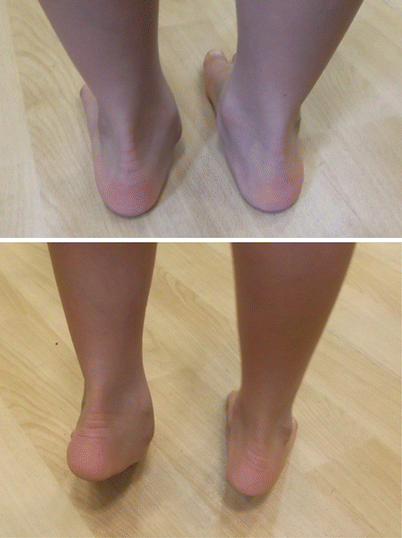
Most patients are asymptomatic. Only 25 % will go on to develop symptoms and this usually happens between 8 and 12 years for children with calcaneonavicular coalitions [5] and between 12 and 16 years for children with talocalcaneal coalitions [7]. Progressive flattening of longitudinal arch and later onset of aching pain, are the main clinical features of tarsal coalition. Symptoms are activity related and are improving with rest. Tip toeing and Jack’s test will not reconstitute the medial arch due to rigidity of subtalar joint (Fig. 20.1). The term “peroneal spastic flat foot” has been misused in the past as a synonym to tarsal coalition, but it describes a rigid flat foot with involuntary contracture of peroneal tendons that may exist without tarsal coalition [3]. Mubarak et al. [8] also noticed that plantar flexion may be decreased in calcaneonavicular coalitions apart from subtalar joint movements.


Fig. 20.1
Clinical photograph of a child with tarsal condition. Heels should move to varus and the medial arches are reconstituted on tip toeing. This does not usually happen when there is a tarsal coalition as in the left of this child
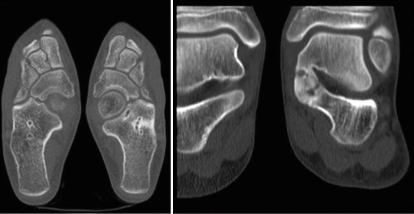
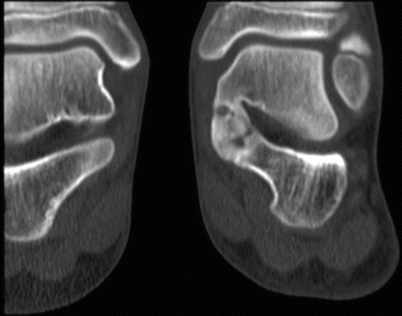
Weight bearing radiographs of the foot anteroposterior, lateral and oblique 45° can diagnose a calcaneonavicular coalition with the oblique being the most helpful view [9]. On oblique and lateral radiographs “anteater’s sign” is diagnostic for calcaneonavicular coalition [10]. In addition to previous radiographs, the Harris axial view can help in identifying talocalcaneal coalitions [11]. An anteroposterior ankle view could show a ball-and-socket type joint in a longstanding tarsal coalition. A C-sign on the lateral view can be indicative of talocalcaneal coalition, but since it has also been found to be positive in flexible flat foot, it is considered neither sensitive nor specific for the diagnosis of talocalcaneal coalition [12]. Dorsal talar beaking is another obvious sign on the lateral view. This is considered a traction spur rather than degenerative disease [13]. The gold standard imaging modality to diagnose tarsal coalition is Computed Tomography which can offer also information regarding the size and location, as well as other concurrent coalitions (Figs. 20.2 and 20.3) [14]. Upasani et al. [15] suggested that a CT scan should be performed in order to assess if there is any concurrent osteoarthritis of adjacent joints as well as for preoperative planning as persistent postoperative symptoms may relate to inadequate resection. MRI scanning has comparable results with CT scan and may be useful for early identification of fibrous coalitions [16, 17].


Fig. 20.2
CT scan showing bilateral calcaneonavicular tarsal coalition (left) and right talocalcaneal tarsal coalition (left)

Fig. 20.3
Talocalcaneal tarsal coalition
Should We Treat Non-symptomatic Coalitions?
Only 25 % of patients with tarsal coalitions will develop symptoms [6]. There is no published evidence to support that painless tarsal coalition will become symptomatic and disabling in the long term. On the other hand, it is unknown if resection of symptom free tarsal coalitions will lead to better outcome in comparison to natural history [3]. For this reason, treatment should be attempted only for symptomatic coalitions.
Is There Any Role for Non-operative Treatment?
It is often suggested that non-operative management should be always used prior to surgical interventions for symptomatic tarsal coalitions. Activity modifications, non steroidal anti-inflammatory drugs and orthotics may provide symptomatic relief. Flat insoles without firm of hard arch have been suggested in order to achieve additional cushioning support for the treatment of talocalcaneal coalitions [3]. A trial of short leg walking cast with hindfoot in neutral position for 4–6 weeks can also provide with symptomatic relief. Thirty percent of patients will remain symptom free after cast immobilization [18]. Calcaneonavicular coalitions are less likely to respond to casting in comparison to talonavicular coalitions [19].
What Is the Best Operative Treatment for Calcaneonavicular Coalitions?
Those patients who have persistent and disabling pain refractory to non-operative measures are usually candidates for operative treatment. The main aim of operative treatment is pain relief [3]. Asymptomatic and minimal symptomatic patients should not be offered an operative intervention [19].
Badgley [20] was the first to describe resection of calcaneonavicular coalitions. Mitchell and Gibson [21] published a case series with simple excision of calcaneonavicular bars with full recurrence of coalition in one third of the patients and partial recurrence in another third of patients. The high recurrence rates led Cowell [22] to modify this technique with interposition of the extensor digitorum brevis (EDB) in the gap of resection. Gonzalez et al. [19] presented their experience with the above procedure and showed excellent results in 77 % with 23 % recurrence rate. Moyes et al. [23] compared their results between patients who had EDB interposition and those who didn’t after calcaneonavicular coalition resection and suggested the interposition of EDB as important part of the operation. Mubarak et al. [8] with a cadaveric study showed that EDB muscle is not enough to fill the gap after resection of calcaneonavicular coalition and presented his results with the use of free fat graft as an interposition material showing lower reossification rates (13 %) and good long term pain relief. Most of other studies have shown encouraging results with resection of calcaneonavicular coalition followed by interposition of either fat tissue or EDB [13, 24, 25].
Recently, a few case reports and small case series have been published suggesting the endoscopic resection of calcaneonavicular coalition as an alternative to open resection, resulting in good outcome with symptomatic relief and improvements in functional scores [26–31]. Larger patient series treated with arthroscopic coalition resection with long-term follow up as well as comparative studies with open resection patient series will be needed to determine the outcome of this method and the true benefit over standard of care.
Significant degenerative arthritis of talonavicular and calcaneocuboid joints are contraindications for the resection of calcaneonavicular coalition and thus preoperative CT scan is imperative [5]. In case of concurrent joint degeneration or failure of complete resection of the coalition that leads to disabling pain, triple arthrodesis has been proposed [5]. Triple arthrodesis has been used as a salvage operation after failure of coalition resection to relief symptoms with satisfactory results [13, 19, 21, 32]. Most case series with primary triple arthrodesis for calcaneonavicular coalition are involving mixed population with adults and show that this operation provides adequate pain relief but has unpredictable functional outcome [13, 14]. The good results from treating calcaneonavicular coalitions with resection and the reluctance of surgeons to perform triple arthrodesis in children due to poor long term outcome made primary triple arthrodesis a rarely used alternative [3] (Table 20.2).
Table 20.2
Treatment of calcaneonavicular coalitions in children and adolescents
Studies | No (feet) | Type of primary surgery | Additional surgery | FU(Y) | Age (Y) | Outcome | Recurrence | Complications | LOE |
|---|---|---|---|---|---|---|---|---|---|
Mitchell and Gibson [21] | 41 | Open resection | Nine feet had revision to triple arthrodesis | 6 | 10–14 | 68 % complete symptoms relief 58 % regained subtalar ROM | 1/3 complete recurrence 1/3 incomplete | IV | |
Mubarak et al. [8] | 96 (78 FU) | Open resection and fat graft interposition | 5 % needed additional osteotomies for deformity correction | 29 m | 7–17 | 87 % excellent 8 % fair 5 % failures | 13 % | IV | |
De Vriese et al. [24] (includes adults) | 18 | Open resection and EDB interposition | 3 (1–10) | 14.2 (9–40) | 10 excellent 4 good 1 fair 3 poor | 16 % | IV | ||
1 | Open resection and sinus tarsi spacer | Removal of spacer | 1 poor | 100 % | |||||
2 | Triple arthrodesis (one adult after failed resection) | ? | |||||||
Gonzalez and Kumar [19] (includes Cowel’s patient series) | 75 | Open resection and EDB interposition | Five feet had triple arthrodesis | 2–23 | 11.2 (8–17) | 77 % excellent – good 16 % fair 7 % poor | 23 % partial recurrence | IV | |
Swiontkowski et al. [13] (includes adults) | 39 | Open resection and interposition of fat graft or EDB | Two had triple arthrodesis and one repeat resection | 4.6 | 12 years (8–54) | 35 improved Four deteriorated | ? | IV | |
5 | Triple arthrodesis | All patients had pain relief | |||||||
Inglis et al. [32] | 16 (11 patients) | Open resection | Three feet had triple arthrodesis and two feet had repeat resection | 23 | Eight patients satisfactory results Three patients unsatisfactory | 25 % | ? | IV | |
Moyes et al. [23] | 10 | Open resection with EDB interposition | Nil | 3.4 | 12 | Nine patients pain free with good ROM | No | One patient pain due to degeneration | IV |
7 | Open resection | Four patients had no symptoms | Three patients | ||||||
Khoshbin et al. [25] | 19 | Open resection with EDP interposition | One concurrent calcaneal osteotomy and one TA lengthening
Stay updated, free articles. Join our Telegram channel
Full access? Get Clinical Tree
 Get Clinical Tree app for offline access

|


