Fig. 10.1
Bryant traction for femoral shaft fracture
Early or Immediate Hip Spica Casting and Traction
Early or immediate hip spica casting is the main treatment for isolated femoral shaft fractures in children 6 months to 5 years with shortening <2 cm [1, 5, 7, 12]. Some form of traction may be used in this age group for 3–4 weeks with or without delayed hip spica cast application in children with excessive shortening or unacceptable angulation in cast [7, 12] (Fig. 10.2). The advantages of hip spica casting are the low cost and high safety with high rate of good results [7, 13].
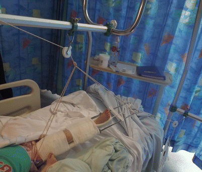
Fig. 10.2
Balanced traction as a temporary or definitive treatment for femoral fracture
In a prospective study by Infante et al. [13], immediate closed reduction and hip spica application either in the emergency room under conscious sedation or in the operating room under general anaesthesia was a safe and effective treatment for isolated femoral shaft fractures in children from birth to 10 years of age who weigh <80 pounds (Level II evidence). After a follow up of at least 2 years, the only complication in 175 children was a re-fracture in a child who fell a week after cast removal. All fractures united within 8 weeks without significant residual angular deformity or limb length discrepancy. In another prospective study by Buehler et al. [14], they tried to identify and predict children who would develop excessive and unacceptable shortening (>25 mm according to their definition) following application of early hip spica cast for isolated femoral fractures (Level II evidence). Fifty children (2–10 years old) were included and 82 % (41 children) had an acceptable outcome. They introduced what they called “the telescope test” in which gentle axial compression was applied under fluoroscopy at the time of reduction and casting. If more than 3 cm of shortening could be demonstrated, traction was used rather than immediate spica casting.
The upper age limit for the use of spica for treating femoral fracture is usually decided on pragmatic basis rather than evidence basis. Although level II evidence does exist to support the use of hip spica casting up to the age of 10 years, patients’ and parent’s convenience is a major drawback. Therefore, the current recommendation of hip spica treatment is limited to children between 6 months and 5 years [5].
Cassinelli et al. [15] retrospectively reviewed the results of immediate spica cast application in the emergency room in 145 children younger than 7 years (level IV evidence). All children younger than 2 years and 85 % of children 2–5 years had acceptable radiographic alignment. Re-reduction in the operating room was required in 11 % of the patients. They found that initial shortening was the only independent risk factor associated with loss of reduction. Mansour et al. [16] retrospectively reviewed two cohorts of children 6 months to 5 years of age who underwent either emergency department (ED) or operating room (OR) application of immediate spica cast (79 children versus 21 respectively) (Level III evidence). There was no significant difference regarding the quality of reduction or the rate of complications between the 2 groups. The hospital charges were significantly higher for the operating room spica casting, $15,983 versus $5150 for the ED casting.
The position of the hip and knee in the spica cast is also controversial [7]. Spica cast can be applied with the hip and knee at 90° flexion (the 90/90 sitting spica cast) [17] or in a more extended hip and knee (about 45° for both) [18, 19]. Illgen et al. [17] advocated the use of early sitting spica cast in a series of 114 femoral fractures in children under 6 years of age (Level III evidence). Loss of reduction was encountered in 20 % of patients and they proposed that a knee flexion angle <50 degrees as a predictive factor for loss of reduction and that >2 cm initial shortening was not a contraindication for early spica casting. However, due to the risk of compartment syndrome and Volkmann’s ischemic contracture after 90/90 spica casting, most orthopaedic surgeons have moved to single or double leg spica casts in a more extended hip and knee position [19, 20]. Flynn et al. [19] studied prospectively the results of using single leg spica cast (walking spica cast) versus traditional spica cast in the treatment of low energy femoral shaft fractures in children 1–6 years of age (Level II evidence). The malunion rate was similar in both groups of children. However, the walking spica cast group were more likely to need cast wedging to treat fracture malalignment. All children with walking spica casts were able at least to crawl and 71 % were able to walk which significantly decreased the care burden for the family. In a more recent prospective randomized controlled trial by Leu et al. [18], 52 children 2–6 years old with diaphyseal femoral fractures were randomly assigned for either single or double leg spica cast (Level I evidence). The orthopaedic outcome was similar in both groups. However, children treated with the single spica cast were more likely to fit in regular car seats and fit comfortably in chairs. Furthermore, the parents of children treated with single leg spica cast took less time off work, which decreases the socioeconomic burden on the family (Fig. 10.3).
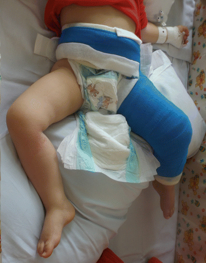
Fig. 10.3
Single leg spica for treating femoral fracture
Operative Treatments
External Fixation
External fixation use in paediatric diaphyseal femoral fractures is a minimally invasive treatment option with no soft tissue dissection and little scarring (Fig. 10.4). Blasier et al. [21] have used external fixation in the treatment of 139 fractures in 132 children with an average age of 8.9 years. The average external fixation time was 11.4 weeks and no cases of nonunion were recorded. The rate of pin tract infection, which required intravenous antibiotic treatment, was 4.5 %. Two re-fractures and one fracture through pin tract were encountered (Level IV evidence). In a randomised controlled trial by Wright et al. [22], external fixation was compared to early spica cast application in children aged 4–10 years (56 patients versus 45 respectively). At 2-years follow up, the malunion rate was significantly higher in the hip spica group [45 % versus 16 %]. Both treatment groups had similar means of RAND physical function health questionnaire, post-hospitalization questionnaire, and for patients satisfaction and for children happiness with treatment (Level I evidence).
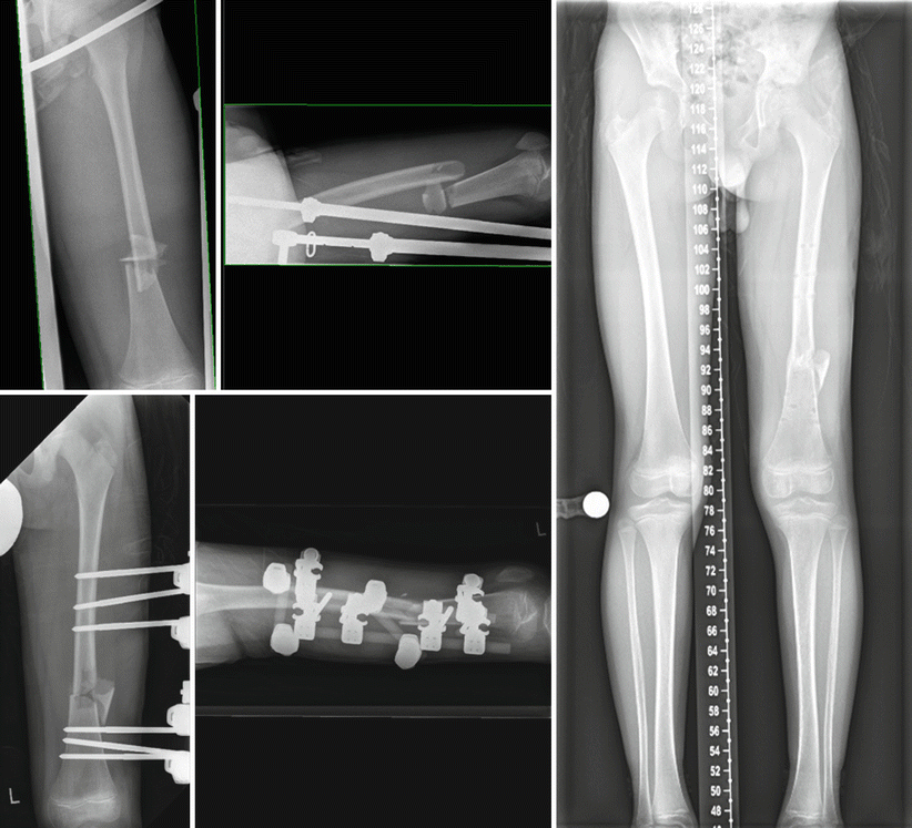
Fig. 10.4
External fixator treatment comminuted femoral shaft fracture
In another randomized controlled trial by Bar-On et al. [23], the use of flexible intramedullary nailing (FIN) was compared to external fixation for the treatment of 20 femur fractures in 19 children. The external fixator group had significantly less callus formation, full weight bearing at a mean of 10 weeks, full range of movement in 16 weeks with return to school within 13 weeks. In the FIN group, full weight bearing was possible in 7 weeks, full range of movement in 9 weeks and return to school at a mean of 5 weeks. Dynamization of the external fixator was thought to decrease stress shielding and promote callus formation with improved healing rates [24]. To study the effect of dynamization on callus formation and healing time, Domb et al. [24] randomized 52 patients to either a dynamic or a static external fixator for treatment of femoral shaft fractures. They found that axial dynamization of the external fixator had no significant impact on healing time and rate of complications (Level II evidence).
Internal Fixation
Implant Choice and Patient Characteristics – Age and Weight
Several implants have been used to stabilise femoral shaft fractures including ESIN, rigid nailing, plates and screws. ESIN is commonly considered for fixing femoral shaft fractures in children 5 years to skeletal maturity [5]. ESIN has also been used in children younger than 2 years [25–27] and patients up to the age of 18 years [25, 28]. Several studies have analyzed the effect of age and weight of patients on complication rates and final outcome using of titanium elastic nails (TEN) [28–31].
Sink et al. [29] compared two cohorts of patients with different surgical treatment algorithms over two different periods of time (Level III evidence). The first period of 2 years represented their earlier experience during which TEN was used regardless of the age of the child or fracture stability (46 children). The higher complication rate (76 %) encountered with this cohort [32] led to a change of practice so that TEN would be used only in children younger than 11 years with a stable fracture. A second cohort of 95 children over the next 3-year period showed a significant decrease in complication rate to 12 %. They treated children 11 years and older who had unstable femoral fractures with submuscular bridge plating or rigid femoral nailing if the femoral canal size was adequate.
In a retrospective study of 234 fractures, (age range 3–18 years) (Level IV evidence), Moroz et al. [28] identified predictors of complications and poor outcome when using TEN. Age above 11 years was associated with poorer outcome (odds ratio = 3.86; P = 0.003). Furthermore, children who weighed >49 kg were 5 times more likely to have a poor outcome. There was a significant association between weight and poor outcome (P = 0.003).
Sagan et al. [33] found that weight was a significant predictor for anterior bow deformity greater than 15° with the use of TEN (Level III evidence). The mean weight of patients with this deformity was 46.5 kg ± 13.5 (n = 11), whereas the mean for those with no deformity was 36.8 kg ± 18.5 (n = 58). On the other hand, they found a trend of increasing frequency of malunion with increasing age, however this was not statistically significant.
Luhmann et al. [30] reported on the complications of TEN in a retrospective series of 43 femoral fractures in 39 children aged 3.8–9.3 years (Level IV evidence). They calculated weight/nail ratio to assess the effect of the patients’ weight on the ability of TEN to control angulation. They found no association between weight/nail ratio and coronal (varus/valgus) angulation, while sagittal (apex anterior/posterior) angulation increased with increasing weight/nail ratio.
Implant Choice and Fracture Characteristics
Fracture Stability
Loss of reduction is a well-known complication following the use of ESIN in unstable femoral fractures causing unacceptable angulation or limb length discrepancy. Length unstable fracture patterns are either spiral or long oblique fractures (the length of the fracture is twice the diameter of the femur at the level of the fracture) or comminuted fractures (Winquist grades III and IV) [29, 32, 34, 35].
Sink et al. [32], in a retrospective comparative analysis (Level III evidence) of complication rates after using TEN in stable and unstable paediatric femoral fractures, found 8 patients (21 %) required further “unplanned” surgery before healing. Six of these eight had unstable fractures and underwent further surgery to shorten or remove extremely prominent or exposed nails or loss of reduction. Two had stable femoral fractures and underwent unplanned surgery to release a thigh compartment syndrome in one and to correct early coronal plane deformity by adding an external fixator in the second. Moreover, fracture shortening and angulation were significantly more prevalent in unstable fracture (10 out of 15 vs. 3 out of 24).
In a study by Lohiya et al. [36], stainless steel Ender’s rods were compared to TENs in 73 children aged 4–15 years (Level III evidence). They reported a significant relationship between angular malalignment and the severity of fracture comminution. Eleven out of 14 patients (71.5 %) with Winquist grades III or IV fractures had malalignment as compared to 21 out of 59 with grades I or II.
In another study by Narayanan et al. [27], 79 femoral fractures in 78 children were stabilized using ESIN (age range 2 to 15 years). Five patients had loss of reduction that required operative correction (n = 2) or resulted in malunion (n = 3). The only two variables that were significantly associated with malunion and loss of reduction were the use of mismatched nail diameters and comminution >25 % of the shaft diameter.
Fracture Site
In a multi-centre North American study of 58 consecutive paediatric femoral shaft fractures (57 children) studying the early results of using TENs, 3 cases lost reduction in the early postoperative period (2 proximal third and one distal third fractures) while 5 of the 9 proximal third fractures healed with more than 5° of angulation. This indicates proximal and distal sites fractures may predict unfavourable outcomes [37].
Pombo et al. [38] reported the results of using TENs in the treatment of subtrochanteric fractures in 13 children 4–17 years (Level IV evidence). They defined subtrochanteric fractures as those within 10 % of the total femur length below the lesser trochanter. All patients had less than 5° of angulation. Eleven patients had excellent results. Two had satisfactory outcome due to limb length discrepancy of 1–2 cm with the fractured limb being longer than the unaffected side. They explained the limb length discrepancy as due to either intra-operative over-distraction across the fracture site or postoperative overgrowth of the femur. They stressed in their technique the importance of advancing the lateral nail into or just distal to the greater trochanter apophysis and advancing the medial nail into the femoral neck until it lies just short of the proximal femoral physis.
Elastic Nailing
Elastic Stable Intramedullary Nailing (ESIN or Nancy nails) were developed and first used at Nancy, France by Ligier et al. [39]. The principle of elastic stable nailing differs from the use of other flexible rods as Ender’s rods, which are stacked to fill the medullary canal. ESIN technique requires balancing forces between two opposing flexible nails of the same diameter. Therefore, it is important to select nails 40 % of the narrowest diaphyseal diameter; contour the nails with a similar gentle curvature, and use medial and lateral starting points that are at the same level in the metaphysis (Fig. 10.5) [27, 37]. Reported advantages are the minimally invasive approach, significantly reduced hospital stay, earlier mobilisation and functional recovery, avoid disruption of family life with its psychological impact on the child and earlier return to school and activities [25, 37, 39–41]. Different names were given for this type of fixation including ESIN (Elastic Stable Intramedullary Nailing), FIN (Flexible Intramedullary Nailing), FTN (Flexible Titanium Nails), TEN (Titanium Elastic Nails), SSEN (Stainless Steel Elastic Nailing) & Nancy nails [25, 33, 37, 39, 42].
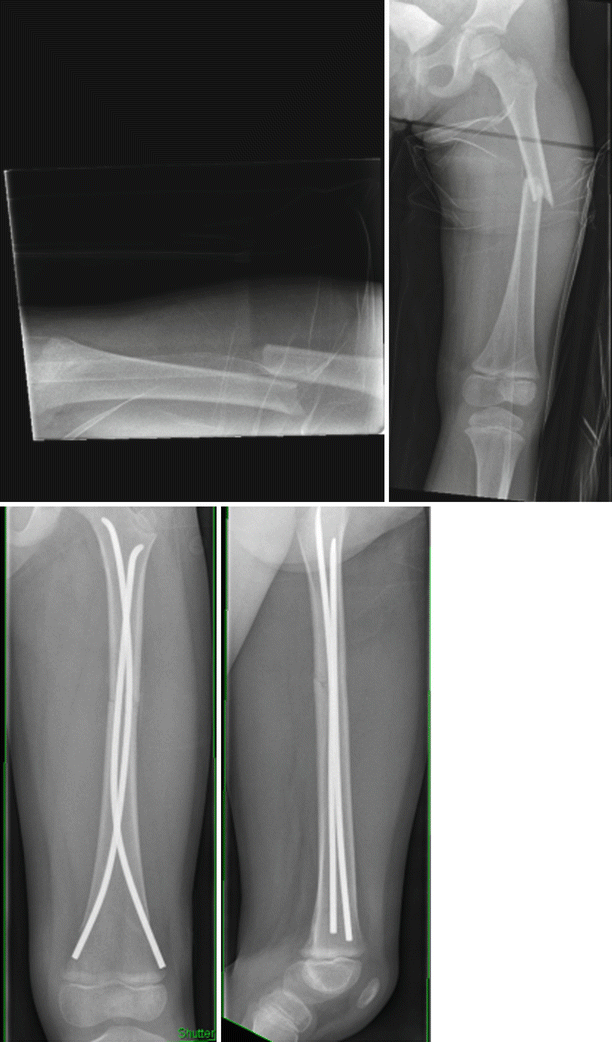
Fig. 10.5
Flexible nailing in femoral fracture
Locked Versus Non Locked ESIN and the Use of End Caps
The advantage of using Ender’s rods over ESIN is that the eyelets of the Ender’s rods add locking capability to the end of the rods. This would in turn decrease symptomatic nail tip irritation and might be beneficial in case of length unstable diaphyseal fractures [34, 43].
Ellis et al. [34] retrospectively compared two groups of paediatric femoral diaphyseal fractures that had either locked Ender’s nail fixation (n = 37) or unlocked Ender’s rods (n = 70) (Level III evidence). They locked the end of the nail to the distal femoral metaphyses using 2.7 mm cortical screws 24 mm in length. Fracture shortening was significantly (p < 0.001) more in the unlocked group (12 mm versus 4.7 mm respectively) and this had caused implant migration, palpable lower ends, knee stiffness and deep infection. Eight patients in the unlocked group had a malalignment in either the sagittal or coronal plane of more than 10° but this was not statistically significant. However, the malalignment was considered unacceptable in only one patient in the locked group and revision was performed.
Self anchoring end caps to lock ESINs have been developed to provide a similar locking principle to the eyelets of the Ender’s rods and tried in 8 femoral fractures (average age 10 years, range 7–15) [44]. The use of end caps eased the removal of nails as they prevented formation of excess bone around nail tips. Furthermore, they protected the skin very well and no nail tip irritation was recorded.
Titanium Rods Versus Stainless Elastic Nails or Rods
ESINs can be made of either titanium or stainless steel alloys. The French originators of this technique, Ligier et al. [39] reported the use of both materials. With the widespread adoption of this technique in North America, there has been a transition towards the exclusive use of titanium nails and several advantages such as closer elasticity to bone, better biocompatibility, better osteointegration and magnetic resonance imaging compatibility have been cited [31, 37]. The modulus of elasticity of 316L stainless steel is 187 GPa, making it 80 % stiffer than the titanium alloy (105 GPa). The lower modulus of elasticity of titanium would ease its intramedullary insertion and limit its permanent deformation. Increased elasticity and better compatibility would also decrease stress shielding and in turn would theoretically increase callus formation and healing rates [37]. The paediatric AO group has shown that a stainless steel nail has the strength of a titanium nail one size larger [45].
Wall et al. [31] compared retrospectively two groups of children (age 4–15, mean 9.4 years) (Level III evidence) who had either titanium ESIN (n = 56) or stainless steel ESIN (n = 48) fixation of femoral shaft fractures. The malunion rate was significantly higher in the titanium group (23.2 %) than in the stainless steel group (6.3 %). The increased flexibility of titanium compared to the stiffer stainless steel has been suggested as the cause of this increased deformation during fracture healing and the higher malunion rate. Furthermore, from an economic point of view, implant price by supplier in 2008 according to Wall et al. [31] was $259–$328 for titanium elastic nails depending on size of the nail and $78 for stainless steel flexible rods. In another retrospective study by Lohiya et al. [36], TENs used in 43 fractures were compared to stainless steel Ender’s nails in 30 fractures (mean age 8.3, range 4–15 years) and the follow up was at least 5 years (Level III evidence). At the final follow up, significant malalignment in the sagittal plane (>15°) was found in 3 patients and minor malalignment in 29 cases and no significant relation could be found between malalignment rates and the material of the nail used. A recent randomized controlled trial was published by Goyal et al. [42] comparing TENs to stainless steel ESIN. Thirty-five children were included, 18 children in the TENs group and 17 children had stainless steel nails (age range 6–12 years). Fracture comminution grades III and IV according to the Winquist Hansen classification [35] were excluded from this comparison. There was no significant difference between both groups regarding degree of fracture site angulation at 6 months follow up (Level I evidence).
Mini-Open Versus Closed Reduction Technique
ESIN are inserted under fluoroscopic assistance with closed reduction of the fracture fragments or with the mini-open reduction technique (small incision blind hand technique). In a retrospective comparative study by Altay et al. [46] (Level III evidence), two groups of children who had TEN insertion with mini-open reduction (n = 42, mean age 8.3 ± 2.7 years) or closed reduction (n = 45 children, mean age 8.8 ± 2.6 years) were compared. Both surgical and fluoroscopy times were significantly longer in the closed reduction group, while there was no significant difference between the two groups in terms of clinical and radiological results. Similar results were reported by Sun et al. [47] who retrospectively compared two groups of children (age range 4–15 years) who had TEN fixation of femoral shaft fractures (Level III evidence). The first group (n = 34, mean age 8.2 ± 2.5 years) had small incision blind hand reduction technique, while the second group (n = 34, mean age 8.6 ± 2.6 years) had closed reduction. Operative time and fluoroscopy time were significantly longer in the closed reduction group. No significant difference between the groups could be found in the complication rates or final outcome using TEN scoring system.
Complications and Nail Tip Irritation
Complication rates with the use of ESIN in the treatment of paediatric femoral shaft fractures might be seen in as many as 62 % of the patients [32]. They can be classified as minor, which resolve without additional surgery or don’t result in long-term morbidity or major when unplanned surgical intervention is required or long-term morbidity ensues [28, 32, 34].
The majority of encountered complications are minor problems and are mostly related to nail tip irritation and pain around the nail insertion sites [28]. The resultant soft tissue irritation around the knee causes delayed recovery of the knee range of motion and might increase the risk of deep infections [27, 28, 32, 34, 37]. Ligier et al. [39] assumed that pin site irritation is mainly caused by early knee motion following surgery.
Narayanan et al. [27] studied specifically the factors that might increase the incidence of pain at the nail insertion sites (Level IV evidence). They found significant correlation with bent nail ends and nails that were prominent more than 10 mm outside the distal femoral cortex. Children who reported pain at nail insertion sites regained functional knee range of motion at a mean of 3 weeks later than patients who didn’t report any symptoms. Their recommendation was not to bend the ends of the nails which should be trimmed short and advanced further with a hollow tamp, so that the unbent nail end lies in close apposition to the supracondylar flare of the distal femoral metaphysis. Ellis et al. [34] compared the results of using flexible stainless steel Ender’s rods in either locked or unlocked configurations to the distal femoral metaphysis (Level III evidence). They found significantly more minor complications in the unlocked group, mostly due to prominent and painful implants. Two of their patients had deep infection which required drainage and both of them were associated with nail migration and prominence >13 mm.
The most commonly reported major complications were loss of reduction, unacceptable angulation exceeding the guidelines [7] and backing out of nails requiring another surgical intervention for trimming. Major complications were more common with the use of ESIN in length unstable femoral fractures [28, 29, 32, 34].
To Remove or Not to Remove ESIN Nails After Fracture Healing
Many authors including the originators of this technique recommended routine removal of ESINs. This can be performed between 3rd and 6th month postoperatively, when solid healing and circumferential callus are evident in plain radiographs [37, 39, 47]. However, whether it is necessary to routinely remove flexible nails implanted in children is still unclear. In a retrospective case series by Narayanan et al. [27], 25 children out of 78 with femoral shaft fracture treated with TEN retained their nails and they reported no symptoms attributable to the implants at the final follow up (mean 3.6 years, range 2–6 years) (Level IV evidence). In another case series by Levy et al. [26], acute complications of femoral flexible nail removal in 163 children were analyzed. The nails were removed due to pain at insertion site in only 54 % (88 children) of the cases. The remainders were removed at the recommendation of the treating surgeon or the request of the family, but not due to symptomatic implants. They couldn’t remove one Ender’s rod in 1 patient in this series as the rod was overgrown with bone and it lied completely within the intramedullary canal. In another 3 children, the insertion sites of 3 additional rods were also overgrown with bone, which resulted in a prolonged extraction time (mean 71 min).
Plating
Submuscular Versus Open Plating
The minimally invasive, submuscular plate osteosynthesis is a favored technique for plating in skeletally immature patients by many surgeons (Fig. 10.6). Abdelgawad et al. [48] reviewed retrospectively 60 femoral shaft fractures in 58 patients with an mean age of 9 years, which were treated with submuscular bridge plating (Level IV evidence). Forty (67 %) were length unstable or complex fracture pattern. Two major complications that required further unplanned surgery were encountered. The first was a deep infection following open femoral fracture, which was managed primarily with debridement and external fixation. The second was a broken 3.5 mm titanium plate, which was replaced by a stronger 4.5 mm stainless steel plate. Minor complications included symptomatic hardware (n = 3), superficial wound infection (n = 2), and one temporary peroneal nerve palsy. In one non-compliant patient there was mild loss of fixation 2 weeks postoperatively which was managed with a long leg cast for 5 weeks. Ten patients had a mean of 9.9 mm LLD (10 mm short to 20 mm long). Abbott et al. [49] compared retrospectively two groups of children who underwent either open (n = 58) or submuscular (n = 22) plating for femoral shaft fractures (Level III evidence). The mean age of both groups was 7.9 ± 3.5 and 8.5 ± 2.4 years respectively. There were no statistically significant differences between both groups regarding operative times, time from operating room to discharge, time to union (full weight bearing), the incidence of LLD of >2 cm or the presence of deep infection (1 case in the open group). On the other hand, estimated blood loss was significantly higher for open plating; however the increased blood loss was not clinically relevant as there was no difference in the need for blood transfusions between groups. The incidence of rotational asymmetry was significantly different with no cases of rotational asymmetry in the open group while 4 out of the 22 submuscular plating patients had clinically detectable rotational differences. Five patients in the open group had unplanned return to the operating room (of no statistical significance), one patient for serial debridement of deep infection, one for periprosthetic femoral fracture 1 year postoperatively and 3 patients for revision of metal failure.
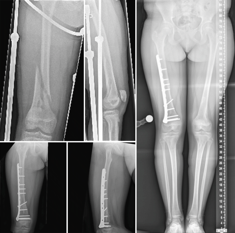
Fig. 10.6
Submuscular plating in a child with femoral fracture
Locked Plating for Paediatric Femoral Shaft Fractures
Hedequist et al. [50] retrospectively reviewed 32 patients with locked plate fixation for femoral fractures (Level IV evidence). Their mean age was 11 years (range 6–15 years). The indications for locking plate fixation were either the presence of comminution (n = 13), pathologic fracture (n = 9), poor bone quality (n = 3) and fracture location (n = 7). Twenty-four patients had submuscular insertion of the plate and 8 patients had open plating. All fractures united uneventfully with anatomic alignment except in one patient who had a severely comminuted distal femoral fracture with development of 12° valgus angulation and 1.5 cm shortening. One patient who had locked plate fixation for osteopenia suffered from fracture at the distal end of the plate 11 months after the index surgery. Plates were removed in 7 patients on discretion of the treating surgeon without complications.
Related posts:
 Evidence-Based Treatment of Flexible Flat Foot in Children
Evidence-Based Treatment for Congenital Dislocation of the Knee
Evidence-Based Treatment for Congenital Femoral Deficiency
Evidence-Based Management of Limb Length Discrepancy
Physeal Injury, Epiphysiodesis and Guided Growth
Evidence-Based Treatment for Feet Deformities in Children with Neuromuscular Conditions
Evidence-Based Treatment of Flexible Flat Foot in Children
Evidence-Based Treatment for Congenital Dislocation of the Knee
Evidence-Based Treatment for Congenital Femoral Deficiency
Evidence-Based Management of Limb Length Discrepancy
Physeal Injury, Epiphysiodesis and Guided Growth
Evidence-Based Treatment for Feet Deformities in Children with Neuromuscular Conditions
Stay updated, free articles. Join our Telegram channel

Full access? Get Clinical Tree


