Evaluation and Management of the Diabetic Foot Wound


Evaluation and Management of the Diabetic Foot Wound


John S. Steinberg

Paul J. Kim


New technologies and evidence-based understanding have led to significant advances in the assessment and management of nonhealing diabetic lower extremity wounds. The implementation of these evidence-based practices has led to more efficient utilization of new technologies and a better understanding of the diabetic foot wound environment. However, the complex care and management of the diabetic wound continues to have a major economic impact on the health care system and, more importantly, on the physical and emotional well-being of those patients suffering from nonhealing chronic wounds. In this chapter, we will review the key principles in the assessment and treatment of problem wounds in the diabetic foot.

Diabetes and diabetes-related complications have become an epidemic level public health concern in the United States and in many other nations around the globe. Worldwide prevalence of diabetes is predicted to reach 366 million people (almost doubling from year 2000 to 2030) if current trends continue (1). Approximately 15% of persons with diabetes will develop a foot ulcer during their lifetime (2). A chronic foot ulcer invokes a tremendous cost economically, mentally, and physically on the diabetic patient and those who surround them (3,4). Some estimates place diabetic patients at 10 to 30 times higher risk of amputation than nondiabetic patients (5,6). Further, there is a significantly increased probability that a lower extremity amputation will be preceded by a foot ulceration (2,7,8). Therefore, early assessment and intervention is vitally important when a member of this patient population is confronted with a nonhealing wound.

Acute wounds of the foot can be readily treated with standard wound care therapies, while chronic wounds may require more advanced treatment modalities. No absolute threshold exists as to when an acute wound becomes a chronic wound; however, the typical wound in a healthy patient is expected to heal either through primary intention or secondary intention within 10 to 14 days with continued remodeling that lasts several months. The diabetic chronic wound becomes stagnant and ceases to shrink and epithelialize due to excesses in wound breakdown or due to other factors that inhibit the wound healing cascade. A wound may be considered chronic in nature when the measurements cease to decrease 10% to 15% per week or 50% over a 1-month period (9). Further, a wound that is larger than 2 cm2, more than 2 months in duration, and a wound that is full thickness or deeper has a 79% decreased likelihood of healing within 20 weeks (10). There are, of course, many reasons that contribute to an acute wound degenerating into a chronic wound. Some of the more common reasons found in the lower extremity chronic wound include ischemia, neuropathy, infection, and pressure.

The proper assessment and treatment of chronic diabetic wounds is essential. The diabetic foot is a complex structure that is obviously disadvantaged when compared with a nondiabetic foot. Keeping this in mind, clinicians must modify wound healing strategies and be ready to engage aggressive and advanced wound care practices when confronted with a nonhealing diabetic foot ulceration. In the following paragraphs, we will discuss the fundamental principles in the assessment and treatment of the chronic diabetic wound in the lower extremity.


THE MICRO- AND MACRODIABETIC FOOT ENVIRONMENT

We begin our discussion with the fundamental changes that occur at the cellular level in a wound of the diabetic foot. It is beyond the limits of this chapter and the capacity of the authors to discuss in detail the complex pathologic mechanisms involved in the diabetic foot. We will limit our discussion to areas of clinical interest where new technologies have been developed to address issues in the micro- and macroenvironment of the diabetic foot.

A basic understanding of the histology and biology of skin and surrounding structures is necessary in evaluating and treating a chronic diabetic wound (Fig. 69.1). There are four important components of skin that should be considered: epidermis, dermis, hypodermis (subcutaneous adipose tissue), and underlying deep soft tissues. The epidermis is the most superficial layer of skin. The majority of the epidermis has no direct vascular supply and the superficial layers are sloughed continuously. Directly below the epidermis, the dermis is often regarded as the most important skin component to wound healing due to the fact that granulation tissue is usually seen at this level. The hypodermis contains adipose tissues where neurovascular bundles are located. Finally, the underlying tissues include fascia, tendon, and bone, all of which have potential for granulation tissue formation. Inflammatory cells and cells that make up the skin contain important growth factors. Specifically, one should note that fibroblasts and keratinocytes are of key importance in the process of tissue regeneration and repair. Keep in mind
the different layers of tissue and their architecture as described above when treating a chronic diabetic wound so that an appropriate plan can be identified and implemented effectively.


Figure 69.1 A histologic cross section of the human plantar skin.


FUNDAMENTAL PRINCIPLES OF WOUND HEALING

A brief review of the normal wound healing cascade is needed to better understand the chronic, nonhealing wound. The classic model of wound healing has been described to involve three phases (11). The first phase is inflammation that begins after the initial insult when platelets adhere to the site of injury and to each other to form a hemostatic plug. Individual platelets contain intracellular structures known as alpha-granules that contain growth factors (cytokines), clotting factors, and other proteins involved in wound healing. After activation by thrombin, platelets aggregate to the area of injury and release the contents of their alpha-granules to initiate clotting. The platelet aggregate (hemostatic plug) serves as a barrier to the external environment. Platelet-derived growth factor (PDGF) is one of the key components released by the alpha-granules. PDGF is also found in macrophages as well as endothelial cells (12). PDGF is involved in the formation of connective tissue, promotion of revascularization, production of granulation tissue, epithelialization, wound contraction, and wound remodeling (13,14 and 15). Further, platelets release transforming growth factorbeta (TGF-β) and platelet-derived angiogenesis factor (PDAF), which play key roles in wound matrix production by promoting collagen production and new capillary formation. PDGF also acts to recruit and activate proinflammatory cells such as fibroblasts, macrophages, monocytes, and neutrophils. These cells, in turn, also secrete growth factors including TGF-β, fibroblast growth factor (FGF), endothelial growth factor (EGF), and vascular endothelial growth factor (VEGF). These cellular interactions and chemotactic communications are critical elements in the wound healing cascade.

The second phase of wound healing is epithelialization. Epidermal cells begin to proliferate and migrate to formulate linkages with each other and begin depositing basement membrane components and also reorganize and degrade the extracellular matrix. Growth factors are intricately involved in this phase as well. Neovascularization occurs at the wound bed, causing the formation of granulation tissue, which infiltrates the temporary matrix. Fibroblasts play a critical role in orchestrating the reorganization of the extracellular matrix into a collagenous matrix through the use of proteases and other enzymes. Growth factors, such as VEGF, contribute to the stimulation of angiogenesis to support wound healing.

The final phase of wound healing involves tissue remodeling. This involves wound contraction facilitated by fibroblasts that have converted to myofibroblasts stimulated by growth factors. Collagen is then continually remodeled through enzymatic degradation by matrix metalloproteinases (MMPs) until final collagen deposition and wound reepithelialization has occurred. The diabetic foot wound displays a vastly altered wound healing cascade from that which we have just described. In the following sections, we will discuss the aberrations in the wound healing cascade that are of particular note to the compromised diabetic lower extremity wound.

It is important to note that MMPs act at every phase of “normal” wound healing. In the chronic, nonhealing wound, MMPs play a significant inhibitory role due to a pathologic imbalance in the amount and proportion of MMPs in the wound base. MMPs are part of a larger class of enzymes known as proteases and are released by keratinocytes, endothelial cells, and inflammatory cells. The expression of MMPs is potentiated by inflammatory factors such as interleukin-1β (IL-1β) and tumor necrosis factor-α (TNF-α). These proteases play an important role in the wound healing process by degrading extracellular matrix proteins, stimulating cell proliferations and migration, and promoting angiogenesis. MMPs are subdivided into collagenases (MMP-1,-8), gelatinases (MMP-2,-9), and stromelysins (MMP-3,-10,-11). Elevated levels of MMPs are thought to have detrimental effects on wound healing. MMPs have been measured at higher levels in chronic wounds as compared with acute wounds, and further, increased concentrations of MMPs have been reported in diabetic wounds compared with nondiabetic wounds (16,17). One of the key regulators of MMPs is the tissue inhibitor of metalloproteinase (TIMPs), which is a glycoprotein produced by fibroblasts. Chronic wounds display decreased levels of TIMPs, suggesting less regulation of MMP levels (18).

Growth factors play an important role in wound healing. Growth factors serve a multitude of functions including recruitment and promotion of inflammatory cells, stimulation of cellular proliferation and regulation, angiogenesis, and the overall stimulation of the wound healing process. Some of the more important growth factors, as previously mentioned, are TGF-β, FGF, EGF, and PDGF (19). The levels of these growth factors have been reported to be decreased in chronic wounds as compared with acute wounds (20,21 and 22). Further, TGF-β appears to be an inhibitor of MMPs and promotes the synthesis of TIMPs (23,24). FGF and EGF play many roles in wound healing, but are particularly important for keratinocyte-induced epithelialization (25,26). PDGF plays a major role in the recruitment and regulation of inflammatory and immunologic cells such as fibroblasts and macrophages, as previously described.


VASCULAR DYSFUNCTION

Vascular compromise also plays a major role in the chronicity of wounds. It is important to remember that a diabetic wound is not always ischemic; however, due to the vascular disease associated with diabetes itself, wounds may progress in this
direction. Vasculopathy associated with diabetes has two major components: macrovascular and microvascular processes. Macrovascular pathologic processes include cerebrovascular, cardiovascular, and, more pertinent to our discussion, peripheral vascular/arterial disease (PVD, PAD). Microvascular dysfunction revolves around issues with the microcirculation at the level of arterioles and capillaries and is more of a dysfunction in physiology rather than anatomy. Again, a detailed discussion of this complicated topic is out of the scope of this chapter; however, fundamental issues associated with the vascular process will be addressed.

PAD is one of the devastating sequelae of diabetes (27). Diabetic patients with PAD have a higher risk of lower extremity amputations (28). The hallmark of the atherosclerotic process is fibrofatty plaque deposits initiated by an inflammatory process within the vessel walls. The plaques may progress to a point at which complete occlusion may occur. Wound care specialists are often the first to identify systemic ischemic disease as manifested by the symptoms of a nonhealing wound. The ischemia discovered will often persist much further systemically and in fact is a common marker for carotid and cardiac vessel disease. “Critical limb ischemia” is a term used to describe the point at which, without intervention, limb amputation is a strong probability (29). Revascularization can be conducted through open or, more commonly now, through endovascular techniques. Open techniques may include the harvesting of the greater saphenous vein to be used as the vessel that bypasses the area of blockage. Endovascular techniques utilize a variety of ablative technologies to remove focal plaques through minimally invasive techniques (30). The analogy of a plumber laying down new pipes around a clog (open) versus the use of a “rotorooter” to remove the clog within the clogged pipe (endovascular) is appropriate. Prior to lower extremity revascularization, an arteriogram or a magnetic resonance angiography (MRI) is performed to identify the areas of occlusion.

The microvascular environment (namely arterioles and capillaries) is also altered in the patient with diabetes (31,32 and 33). Generally, there appears to be two components in the diabetic microvascular system that are altered: first, there is an increase in vascular permeability, and second, there is an impairment in the regulation of vascular tone and local blood flow (34). This, in part, has to do with a thickening of the capillary basement membrane and a decrease in capillary size (35,36). There are numerous factors involved in the pathologic microvascular environment in the diabetic foot, so we will limit our discussion to those associated with currently available clinical interventions.

Endothelial function, or more accurately, dysfunction, has significant effects in the microvasculature. Specifically, the role of nitric oxide (NO) in regulating vascular tone in the diabetic patient has been a major focus of research. NO is an endogenous gas produced by cells with many diverse physiologic effects. The substrate arginine is converted by the enzyme nitric oxide synthase (NOS) to citrulline with the liberation of NO. After release, NO has a half-life of seconds with subsequent binding to receptors on or within the cell causing a second messenger cascade. The result of this cascade depends on the type of NOS. There are many subtypes of NOS, including nNOS, which is found in neurons; iNOS, which is an inducible form found throughout the body; and eNOS, which is found in vascular endothelial cells. Of particular interest are iNOS and eNOS, which have implications in the lower extremity and wound healing cascade.

Endogenous NO acts upon endothelial cells causing dilatation of vessels (both arterial and venous). Contrarily, inhibition of NO synthesis causes vasoconstriction and hence hypertension (37). Evidence also suggests that there is decreased NO activity with an increase in activity levels by vasoconstrictors in the diabetic rat model (38). Further, the vessels of insulin-dependent patients with diabetes demonstrate less responsiveness to NO and potentially a decrease in availability of NO (39,40 and 41). NO has also been shown to increase blood flow to the microcirculation adjacent to wound sites (41).

There have been a variety of clinical strategies employed in an attempt to enhance or augment NO-related endothelial function. One strategy involves the use of folic acid, vitamin B6, and vitamin B12. Supplementation with folic acid has been shown to improve NO-mediated endothelial function in diabetic patients (42,43). Folate has also been used in combination with B6 and B12 in an attempt to decrease homocysteine levels (44,45 and 46). Elevated homocysteine levels have been implicated in endothelial dysfunction (47,48,49 and 50). Another strategy involves the use of NO liberators including topical applications of nitroglycerin and oral formulations of L-Arginine. Both have been reported to promote wound healing (51,52,53,54,55,56,57,58 and 59). However, others have suggested that NO augmentation may not be beneficial or effective in a chronic wound environment (60,61,62,63 and 64).

NO also has more direct implications on the wound environment itself. NO has been measured at higher levels in proximity to wound sites (60). NO appears to be maximally expressed early in the wound healing process, with sustained decreased levels for the first couple weeks after injury (65). Cytokines stimulate macrophages and fibroblasts to produce NO (66 and 67). NO appears to have cytotoxic properties, which suggests some level of antimicrobial activity (68). Further, inhibition of NO synthesis retards collagen synthesis and deposition, which we know are key components in providing principal strength characteristics of wounds (69).

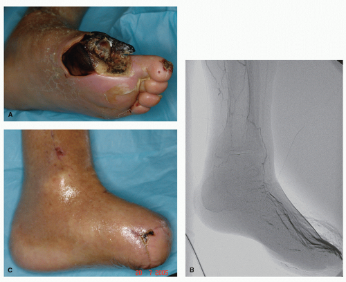
Proper assessment and treatment of vascular dysfunction must be addressed prior to or in conjunction with wound care modalities (Fig. 69.2). If blood flow is compromised to the affected extremity, the wound will not heal or will be severely retarded in healing. The wound is entirely dependent on adequate nutrient and oxygen delivery. Hence, vascular evaluation and possibly consultation is necessary in the treatment of a chronic diabetic wound.


PERIPHERAL NEUROPATHY

Peripheral neuropathy has been cited as a pivotal process that contributes to the chronicity of diabetic wounds and can affect up to 66% of patients with diabetes (70,71,72 and 73). Peripheral neuropathy can be generally defined as a progressive loss of peripheral nerve fibers. In the diabetic population, this typically presents in a symmetrical fashion and involves sensory, motor, and autonomic neuropathy. The etiology of peripheral neuropathy is most attributable to hyperglycemia (74). Hyperglycemia contributes to metabolic disturbances that negatively impact nerve function. Other contributing factors include focal areas of nerve entrapment, which can lead to degeneration of the nerve fiber. Nerve entrapment may benefit from nerve surgical decompression (75).

Sensory neuropathy can often present as a painful process, with patients reporting symptoms of “pins and needles” or
“burning” sensation in its early stages. Peripheral neuropathy may progress to a point in which complete loss of sensation occurs. The typical insensate process begins distally at the toes and advances proximally and is said to be in a “stocking and glove” distribution. The critical aspect of peripheral neuropathy is that patients are unaware of focal areas of trauma, which may lead to the development of a wound (72). This wound continues to be traumatized and inadequately treated and hence becomes chronic in nature.


Figure 69.2 A: Patient with DM and PAD presents with failing wound site following hallux amputation. B: Angiogram shows minimal arterial flow below the knee and only a faintly visible anterior single vessel below the ankle. C: Following revascularization of the extremity, a limb-sparing transmetatarsal amputation is performed with closure.

Motor and autonomic neuropathies also contribute to the chronicity of a wound. Motor neuropathy may lead to an imbalance of muscles or muscle atrophy in the lower extremity. This may lead to areas of increased pressure that may subsequently lead to tissue breakdown. Autonomic neuropathy involves the denervation of the sympathetic nervous system, leading to arterial blood flow being shunted away from the nutrient capillaries. This arterial to venous shunting diverts the nutrients and oxygen away from the underlying soft tissue structures of the plantar aspect of the foot. Hence, tissues become less tolerant to stresses leading to tissue breakdown. This process may also explain loss of sweat production and fat pad atrophy.

NO also plays a role in autonomic neuropathy. Human studies have demonstrated decreased NO production in diabetic patients with peripheral neuropathy (76). Associated with this fact, homocysteine levels are elevated in persons with type 2 diabetes and peripheral neuropathy (77). Peripheral neuropathy is associated with a decrease in perfusion to peripheral nerves, causing hypoxia to the nerves (78,79). Several different clinical strategies have been developed to offset the endothelial dysfunction associated with peripheral neuropathy. Technologies utilizing monochromatic nearinfrared photoenergy therapy have been postulated to induce the release of NO through photoenergy, thereby increasing blood flow to the peripheral nerves (80,81,82 and 83). Topical application of NO in the form of a spray has also been used for the treatment of painful peripheral neuropathy with good success (84). Further, oral formulations utilizing the combination of supplements (folate, B6, B12), as described above, have been used to manage peripheral neuropathy (42,43,44,45 and 46).
Other oral medications targeting symptomatic management of peripheral neuropathy that is potentially unrelated to NO have been utilized including tricyclics, selective serotonin reuptake inhibitors, anticonvulsants, antiarrhythmics, narcotics, and nonsteroidal anti-inflammatories (85).

Peripheral neuropathy is a significant contributing factor to the development and chronicity of a diabetic wound (86). Proper management of peripheral neuropathy with early intervention utilizing some of the treatment options discussed above may prevent the onset of a diabetic wound and also retard the progression of neuropathy that may lead to an insensate foot (Fig. 69.3).


EVALUATION OF THE DIABETIC WOUND

Proper and timely assessment is critical in healing a diabetic wound. The first critical step in diabetic wound evaluation is to assess the vascularity to the wound site. The next step is
to establish the level of infection or contamination. Further, evaluation of any gross mechanical foot deformities should be conducted. Finally, a thorough evaluation of the quality of the wound including wound measurements is vitally important.


Figure 69.3 A: Neuropathic plantar hallux ulceration in patient with diabetes and hallux limitus. B: Offloading, débridement, and moist healing yield a greatly improved wound base but mechanical etiology prevents complete closure. C: Long-term closure and healing with ambulation in custom-molded insoles following Keller arthroplasty with application of living BAT graft.


VASCULAR EXAM

A wound requires oxygenation and nutrient delivery for healing to take place. The vascular patency can be assessed at the wound site through both noninvasive and invasive methods. Initial evaluation of skin color (pallor), hair distribution (hair absent on the dorsal aspect of the toes), atrophy of skin and nails, and decreased temperature to the affected limb can raise the clinical suspicion for ischemia. Palpation of pulses is also a sensitive measure of blood flow about a wound site. The dorsalis pedis (DP) artery, posterior tibial (PT) artery, and the perforating peroneal artery are the major large vessels that feed the foot. The absence of a palpable DP or PT artery warrants immediate further investigation. If one or both are absent, a handheld Doppler should be used to discern between triphasic, biphasic, or monophasic signals. If anything less than a triphasic signal is found, then further evaluation utilizing an ankle brachial index (ABI) is warranted. An ABI is a measure of systolic pressure differences between the ankle artery pressure divided by the brachial artery pressure. Although there is no absolute threshold that predicts successful wound healing, some general guidelines are helpful. An ABI of 0.90 or less is considered abnormal, 0.71 to 0.90 indicates mild PAD, 0.41 to 0.70 indicates moderate PAD, and 0.40 or less indicates severe PAD (87). In the chronic wound environment, patients are more likely to require an amputation with an ABI of less than 0.50 (88). It is important to note that noncompressible, calcified vessels will falsely elevate the ABI and provide unreliable readings (87). Transcutaneous oxygen tension/pressure (TcPO2) is another noninvasive option for evaluating the level of ischemia. This is a sensitive measure of assessing blood perfusion to the skin and can predict successful wound closure (89). There is a high risk of a wound healing failure at a TcPO2 of less than 30 mm Hg (89,90,91 and 92). More advanced and invasive imaging techniques may be necessary for further evaluation (arterial duplex ultrasound, arteriogram, MRA), as previously described.


INFECTION IDENTIFICATION

Infection is a direct impediment to wound healing. An infected wound must be addressed early and aggressively. However, with the rise in antibiotic resistance, it is unrealistic, irresponsible, and unacceptable to treat all bacterial infections of the lower extremity with a single large spectrum antibiotic. The appropriate and judicious use of antibiotics specifically targeting the offending organism is necessary to slow down the growing concerns of bacterial resistance (93,94,95 and 96).

Proper assessment and interpretation of clinical signs and symptoms is the obvious starting point to diagnose an active infection. For soft tissue infections, the classic signs include rubor (redness), tumor (swelling), dolor (pain), calor (heat), and functio laesa (loss of function). The assessment of a superficial infection (cellulitis) should include drawing a line of demarcation as a reference to advancing or receding infection. Wound depth should be probed utilizing a sterile instrument. This will enable the clinician to fully examine the extent of the wound and may also aid, although not definitively, in the diagnosis of bone infection (97,98). Any odor or drainage emanating from an open wound should be noted. In the presence of aggressive infection, the patient may report fever, flu-like symptoms, malaise, nausea, vomiting, or diarrhea. However, in many immunocompromised patients, these systemic factors present with a lesser frequency as the patient is unable to mount a significant systemic response to the infection.

Gram stain and culture and sensitivity are valued tools to aid in the diagnosis and treatment of infection. If a culture is warranted, both aerobic and anaerobic cultures should be taken. Culture and sensitivity reports will guide the clinician to prescribe appropriate antibiotics. Deep tissue and bone cultures are best taken in the operating room. Swab cultures of drainage, purulence, or open wound surfaces should be reserved for those wounds with a high index of suspicion for infection. This selective use of cultures is of key importance as superficial swab cultures often grow normal skin flora and nonpathogenic wound contaminants. Patients who are immunocompromised are often more likely to have infections, which are polymicrobial or contain atypical bacterial pathogens.

Ancillary tests can also assist the clinician in diagnosing an infection. Laboratory tests are of particular importance in patients with soft tissue infections that show signs and symptoms of systemic toxicity including fever or hypothermia, tachycardia (heart rate, >100 beats/min), and hypotension (systolic blood pressure, <90 or 20 mm Hg below baseline) (99). These laboratory tests include blood cultures, complete blood cell count with differential, creatinine, bicarbonate, creatine phosphokinase, and C-reactive protein (CRP) levels (99). An elevated white blood cell (WBC) count can indicate an aggressive infectious process. Immature polymorphonuclear leukocyte (PMN) bands may also be an important indicator of infection. An increase in bands occurs with acute infections when the production of mature WBCs cannot keep up with the demand. This process is referred to as a “left shift.” When a high WBC and/or fever is present, blood cultures should be ordered. Blood cultures (two sets) are drawn 20 minutes apart and from different sites. Erythrocyte sedimentation rate and CRP may also be markers of infection; however, these values are nonspecific to infection and are more reflective of general inflammatory conditions.

Radiographic modalities may also be helpful in the diagnosis of infection. Standard plain film radiographs can assist in the diagnosis of bone infection (osteomyelitis) or the presence of gas in the soft tissue planes. Some radiographic markers of bone infection include periosteal elevation, cortical erosions, sequestrum, and involucrum. There may be a delay in these radiographic findings of bone infection; hence, follow-up radiographs over several weeks may be helpful. If there is suspicion of osteomyelitis on plain film radiographs, MRI may be used to evaluate for a more definitive diagnosis. An MRI may also be used to look for soft tissue infections such as an abscess located in the deeper layers of tissue. Computed tomography (CT) may be used to visualize changes in the cortical bone due to infection. Nuclear medicine bone scans reflect changes in metabolic activity of bone and these diagnostic modalities are very sensitive, but often nonspecific for bone infections. The indium-111 WBC-labeled and the technetium-99 WBC-labeled (HMPAO-hexamethylpropyleneamine) scans are more specific for infection than traditional bone scans. When late images are used, these types of scans have been reported to yield comparable levels of sensitivity and specificity as histopathologic evaluations of bone specimens for the diagnosis of osteomyelitis (100).

Once the diagnosis of an infection is made, the specific bacteria are identified through culture and sensitivity, and
therapeutic intervention is started. Laboratory markers become important to monitor efficacy and side effects of systemic antibiotic treatment. Renal function, as measured by creatinine clearance; hepatic function, as measured by AST, ALT, alkaline phosphatase, and bilirubin; and bone marrow function, as measured by CBC are all useful measures to adjust antibiotic doses and to monitor for antibiotic-related toxicities.

Although the antibiotic choices available appear to be plentiful, a cautious and systematic evaluation of the infectious process with the isolation of the offending organism(s) is necessary to curtail growing bacterial resistance. Selective antibiotic use is absolutely essential for the appropriate treatment of infections. This is of particular importance given the number of years that it takes for new drug research and development. Appropriate consultation with an infectious disease specialist will help ensure that the best possible treatment plan is implemented and at the same time will help to maintain antibiotic efficacy into the future through lowered resistance.


FOOT DEFORMITY

A subtle or obvious bony foot deformity may play a large causal role in wound development and chronicity (86,101). Hallux limitus, hammer toes, and Charcot collapse of the medial and lateral columns are some of the deformities that potentiate ulcer development and chronicity (101,102 and 103). Areas of erythema and/or callous formation may indicate an underlying bony deformity. Radiographic evaluation is a must when a wound is identified, particularly if the wound is located on a weight-bearing surface, although a bony irritation elsewhere on the foot can cause an ulceration to occur as well. Sometimes, it is helpful to place a metallic marker in the area of irritation to correlate with a bony prominence. An underlying bony deformity can also be identified utilizing a plantar pressure measuring device that can isolate areas of increased peak plantar pressures. A foot deformity in itself may not produce a wound; however, in conjunction with processes such as peripheral neuropathy and PAD, it places the diabetic foot at risk.


SOFT TISSUE

Soft tissue changes can predispose the lower extremity to ulceration and should also be assessed. Diabetic patients experience decreased soft tissue density and fat pad atrophy (104,105 and 106). Therefore, a careful examination and palpation of any bony prominences with a perceptible decrease in soft tissue overlay is an important component of the physical exam. Further, diabetic
patients experience collagen glycosylation, which is demonstrated to decrease in the mobility of joints (107,108,109,110 and 111). When possible, each joint should be assessed for available range of motion.


Figure 69.4 Treatment of equinus can be essential for successful management of plantar foot pressures in the patient with diabetes. A: Perioperative exam showing ankle equinus. B: A percutaneous triple hemisection of the tendo Achilles can provide relief from equinus deformity. C: Postoperative exam demonstrates the improved ankle range of motion following the tendo Achilles lengthening.

The limitation of joint mobility at the ankle joint caused by a contracted Achilles tendon has been defined as an equinus deformity. Equinus has been implicated in being a major deforming force in the development and chronicity of plantar diabetic wounds and has also been thought to play a role in midfoot Charcot collapse (Fig. 69.4) (112,113,114,115,116,117,118 and 119). Hence, an evaluation for, and an identification of, an equinus deformity should be a part of the initial evaluation of a wound. This involves measuring the maximal passive dorsiflexion available at the ankle joint with the knee extended and with the knee flexed. If the patient cannot dorsiflex beyond neutral with the knee extended, but can pass neutral with the knee flexed, the equinus is defined as “gastrocnemius equinus.” If the patient cannot dorsiflex past neutral with the knee extended or with the knee flexed, the equinus is defined as “gastrocnemius-soleus equines.” This difference is attributed to the anatomical origin site of the gastrocnemius across the knee joint on the femoral condyles. With flexion at the knee joint, the gastrocnemius component has been eliminated. This distinction becomes significant when considering surgical lengthening of equinus, which is discussed in another section of this book.


DIRECT WOUND EXAM

A direct and thorough evaluation of the wound and the surrounding tissues is absolutely critical. This involves both qualitative and quantitative measures with the qualitative assessment including the evaluation of the odor and visual inspection of the wound. The quantitative assessment of the wound includes serial wound measurements (including exploration of sinus tracts), wound fluid cultures, and wound biopsy.

An important qualitative assessment involves smelling the wound. Odor can be a powerful indicator of a bacterial infection and the overall health of the wound. For example, pseudomonal infections have been described as emitting a “fruity” odor such as that of a grape (120). Visual qualitative assessment is also important and the tissue surrounding the wound should be assessed for erythema and edema. This may indicate a superficial infection or deep abscess. The surrounding tissues should also be palpated to assess for “bogginess,” which may indicate an abscess, or induration, which may indicate a more consolidated infectious process. The next area of visual inspection includes the wound edges. Typically, the rim of the wound is enveloped by hyperkeratotic tissue that also may overlie the entirety of the wound, thereby disguising the true extent of the defect. Hyperkeratotic tissue can also be a precursor to a chronic wound in the form of a small callous. The hyperkeratotic tissue is the body’s response to areas of higher tissue stresses. This includes areas of focal pressures and shear forces usually found on the plantar aspect. The reduction of these forces is necessary for wound healing. Hyperkeratotic tissue retards full closure of a wound through several different mechanisms. First, hyperkeratotic tissue acts as a physical barrier to complete wound healing. Next, hyperkeratotic tissue may prevent drainage from the wound or entrap foreign debris by acting as a wound cover, which creates an ideal milieu for bacteria. The importance of débridement of this tissue will be discussed in another section of this chapter.

Three types of tissue are often encountered in the wound base. The first type is necrotic tissue. Necrotic tissue is also found along the wound edges, is identified by a black or gray appearance, and is nonviable. This tissue has become necrotic as a consequence of infection or lack of perfusion. Regardless of the etiology of the necrosis, this tissue must be removed because this is a nidus for infection. The second type of tissue is fibrotic tissue, which can be identified by a whitish or yellowish color with a tough, stringy, and/or shiny appearance. The fibrotic tissue is composed primarily of collagen laid down in a disorganized fashion, which will create an impenetrable wall for granulation tissue to invade. The third type of tissue encountered at the base is granular tissue, which can be identified by a beefy red appearance with a goose pimple texture. Granular tissue is composed of vascular buds and indicates a positive healing potential. Typically all three tissue types are encountered in the wound base.

A wound may track to deeper layers. The extent of any sinus tracks should be explored utilizing a sterile blunt probe. Typically, the sinus track will follow tissue planes, although all directions of the sinus track should be explored. For example, an ulceration on the plantar aspect of the first metatarsal head could track along the abductor hallucis muscle or the adductor hallucis muscle causing liquefaction of the tendons and muscle belly. A sinus track may also indicate a deeper infection, possibly involving bone. Drainage may emanate from this area. This drainage may be frank pus, serous, or serosanguineous, which should be cultured. Again, an odor may be present and should be noted.

Finally, quantitative evaluation of the wound should be conducted. This begins with wound measurements taken at every patient visit with the use of a metric disposable ruler or transparent measurement template. Wound dimensions should include length, width, and depth, which allows for total volume measurement. This allows for tracking of wound contracture over time. The preciseness of this type of measurement is open to academic debate. However, the important point is the consistency and continuity of the wound measurement. Digital photographic documentation can also assist in the assessment of the diabetic wound and should also be done in a serial manner. If a wound persists despite being infection free, appropriate offloading, and an adequate blood supply, other causes including collagen vascular disease require investigation via a tissue biopsy and laboratory markers.

Multiple classification systems have been devised to guide assessment and treatment of the diabetic wound. The Wagner and Meggitt classification for diabetic ulcers is widely accepted and utilized (121,122 and 123). This classification system focuses on assessing the depth of the wound and its correlation with the structures encountered (Table 69.1). The University of Texas
Diabetic Wound Classification System also assesses for wound depth but additionally accounts for infection and ischemia (Table 69.2) (124). The importance of classification systems may be the subject of debate; however, their use can guide the clinician in treatment and can often be predictive of outcomes.


TABLE 69.1 Diabetic Foot Classification by Wagner and Meggitt


Grade 0


Intact skin; hyperkeratotic lesion around or under bony deformity


Grade 1


Superficial ulcer; base may be necrotic or viable with early granulation tissue


Grade 2


Deeper lesion extending to bone, ligament, ten don, joint capsule, or deep fascia; no abscess or osteomyelitis


Grade 3


Deep abscess, osteitis, or osteomyelitis


Grade 4


Portion of the toes or forefoot is gangrenous (moist or dry)


Grade 5


Complete involvement of foot; no foot healing or local procedure is possible


TABLE 69.2 The University of Texas at San Antonio Ulcer Classification System


Grade 0


Grade 1


Grade 2


Grade 3


Stage A


Pre- or postulcerative lesions completely epithelialized


Superficial wound not involving tendon, capsule, or bone


Wound penetrating to tendon or capsule


Wound penetrating to bone or joint


Stage B


Infected


Infected


Infected


Infected


Stage C


Ischemic


Ischemic


Ischemic


Ischemic


Stage D


Infected and Ischemic


Infected and ischemic


Infected and ischemic


Infected and ischemic


The proper evaluation of a diabetic wound is absolutely critical. It is the first and most important step in the treatment of a chronic diabetic wound. Proper evaluation will guide the clinician toward an appropriate treatment plan.


TREATMENT OF A DIABETIC WOUND

A variety of treatment options are available to wound care specialists for healing a chronic diabetic foot wound. The science of medicine helps guide the clinician in the effectiveness of treatment therapies; however, the art of medicine assists the wound care specialist in the proper treatment of a specific chronic diabetic wound. A flexible treatment algorithm is necessary to effectively heal a chronic wound as the findings and wound presentation can change from week to week. A stalled wound must be quickly recognized and an appropriate change in the treatment plan should be implemented.


DÉBRIDEMENT

Débridement is a fundamental first step in the treatment of a chronic diabetic wound (125,126). Débridement involves the removal of necrotic and fibrotic tissue (which may be spaceoccupying and have a high bacterial bioburden) and inhibitory factors (including MMPs). Débridement alone may not be sufficient in healing a chronic wound, but should always be conducted prior to the use of any adjunctive wound healing modality. There are multiple débridement techniques including enzymatic, mechanical (wet to dry dressing change, saline irrigation), autolytic, biologic, and surgical (127,128).

Only gold members can continue reading. Log In or Register to continue

Stay updated, free articles. Join our Telegram channel

Jul 26, 2016 | Posted by in MUSCULOSKELETAL MEDICINE | Comments Off on Evaluation and Management of the Diabetic Foot Wound

Full access? Get Clinical Tree

Get Clinical Tree app for offline access