Fig. 50.1
(a, b) Intraosseous phalangeal epidermoid cyst. The anteroposterior and lateral radiographs showing the characteristic osteolytic lesion
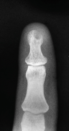
Fig. 50.2
Well-defined round osteolytic lesion in a most common location
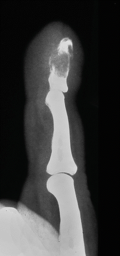
Fig. 50.3
In growing lesions, the cortex is expanded and thinned. Fractures may be observed in the phalanges
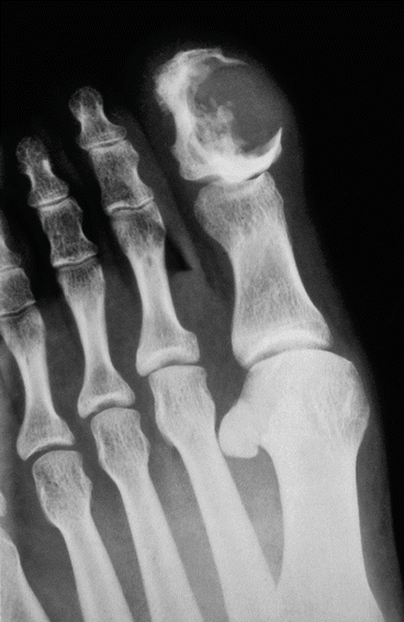
Fig. 50.4
Epidermoid bone cyst in the terminal phalanx of the hallux. The lesion destroyed the cortex
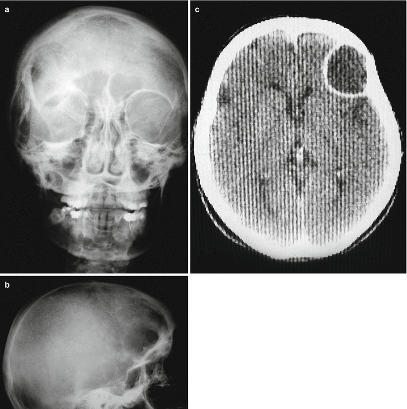
Fig. 50.5
(a, b) Anteroposterior and lateral view of a parietal bone showing a well-defined round osteolytic lesion with absence of trabecular pattern. (c) CT scan shoes a well demarcated lytic lesion with a sclerotic rim. Well-demarcated lytic lesion
Related posts:


Stay updated, free articles. Join our Telegram channel

Full access? Get Clinical Tree


