Fig. 29.1
Medial collateral ligament and ulnar nerve. E medial epicondyle, Ab anterior bundle, Pb posterior bundle, U ulna, Un ulnar nerve
Lateral ligament complex, made up of lateral ulnar collateral, radial collateral, annular ligament, and the accessory collateral ligament described by Martin [17]. This complex is the primary restraint to posterolateral rotatory instability and varus forces. To create functional posterolateral instability are necessary combined injuries. In fact isolated lateral ulnar collateral or radial collateral ligament injuries do not result in instability [18, 19].
The secondary stabilizers are represented by:
Radial head: an important secondary restraint [10] because 60 % of axial loads are imparted through radiocapitellar joint [20]. Radial head injuries compromise lateral and medial stability by decreasing tension in the lateral ligament complex and because it is a secondary restraint to valgus load, respectively.
Capsule: surrounding entirely the joint and gives better contribution in extension.
Muscular support: anterior and posterior muscles that travel across the elbow and enable flexion and extension mobility. The elbow is also the site of origin for the flexor/pronator and extensor/supinator musculature of the forearm located medially and laterally, respectively. Anconeus muscle is anatomically oriented to provide restraint to posterolateral rotatory instability. The extensor/supinator musculature provides dynamic lateral stability; it is often avulsed with lateral ligaments. The flexor/pronator musculature provides dynamic valgus stability. These musculature groups have compression effect on ulnohumeral joint, augmenting bony stability [21].
29.3 Injury Mechanism
The mechanisms of injury are represented by subluxation and dislocation events.
The instabilities can be divided into simple and complex. The simple elbow instability indicates a dislocation with soft tissue lesions without associated fractures that can compromise joint stability [6, 22]. The most frequent form is a posterior dislocation produced by a posterolateral rotation mechanism (PLRI) as described by O’Driscoll [23].
The term complex elbow instability, which replaces the former “fracture–dislocation” and “transolecranon fracture,” means on the other side the association of ligaments and bony lesions.
Simple dislocations without any secondary injuries to the bone occur more often than complex dislocation [6]. The latter account for 15–20 % of all elbow dislocations [24, 25].
Referring to direction and mechanism of dislocation, we can distinguish PLRI and valgus stress (which can be post-traumatic or due to overuse).
So dislocations can be simple (with only soft tissue injuries) and complex (with bone and soft tissue injuries).
Simple dislocations are classified as anterior, posterior (direct posterior, posterolateral, and posteromedial), and divergent (extremely rare in which the humerus is jammed between the radius and ulna and the interosseous membrane is destroyed) [26, 27].
Complex dislocations can be anterior and posterior.
A posterior dislocation is caused by a fall on the wrist while the elbow joint is extended and the wrist is pronated. The impact of the tip of the olecranon on the olecranon fossa has a leverage effect, while the coronoid process slips in a dorsal direction over the trochlea of humerus [23, 28].
A posterior dislocation is also caused by a fall on hand with a flexed elbow joint where the force, acting in a direct axial direction, makes the olecraon slip out [29, 30].
An anterior dislocation occurs through a combination between a flexed elbow and a force acting dorsally.
The most common mechanism is fall on outstretched hand generating axial load through the elbow.
29.3.1 PLRI
In all posterior dislocations, there is a lateral ligament disruption generating the so-called posterolateral rotatory instability (PLRI) [31, 32].
PLRI consist in three stages of instability that correlates with the severity of soft tissue injury:
Stage 1 – injury to lateral ligaments and extensor origin so this causes posterolateral shift of ulnohumeral and radiocapitellar joints.
Stage 2 – injury propagates to the anterior and posterior capsules, so this causes posterolateral subluxation with perching of the coronoid under the trochlea.
Stage 3 – posterolateral dislocation:
3a: anterior bundle of MCL is intact, so there is pivoting around intact ligament.
3b: anterior bundle of MCL is disrupted with complete dislocation (the most common injury pattern).
3c: complete stripping of all soft tissue from the distal humeral. It is grossly unstable unless flexed >90°.
O’Driscoll et al. [23] described a circle strategy for the sequence of injuries to soft tissue regarding to a simple posterior dislocation. The force generated flows from lateral to medial. This leads to rupture of the LCL, then the anterior and posterior capsules, and lastly the MCL.
29.3.2 Valgus Injuries
These injuries can be determined by falling with severe valgus moment or combined with direct contact at lateral elbow, often seen in contact athletes.
Repetitive microtraumas represent a common mechanism for valgus injuries in throwers.
Most common clinical pictures are represented by medial ligament injury, avulsion of flexor/pronator mass, and radial head/neck compression fractures.
29.3.3 Varus Injuries: Posteromedial Dislocation
Frequently due to a fall, a lateral ligament injury is often associated with medial facet coronoid fractures.
29.3.4 Complex Injuries
The mechanism of injury is similar to simple dislocation. Loading pattern and arm position determine associated bony lesions.
The timing of the lesions can be acute (post-traumatic) or chronic (post-traumatic or due to overuse).
29.4 Clinical Evaluation
First of all, it is necessary to have a complete medical history of the patient to locate the seat of the pain and the mode of onset, acute or chronic, and the cause, traumatic or not.
The patient may report a lateral elbow pain; a feeling of popping, snapping, or shifting; or recurrent subluxation or dislocations.
Patient may have apprehension while performing activities requiring forced extension of the elbow. The patient who practices sport launch may experience pain in the medial elbow during the acceleration phase.
Atraumatic onset is uncommon with PLRI.
Throwers may have changed their training; they can report loss of velocity and control.
The examiner should assess any predisposing factors such as surgical procedures performed in the lateral region as aggressive tennis elbow release, radial head resection, multiple lateral elbow injection (due to the possible weakening of the ligaments) and prior injury as cubitus varus for pediatric supracondylar humerus fracture malunion.
Anatomical deformity of the profile can hide bone injury or dislocation of the joint.
Before any clinical maneuver, it is important to rule out bony, nervous, and vascular disorders [24].
Clinical examination must include an accurate assessment of the ipsilateral shoulder, elbow, and wrist to exclude the presence of previous injuries or pathologies.
An evaluation of the ipsilateral distal radioulnar joint and the interosseous membrane for the presence of an Essex–Lopresti injury would also appear to be important [33, 34].
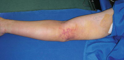
An injured elbow may be swollen due to the presence of periarticular edema or hematoma (Fig. 29.2). It is important to exclude the presence of a compartmental syndrome which rarely can develop from the beginning of the trauma. Therefore, clinical monitoring during the early hours is necessary [23].


Fig. 29.2
MCL acute tear and elbow bruise
Excluding bone and neurovascular lesions, stability tests must be performed.
Sometimes these maneuvers are performed after sedation (because of the pain) and under radiological control for better assessment.
Some patients arrive in the emergency department with joints which have already been spontaneously reduced.
In such cases, the diagnosis is derived from the case history and any possible instability, which may be present.
Several clinical maneuvers have been described to highlight different types of elbow instability.
29.4.1 Valgus Instability
Patients with this kind of instability usually report medial elbow pain and decreased strength during overhead activity; there may be symptoms of ulnar neuropathy from either acute or chronic UCL injury caused by edema/hemorrhage of the medial elbow or excessive traction on the nerve.
Patients with isolated UCL injury often have point tenderness 2 cm distal to the medial epicondyle, slightly posterior to the common flexor origin. The UCL stability can be assessed with specific physical exam tests. The “milking maneuver” involves having the patient apply a valgus torque to the elbow by pulling down on the thumb of the injured extremity with the contralateral limb providing stability [35]. With the modified milking maneuver, the examiner provides stability to the patient’s elbow and pulls the thumb to create a valgus stress on the UCL [36]. These tests result in pain and widening at the medial joint line if the UCL is insufficient. O’Driscoll and coworkers described the moving valgus stress test, in which the valgus torque is maintained constantly to the fully flexed elbow and then quickly extends the elbow [37]. This test is positive if medial elbow pain is elicited and has a 100 % sensitivity and 75 % specificity. The abduction valgus stress test is performed by stabilizing the patient’s abducted and externally rotated arm with the examiner’s axilla and applying a valgus force to the elbow at 30° of flexion. Testing with the forearm in neutral rotation has been shown to elicit the greatest valgus instability [38]. A positive test results in medial elbow pain and widening along the medial joint line. Even so, valgus laxity can be subtle on physical exam, and the range of preoperative detection is between 26 and 82 % of patients [39, 40]. Furthermore, Timmerman and colleagues found valgus stress testing to be only 66 % sensitive and 60 % specific for detecting abnormality of the anterior bundle of the UCL [41].
29.4.2 PLRI
PLRI is first described in 1991 by O’Driscoll and colleagues in a series of five patients [32].
The clinical mechanism of injury to the lateral stabilizers of the elbow that results in PLRI has been hypothesized to consist of supination of the forearm, combined with a valgus and axial load to the elbow [23]. The presentation is variable and can include lateral elbow pain; mechanical symptoms such as snapping, clicking, catching, or locking; and recurrent episodes of instability. Patients often report their elbow feels loose or like it is sliding out of place, especially when loading it in a slightly flexed position with a supinated forearm, as when pushing off an armrest while standing from a chair.
On physical exam, patients often have normal upper extremity strength and elbow range of motion and minimal to no tenderness around the LCL complex. Several provocative maneuvers have been developed to elicit instability symptoms. The posterolateral rotatory instability test is performed by supinating the forearm and applying valgus and axial forces to the elbow while flexing the elbow from full extension [32]. A positive test is demonstrated by reduction of a subluxated radial head when the patient is under general anesthesia or apprehension during testing when the patient is awake [32]. More recently, Regan and Lapner described two other apprehension tests, the chair sign and push-up sign [42]. The chair sign is performed by having the patient actively push off the armrests of a chair with the forearms supinated and the elbows at 90°. The test is considered positive with reluctance to fully extend the elbow during push off. The push-up sign is conducted by having the patient push off from the ground with the forearms supinated, elbows at 90°, and arms abducted to greater than shoulder width. A positive test results in apprehension and guarding as the elbow is terminally extended. These apprehension tests have been determined to be more sensitive than the posterolateral rotator instability test in awake patients. The table-top relocation test has been recently described by Arvind and Hargreaves [43]. The patient is asked to stand in front of a table. The hand of the symptomatic arm is placed over the lateral edge of the table. The test involves three parts. The patient is initially asked to perform a press-up with the elbow pointing laterally. This maintains the forearm in supination. Pressure is pushed down through the hand onto the table, as the elbow is allowed to flex (bringing the chest toward the table). In the presence of posterolateral rotatory instability (PLRI), positive apprehension and a reproduction of the patient’s pain occur as the elbow reaches approximately 40° of flexion. The maneuver is then repeated, using the thumb of the examiner placed over the radial head, giving support and preventing posterior subluxation while the press-up is performed. Patients with posterolateral rotatory instability find that their symptoms of pain and instability are relieved by this second maneuver, which is similar to the relocation test of the shoulder. Finally, removal of the examiner’s supporting thumb from the weight-bearing, partially flexed elbow reproduces the pain and apprehension. The relief and recurrence of pain during the second and third maneuvers helps to exclude articular pathology as the cause of pain and reinforces the diagnosis of instability.
29.5 Diagnostic Imaging
Conventional plain anteroposterior and lateral radiographs should be taken before any clinical maneuver. Oblique views may be necessary for a better assessment of the coronoid process and the radial head [6, 33]. Depending on the symptoms and pain experienced, radiographs should also be made of the adjacent joints. Similar to medial elbow instability, plain radiographs of the elbow are used to identify an avulsion fragments or associated fractures (e.g., coronoid, radial head) that can contribute to instability. Associated arthritic changes or loose bodies may also be seen. Widening of the ulnohumeral joint space after reduction of an acute dislocation, the so-called drop sign, has been associated with significant ligamentous injury and increased risk of recurrent instability [44].
Stress radiographs can be taken at the point of maximum rotatory subluxation during the pivot-shift test and may show widening of the ulnohumeral joint space on the lateral and anteroposterior views and posterior subluxation of the radial head on the lateral view.
MRI/arthroMRI is useful in case of chronic instabilities evidencing chondral associated lesions and possibly showing a leakage of contrast fluid in case of lateral or medial collateral lesions [45].
Stay updated, free articles. Join our Telegram channel

Full access? Get Clinical Tree


