Fig. 29.1
Important neurovascular structures within the antecubital fossa
This chapter will provide a summary of the key anatomy, portal placement, and basic surgical setup and technique for elbow arthroscopy.
Anatomy
A comprehensive understanding of the anatomy of the elbow is essential before proceeding with elbow arthroscopy. Important bony anatomic landmarks include: the medial and lateral epicondyles, the olecranon process, and the radial head. Anatomic landmarks should be palpated and marked prior to portal placement.
The soft spot, also known as the anconeus triangle, is located in the center of the triangle formed from the lateral epicondyle, radial head, and olecranon process. The soft spot can be used to insufflate the joint prior to portal placements, and it can also be used as a direct lateral portal (Fig. 29.2).
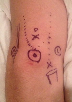
Fig. 29.2
Surface landmarks of elbow. Posterior view. Medial epicondyle, ulnar nerve, olecranon process are outlined in relation to elbow joint. X marks soft spot portal. P marks posterocentral portal
Several sensory nerves surround the elbow, including: the medial antebrachial cutaneous, the medial brachial cutaneous, the lateral antebrachial cutaneous, and the posterior antebrachial cutaneous nerves [4]. The medial antebrachial cutaneous nerve provides sensation to the medial aspect of the forearm and elbow. The medial brachial cutaneous nerve supplies sensation to the posteromedial aspect of the arm, to the level of the olecranon. The lateral antebrachial cutaneous nerve, supplies sensation to the elbow and lateral aspect of the forearm. It is a branch of the musculocutaneous nerve, and exits between the brachialis muscle and the biceps. The posterior antebrachial cutaneous nerve supplies sensation to the posterolateral elbow and posterior forearm. It is a branch of the radial nerve and courses down the lateral aspect of the arm [5].
The median, radial, and ulnar nerve and brachial artery are the main neurovascular structures around the elbow [4].
Indications and Contraindications
The indications for elbow arthroscopy are numerous and include both diagnostic and therapeutic indications. Diagnostic indications include septic arthritis, traumatic and degenerative arthritis, and intra-articular fractures [6, 7]. Therapeutic indications include the removal of loose bodies, synovectomy, capsular release, plica excision, treatment of osteochondritis dessicans, and tennis elbow release. New evolving indications include olecranon bursectomy and arthroscopic assisted fracture management [8, 9].
Contraindications for elbow arthroscopy include any conditions that distort the normal soft-tissue or normal bony anatomy, which prevents making accurate portal placement [10]. In patients with prior ulnar nerve transposition, or a subluxing ulnar nerve, the nerve should be identified prior to portal placement to prevent iatrogenic injury. Extensive heterotrophic ossicification, prior skin grafts or flaps and burns preclude safe joint access and should be avoided [11].
Surgical Technique
Anesthesia
General or regional anesthesia may be used for elbow arthroscopy. Most surgeons prefer general anesthesia for elbow arthroscopy because of patient comfort and complete muscle relaxation. The use of regional anesthesia can complicate the postoperative neurologic assessment and can be compromised by the use of supraclavicular and extended axillary blocks [5].
Instrumentation
A standard 4.0-mm, 30° arthroscope provides exceptional visualization of the elbow joint. Sometimes a smaller 2.7 mm arthroscope may be useful for visualization in adolescent patients, and in smaller viewing spaces, such as the direct lateral portal and in the posterior compartment [5, 8]. Cannulas are utilized to allow ease in switching working and viewing portals, without repeated joint capsule trauma. In addition, the risk of neurovascular injury is minimized when fewer portals are established. It is vital to maintain the arthroscopy portals with cannulas in order to decrease fluid extravasation into the soft tissues and swelling [5]. Maintaining capsular distention is key to successful elbow arthroscopy. If there is fluid extravasation into the soft tissues, the capsule will collapse, and prevent further elbow arthroscopy.
In elbow arthroscopy, side-vented inflow cannulas should be avoided to prevent fluid extravasation into the soft tissues [12]. Only blunt-tipped and conical trocars should be used, to decrease the possibility of articular cartilage or neurovascular injury [8]. Specialized arthroscopic instruments: forceps, probes, shavers, and burrs are utilized in elbow arthroscopy [5].
Gravity inflow or a mechanical pump can be used in elbow arthroscopy. Some surgeons think gravity provides for enough joint distention while minimizing fluid extravasation. A mechanical pump can safely be used, but the inflow pressure should be minimized, no greater than 35 mmHg, in order to lessen fluid extravasation [5].
Patient Positioning
Supine Position
Andrews first described supine positioning for elbow arthroscopy in 1985 [2]. After adequate anesthesia has been obtained, care is taken to pad all bony prominences and place the shoulder at the edge of the operating table. The patient is positioned with the shoulder in 90° of abduction, and 90° of elbow flexion. The arm is then secured using an overhead traction device and a non-sterile tourniquet is applied.
Supine positioning offers numerous advantages [13]. The supine setup is simple and allows for easy airway access for anesthesia. The anatomy is clearly defined and oriented in this familiar anatomic position. Furthermore, if the procedure needs to be converted to open, it’s a simple task.
The weakness of the supine position includes the difficulty working in the posterior compartment and the need for an additional traction device.
Prone Position
Poehling first described the prone position for elbow arthroscopy in 1989 [3]. After adequate anesthesia has been obtained, the patient is rolled onto chest rolls. The nonoperative extremity is placed onto a well padded arm board, with the shoulder in 90° of abduction and the elbow in 90° of flexion (Figs. 29.3 and 29.4). The operative extremity should be supported appropriately to allow the shoulder to be abducted 90° and the elbow hanging freely at 90° of flexion. This can be achieved by either a padded bolster, or several rolled towels positioned on an arm board.
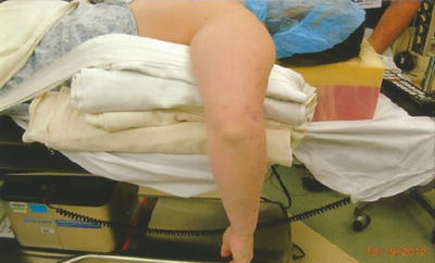
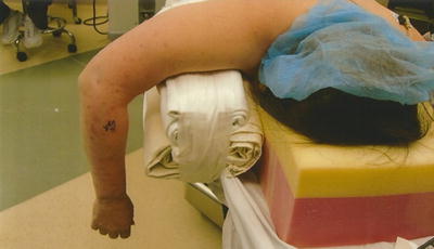
Fig. 29.3
Prone positioning with shoulder flexed and abducted 90° over a padded arm table with folded blankets
Fig. 29.4
Prone positioning for elbow arthroscopy
There are several advantages to the prone position. The posterior compartment is easily accessed without the need for traction. In addition, the arm can be effortlessly manipulated from full extension to full flexion. “Flexion of the elbow allows the neurovascular structures to sag anteriorly, providing a greater margin of error, when establishing anterior portal sites” [14].
The main disadvantage of the prone position is the general anesthesia requirement and poor airway access by anesthesia. Furthermore, conversion to an open procedure is much more difficult as compared to the supine position. Therefore, if anterior open procedures are necessary, this will require repositioning to a supine procedure.
Lateral Decubitus Position
O’Driscoll and Morrey first described the lateral decubitus position in 1993 [15].
After the patients are placed under general anesthesia on a bean bag, the patient is turned and secured in a lateral decubitus position. An axillary roll and non-sterile tourniquet is applied. The shoulder is flexed and internally rotated 90°. The arm is positioned on a padded arm holder, positioning the elbow in 90° of flexion (Fig. 29.5).
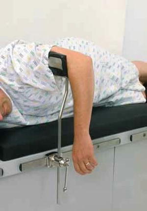
Fig. 29.5
Picture of arm holder for lateral position
The lateral decubitus position allows for the benefits of prone positioning, without the airway difficulty of the prone position. The disadvantage of the lateral decubitus position is the need for a padded arm holder and difficulty with converting to an open procedure.
Portals
Numerous portals have been described for elbow arthroscopy. The most common portals utilized are the anterolateral, proximal lateral, midlateral, anteromedial, proximal medial, and straight posterior [11].
Related posts:
 Arthroscopic Management of Distal Radius Fractures
Management of Ulnar Impaction
Wrist Arthritis: Arthroscopic Techniques of Synovectomy, Abrasion Chondroplasty, Radial Styloidectomy, and Proximal Row Carpectomy of the Wrist
Suture-Button Suspensionplasty for the Treatment of Thumb Carpometacarpal Joint Arthritis
Arthroscopic and Open Radial Ulnohumeral Ligament Reconstruction for Posterolateral Rotatory Instability of the Elbow
Arthroscopic Arthrolysis
Arthroscopic Management of Distal Radius Fractures
Management of Ulnar Impaction
Wrist Arthritis: Arthroscopic Techniques of Synovectomy, Abrasion Chondroplasty, Radial Styloidectomy, and Proximal Row Carpectomy of the Wrist
Suture-Button Suspensionplasty for the Treatment of Thumb Carpometacarpal Joint Arthritis
Arthroscopic and Open Radial Ulnohumeral Ligament Reconstruction for Posterolateral Rotatory Instability of the Elbow
Arthroscopic Arthrolysis
Stay updated, free articles. Join our Telegram channel

Full access? Get Clinical Tree


