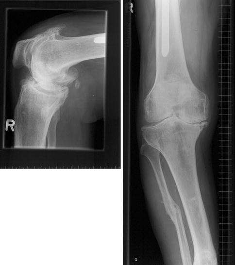
Fig. 1
(a) Long-leg radiographs of the right knee with a non-locked femoral nail in situ. (b) Anteroposterior and lateral lower-leg radiographs showing healed tibial and fibular fractures after a motorcycle accident 50 years ago

Fig. 2
Anteroposterior and lateral radiographs of right knee presenting a severe posttraumatic varus type OA
Questions
1.
What possible problems do you expect in this case when considering a TKR?
2.
How would you address the malalignment in this case?
It was decided to remove the femoral nail and perform a combined supracondylar valgisation osteotomy to correct the considerable extra-articular deformity (closed wedge of 8°) and a TKR using a custom-made femoral component (balanSys®, Mathys Medical, Bettlach, Switzerland). The osteotomy was stabilized with a long uncemented press-fit intramedullary rod connected to the femoral component (Fig. 3).


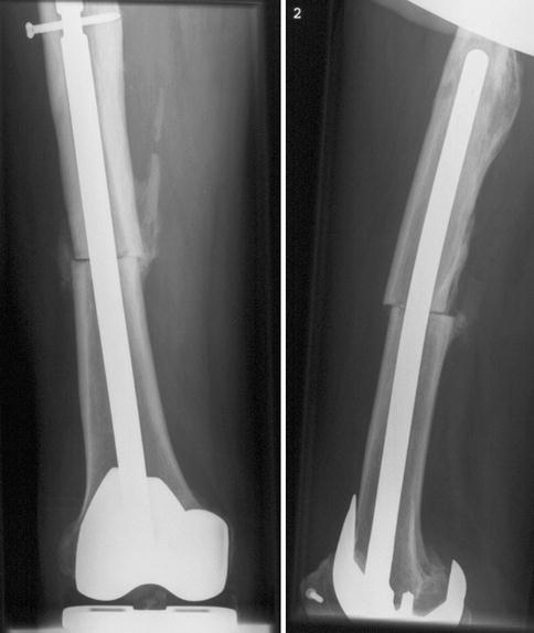
Fig. 3
Anteroposterior and lateral radiographs showing the dynamically locked intramedullary rod, which was necessary due to delayed union of the osteotomy site
Questions
3.
Would you have locked the intramedullary rod?
4.
Do you expect healing of the osteotomy site?
5.
Would plating be an option in such a case?
Due to a delayed union of the osteotomy site at 4 months after surgery, the femoral rod was dynamically locked at this time (Fig. 3). At TKR surgery, it was decided not to lock the rod as it presented with good press-fit stability.
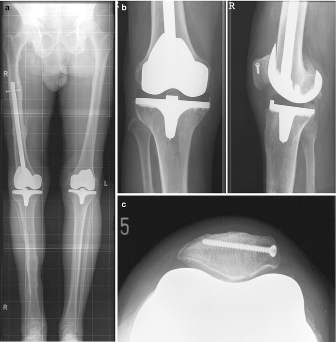
The further initial follow-up was uneventful. The patient recovered with an excellent functional result. He was able to walk with his dogs through the forest and even returned to alpine skiing (Fig. 4).


Fig. 4
Standard radiographs 1 year after surgery showing completely healed femoral and patellar osteotomy sites. (a) Long leg radiographs, (b) AP and lateral and sunrise (c) view
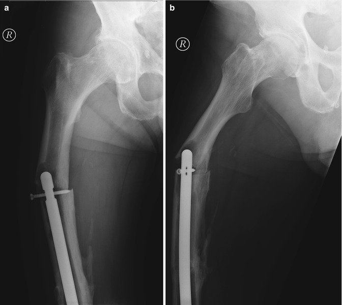
Eight years after index surgery, the patient slipped while walking his dog and fell on his right side. A periprosthetic femoral fracture at the proximal tip of the intramedullary rod was diagnosed (Fig. 5) [1–7].


