Fig. 1
AP (a) and lateral (b) view of the right knee. Femoral and tibial TKR components are uncemented. The lateral view (b) showed a patella baja and aseptic loosening of the patellar component
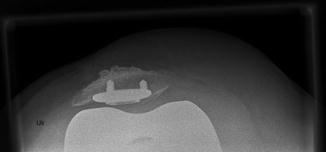
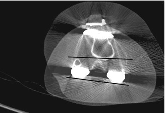
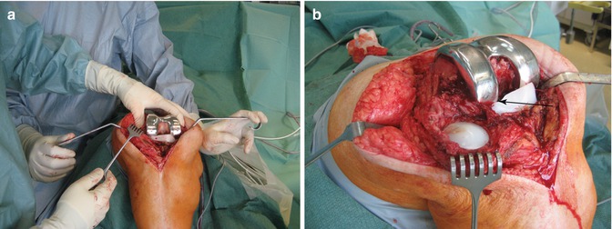
The metal-back patella button showed maltracking and was riding over the lateral condyle (Fig. 2). No radiolucent lines were seen the tibial and femoral component but around the pegs of the patella component. The CT scan showed significant internal rotation of the femoral component (Fig. 3). Second revision surgery was considered. The thumb and index finger in Fig. 4 mark the medial and lateral epicondyle during surgery. The femoral component was significantly internally rotated (Fig. 4). Furthermore, a lateral liftoff of the femoral condyle in flexion was noted. Increased stress in the patellofemoral joint may have caused early aseptic loosening of the patella component.


Fig. 2
The Merchant’s view shows lateral maltracking of the patella. This might be due to incorrect placement of the patella component or femoral malrotation

Fig. 3
The CT scan shows the internal rotation of the femoral component in regard to the transepicondylar line

Fig. 4
Intraoperative view. The transepicondylar line is marked with the thumb and index finger and shows the internal rotation of the femoral TKR component (a). Note an evident lateral femoral liftoff (b)
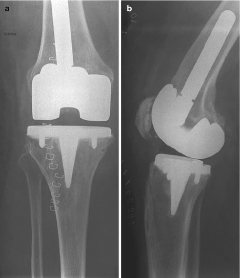
The femoral component was revised using an intramedullary stem (Fig. 5a, b). The patella component was only removed due to limited available bone stock. Clinical follow-up after 4 years showed a sufficient range of motion (0–120°) without pain during climbing up and down stairs.


