Fig. 1
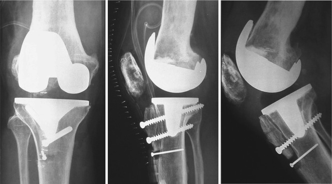
(a) One-year follow-up after fenestration (arrow) of the distal femur and medullary reaming of the whole femur. Patella baja resulting from the accident in the childhood and a later fracture. (b) Serious osteoarthritis 10 years later, prior to TKR
Growing up the patient became a farmer in a Swiss mountain area. Up to the age of 46 years, he frequently suffered from a sinus tract infection with intermittent painful pus retentions. At first presentation to our clinic, a central sequestra in the distal femur was found and eliminated by a lateral fenestration of the distal femur. This procedure was combined with central medullary reaming of the whole femur [2, 3]. No communication with the knee joint was noted at this time. In the biopsies S. aureus was identified and subsequently treated with 2 weeks of intravenous flucloxacilline (4 × 2 g) followed by 6 months of oral ciprofloxacine (2 × 750 mg) and rifampicin (2 × 450 mg) [4]. The follow-up was uneventful with no signs of recurrence for the following 10 years (Fig. 1).
Then the patient experienced progressive weight-bearing right knee pain. Due to his limitations in daily activity, he presented to a local orthopaedic surgeon. The orthopaedic surgeon recommended a TKR.
Questions
1.
Would you also recommend a TKR for this patient? Any alternative?
2.
If so, what would be your diagnostic approach before performing the TKR?
However, as the orthopaedic surgeon feared a recurrence of the previous infection, he primarily performed a diagnostic arthroscopy and took three biopsies. These showed no bacterial growth.
A CT and an anti-granulocyte bone scan did not reveal any signs of infection. There was extensive scar tissue in the anterior knee compartment. Finally a non-hinged TKR was performed using a lateral approach including an osteotomy of the tibial tubercle (Fig. 2).


Fig. 2
Postoperative anterior-posterior and lateral radiographs 4 months after proximal screw removal showed no healing of the tibial tubercle fragment, which was initially proximalised in order to reduce the patella baja. There were no clear radiolucent lines along the cement bone interface
Questions
3.
Would you have also used this approach? If not, which one would you have used?
4.
Would you have also used a posterior stabilised TKR?
5.
Would you use intra- or extramedullary referencing for TKR orientation
6.
Would you use a specifically extended antibiotic prophylaxis in these cases at risk?
Approximately 2 weeks later, the patient presented with a persistent pus secretion and wound dehiscence over the patellar tendon, which was right in the centre of the pre-existing anterior scar area.
Questions
7.
Would you do the debridement with or without exchange of the PE inlay?
Open debridement with an exchange of the polyethylene inlay was performed. Staphylococcus aureus was found in 8 of 8 samples, coagulase-negative staphylococcus (CNS) in 2 of 8 samples. The latter were resistant against flucloxacilline, but both were sensible for rifampicin. Due to earlier allergic reactions to penicillin treatment, the patient was started with daptomycin.
After a second (arthroscopic) revision, vacuum treatment was initiated. The treatment was continued for a total of 4 months, but the sinus persisted. The antibiotic treatment was continued with oral levofloxacin (2 × 500 mg) and rifampicin (2 × 450 mg). Neither healed the sinus (Fig. 3) nor did the C-reactive protein (CRP) decrease below 15 mg/l.


Fig. 3
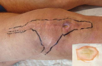
Five months after TKR. Extended scar area with sinus in its centre
Questions
8.
Would you use the vacuum pump treatment in this case?
9.
If so, how long would you continue it?
At this stage the patient was again referred to our clinic. The knee was swollen, warm, and red.
The radiographs did not show any osseous integration of the tubercle fragment (Fig. 2). An anti-granulocyte bone scan revealed increased tracer activity around the prosthesis, which indicated a periprosthetic joint infection. The open fistula penetrated the patellar ligament partially destroying it (Fig. 3).
Questions
10.
What would you propose now as the treatment plan?
The following plan for treatment was proposed to the patient:
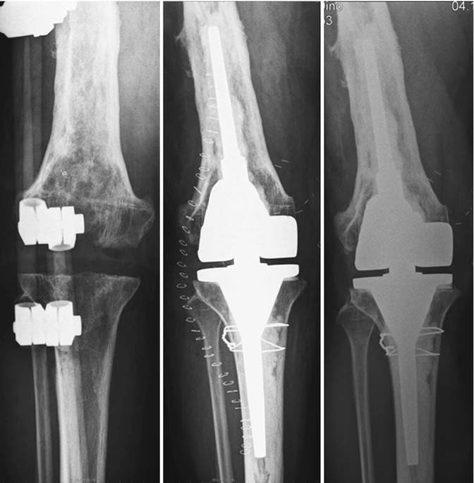
Removal of the TKR, bridging of the knee with external fixation, curative intravenous antibiotic treatment for 6 weeks; 1 week after removal of the TKR, replacement of the scar area by a free flap; reimplantation 8 weeks after removal of the TKR and an antibiotic-free interval of 2 weeks (Fig. 4) [5].


Fig. 4
Anterior-posterior radiographs with bridging external fixation and after reimplantation of a hinged TKR (postoperative and 1 year control). No radiolucent lines
Eleven tissue samples – equal portions for microbiologic and histologic examination – were harvested. The implants were sent for sonication [6]. Antibiotic treatment was started empirically, first with amoxicillin and clavulanic acid but then had to be changed to vancomycin (2 × 1 g) and levofloxacin (2 × 500 mg). Eight days later, the plastic surgeon performed a free lateral upper arm flap including an anastomosis of a skin nerve. Part of the aponeurosis of the M. triceps brachii was taken with the graft and integrated in the deficient patellar tendon.
The microbiologic analysis of the samples revealed a complex situation of six different bacteria, all three strains of staphylococci being resistant against rifampicin. Doxycycline was then added to the antibiotic therapy. After final microbiological results, the antibiotic treatment was changed to vancomycin 2 × 1 g i.v. and oral doxycycline (Table 1).
Table 1


Bacteriology at the moment of removal of TKR
Stay updated, free articles. Join our Telegram channel

Full access? Get Clinical Tree


