Fig. 1
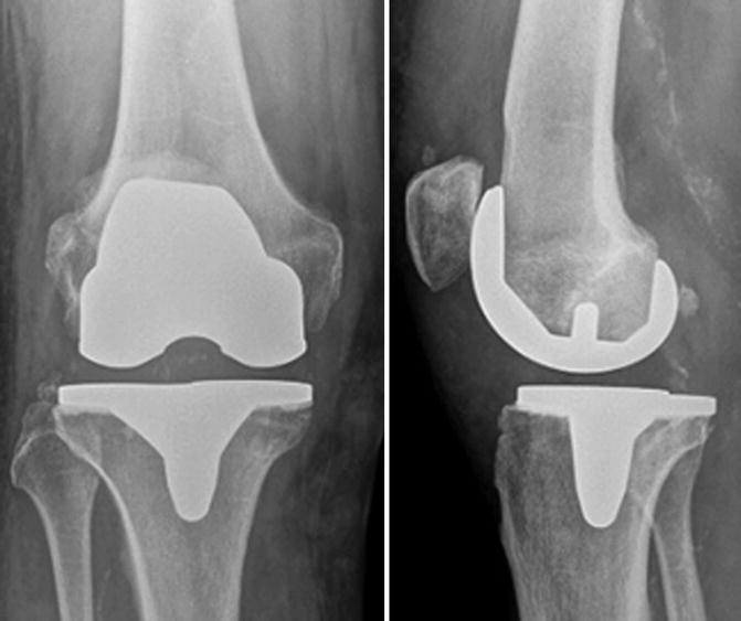
Anterior–posterior and lateral weight-bearing radiographs of the right knee indicating osteoarthritis Kellgren–Lawrence grade 3 in several knee compartments
A cruciate-retaining total knee replacement (PFC Sigma RP, DePuy, Warsaw, Illinois, USA) was implanted, and the perioperative and early postoperative follow-up was uneventful. The femoral component was uncemented and the tibial cemented.
At 6 weeks follow-up, the patient complained about a limited flexion of 80°. However, being pain-free, he was overall satisfied. The routine anterior–posterior and lateral weight-bearing radiographs are presented (Fig. 2).


Fig. 2
Anterior–posterior and lateral weight-bearing radiographs after TKR
Questions
1.
Would you prefer primary patellofemoral resurfacing?
2.
How would you comment on these postoperative radiographs?
3.
Do you think the implants are correctly placed?
4.
Is there anything remarkable on the radiographs?
Despite an intensive physiotherapeutic training, the flexion deficit persisted. No signs of infection were present.
Questions
1.
What would you recommend to this patient?
2.
Would you do a manipulation under anaesthesia and arthroscopic or open arthrolysis?
An arthroscopic arthrolysis was performed, and intraoperatively a flexion of 115° was documented.
Although intensive CPM training as well as physiotherapy was initiated 6 weeks later, the flexion again decreased to 90°.
Four months later the flexion was still limited to 90°, and the patient was unhappy as he could not cycle and walk pain-free. The orthopaedic surgeon decided to do a second arthroscopic arthrolysis. Here extensive scar tissue in the whole knee, but in particular in the anterior knee and in the superior recess, was found and debrided. The patient was discharged without complications and with a postoperative flexion of 115°. At 6 weeks and 6 months follow-up, the flexion already decreased to 105°, but the patient was pain-free and satisfied.
However, 3 months later the flexion had again decreased to 90°, and the patient complained about end-range anterior knee pain. His knee felt stiff and tight.
Questions
1.
What would you do now (diagnostics, treatment)?
2.
Would you do a manipulation under anaesthesisa arthroscopic or open arthrolysis?
3.
Anything else?
For further diagnostic a 99mTc-HDP-SPECT/CT was performed. Two major findings were observed:
Questions
1.
What are the two major findings?
2.
What is your treatment recommendation now?
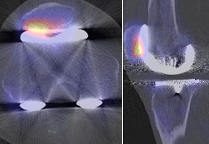
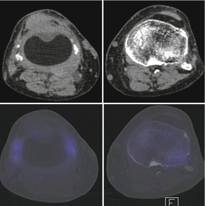
The SPECT/CT (Figs. 3 and 4) revealed:


Fig. 3
99mTc-HDP-SPECT/CT of the patient’s right knee (coronal and sagittal views)


