Fig. 1
Radiographs (anteroposterior, lateral) of the left knee showing a cruciate-retaining fixed-bearing TKR (Triathlon, Stryker, Switzerland). The patella was unresurfaced. The tibial TKR was slightly oversized in the mediolateral direction. In the anterior-posterior direction, there was no overhang. There were no signs of loosening
Questions
3.
What is your differential diagnosis now?
4.
What are your next steps in diagnostics?
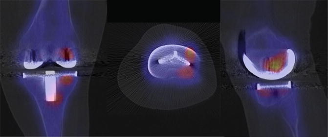
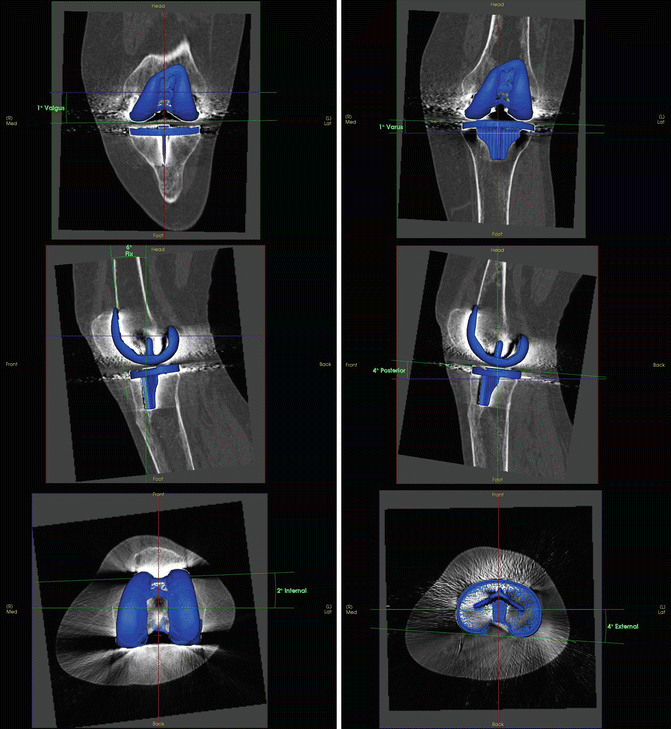
SPECT/CT was performed to accurately determine TKR component position and to assess for loosening or infection (Figs. 2 and 3). It showed no loosening or infection. Using a customised software (Orthoexpert v1.15©), 3D-CT revealed an overall correct TKR position (2° internally rotated, 6° flexed and 1° valgus femoral and 4° external rotated, 4° posterior and 1° varus positioned tibial TKR components) but an oversized laterally overhanging tibial TKR component.


Fig. 2
99mTc-HDP-SPECT/CT images of the left knee showing only minor tracer uptake around the femoral and tibial TKR component, indicating a well-fixed TKR

Fig. 3
Using a customised software (Orthoexpert v1.15©), 3D-CT revealed an overall correct TKR position but an oversized laterally overhanging tibial TKR component
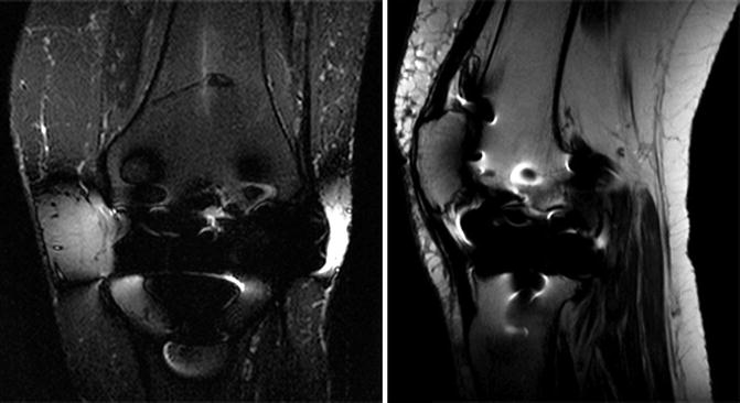
MRI showed no signs of loosening or bone oedema (Fig. 4). However, due to metal artefacts, the lateral overhanging tibial TKR component could not be evaluated in terms of soft tissue irritation.


Fig. 4
MRI of the left knee. No signs of loosening or bone oedema. Due to metal artefacts, a possible impingement of the lateral ligament structures could not be excluded
Questions
4.
What is your diagnosis and proposed treatment?
5.
How would you address the tibial component malposition?
6.
What else would you do?
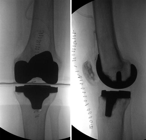
Revision surgery of the tibial TKR component with secondary resurfacing of the patella was performed (Fig. 5).


Fig. 5


Intraoperative fluoroscopy (anteroposterior, lateral) of the left knee after tibial component TKR revision surgery and secondary resurfacing of the patella
Stay updated, free articles. Join our Telegram channel

Full access? Get Clinical Tree


